

A Huge Conjunctival Atypical Fibroxanthoma
- PMID: 39525205
- PMCID: PMC11544678
- DOI: 10.7759/cureus.71130
A Huge Conjunctival Atypical Fibroxanthoma
Abstract
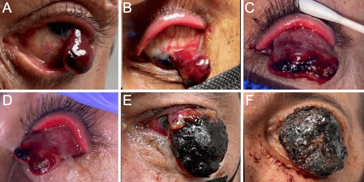
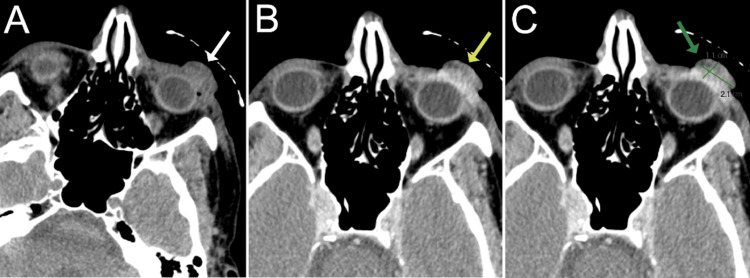
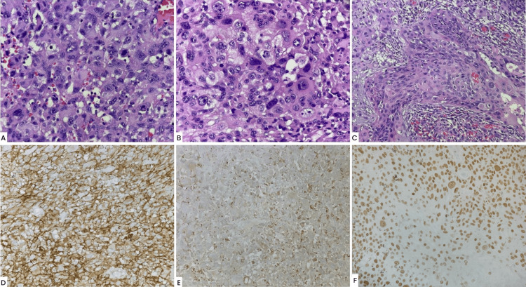
We report the case of a Hispanic male whose conjunctival atypical fibroxanthoma (AFX) grew very large, with intraocular as well as extraocular muscle extensions, and was treated with exenteration. A 50-year-old male presented with a one-month history of foreign-body sensation in his left eye. The initial examination revealed an erythematous, vascularized, pedunculated lesion arising from the left eye conjunctiva. A tissue biopsy from the lesion confirmed the diagnosis of an AFX with a small component of squamous cell carcinoma. The patient was intermittently lost to follow-up until, one-and-a-half years after his initial visit, he underwent an exenteration of the left eye as the tumor had intraocular and extraocular muscle extensions. Subsequently, he was scheduled for ongoing monitoring.
Keywords: atypical fibroxanthoma; conjunctiva; cornea; exenteration; tumor.
Copyright © 2024, Ramirez Marquez et al.
Conflict of interest statement
Human subjects: Consent was obtained or waived by all participants in this study. Conflicts of interest: In compliance with the ICMJE uniform disclosure form, all authors declare the following: Payment/services info: All authors have declared that no financial support was received from any organization for the submitted work. Financial relationships: All authors have declared that they have no financial relationships at present or within the previous three years with any organizations that might have an interest in the submitted work. Other relationships: All authors have declared that there are no other relationships or activities that could appear to have influenced the submitted work.
Figures
References
-
- Combined intraepithelial squamous neoplasia and atypical fibroxanthoma of the cornea and limbus. Engelbrecht NE, Ford JG, White WL, Yeatts RP. Am J Ophthalmol. 2000;129:94–96. - PubMed
-
- Case of atypical fibroxanthoma in the palpebral conjunctiva. Mochizuki K, Yamada T, Mori Y, Sawada A, Mori I, Ohnishi Y. Jpn J Ophthalmol. 2008;52:404–406. - PubMed
-
- Atypical fibroxanthoma of the bulbar conjunctiva. Shieh C, Daluvoy MB, Ellington KS, Proia AD. Cornea. 2015;34:1620–1622. - PubMed
Publication types
LinkOut - more resources
Full Text Sources