. 2024 Sep 4;34(36):2401952.
doi: 10.1002/adfm.202401952. Epub 2024 Apr 18.

Photo-responsive decellularized small intestine submucosa hydrogels

Affiliations

Photo-responsive decellularized small intestine submucosa hydrogels

Van Thuy Duong et al. Adv Funct Mater. .

Abstract

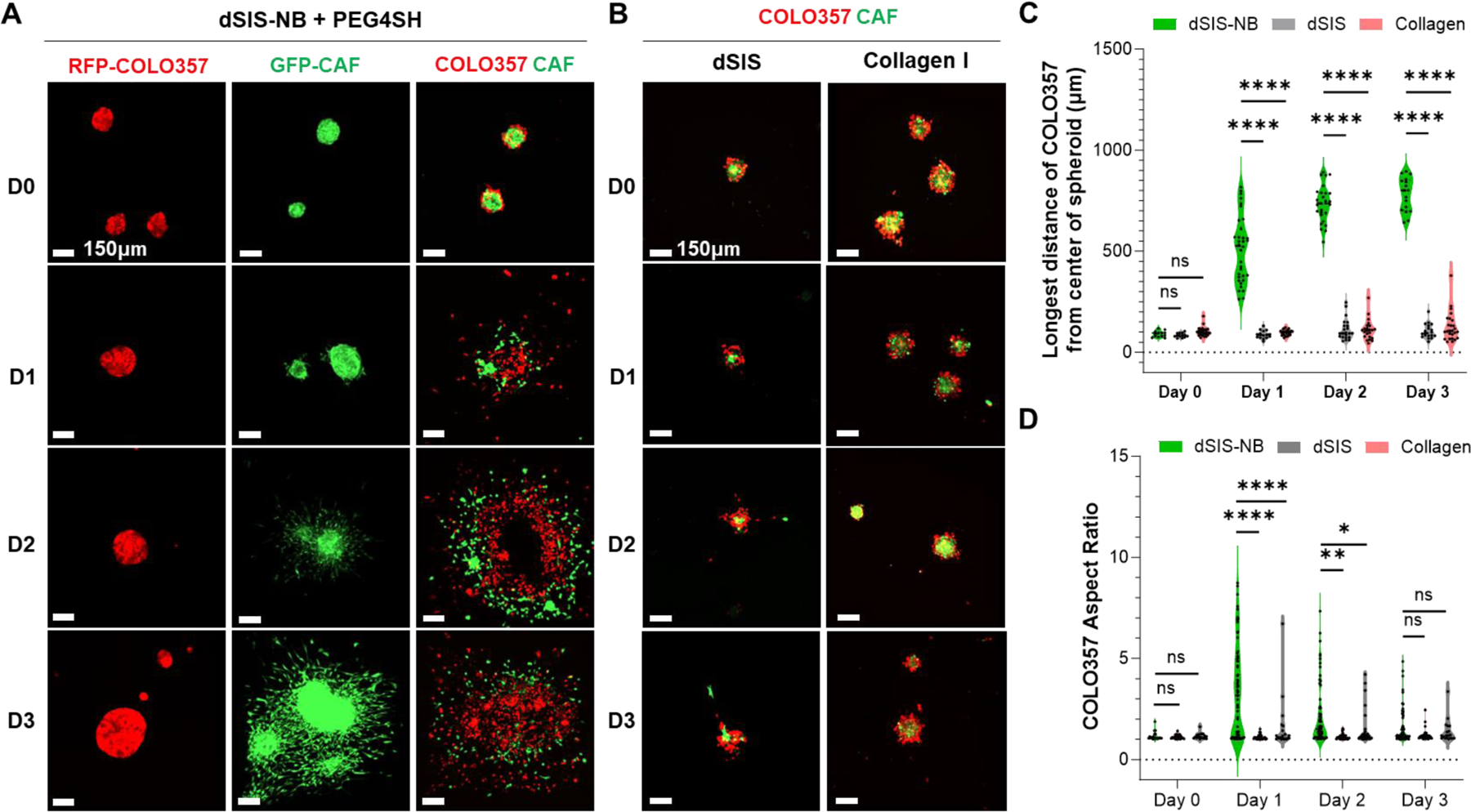
Decellularized small intestine submucosa (dSIS) is a promising biomaterial for promoting tissue regeneration. Isolated from the submucosal layer of animal jejunum, SIS is rich in extracellular matrix (ECM) proteins, including collagen, laminin, and fibronectin. Following mild decellularization, dSIS becomes an acellular matrix that supports cell adhesion, proliferation, and differentiation. Conventional dSIS matrix is usually obtained by thermal crosslinking, which yields a soft scaffold with low stability. To address these challenges, dSIS has been modified with methacrylate groups for photocrosslinking into stable hydrogels. However, dSIS has not been modified with clickable handles for orthogonal crosslinking. Here, we report the development of norbornene-modified dSIS, named dSIS-NB, via reacting amine groups of dSIS with carbic anhydride in acidic aqueous reaction conditions. Using triethylamine (TEA) as a mild base catalyst, we obtained high degrees of NB substitution on dSIS. In addition to describing the synthesis of dSIS-NB, we explored its adaptability in orthogonal hydrogel crosslinking and used dSIS-NB hydrogels for cancer and vascular tissue engineering. Impressively, compared with physically crosslinked dSIS and collagen matrices, orthogonally crosslinked dSIS-NB hydrogels supported rapid dissemination of cancer cells and superior vasculogenic and angiogenic properties. dSIS-NB was also exploited as a versatile bioink for 3D bioprinting applications.

Keywords: 3D bioprinting; Decellularized extracellular matrix; cancer engineering; functional vascularization; small intestine submucosa; thiol-norbornene.


Conflict of interest statement

Conflict of interest statement The authors have no conflicts of interest to declare.

Figures

Figure 1.
Figure 1.. Synthesis and characterization of dSIS-NB and photo-crosslinking of thiol-norbornene dSIS-NB hydrogels.
(A) Representative images of dSIS and dSIS-NB: i) fresh bovine SIS after removal of mesenteric tissue, mucosal epithelium, and lamina propria, ii) smooth white SIS strips after washing with SDS and Triton-X, iii) lyophilized dSIS powder, iv) dSIS dissolved in an acidic pepsin solution, v) freeze-dried dSIS-NB. (B) 1H NMR spectra of dSIS and dSIS-NB. Peak a: alkene protons (HC=CH), Peak b: ethyl protons (CH2), Peaks c and d: methine protons (C3CH) (C) dsDNA contents of pristine SIS, dSIS, and dSIS-NB. (D) SDS-PAGE image of dSIS and dSIS-NB. (E) Schematic of thiol-norbornene photo-crosslinking. (F) In situ photo-rheometry of thiol-norbornene crosslinking of 0.8 wt% dSIS-NB and 0.8 wt% PEG4SH (5 mM LAP, 365 nm light at 2.9 mW/cm2). (G) Star-shaped dSIS-NB hydrogels (top) and hydrogel fiber (bottom). (H) Scanning electron microscope images of dSIS and thiol-norbornene dSIS-NB hydrogels with different PEG4SH concentrations.
Figure 2.
Figure 2.. dSIS-NB hydrogel supported growth and rapid invasion of cancer cells.
(A) dSIS-NB hydrogels encapsulated with spheroids containing either RFP-labeled COLO357 (a PCC line), GFP-labeled human pancreatic CAFs, or a combination of both. 0.8 wt% dSIS-NB, 0.4 wt% PEG4SH, 5 mM LAP, 365 nm light at 2.9 mW/cm2. (B) Physically crosslinked dSIS (0.8 wt%) and collagen I (0.4 wt%) gels with COLO357/CAF mixed spheroids. (C) The longest distances of COLO357 cells migrated from the center of the spheroids. (D) The aspect ratio (length/width) of COLO357 cells. Data in C and D were quantified by confocal images from COLO357/CAF mixed spheroids.
Figure 3.
Figure 3.. dSIS-NB hydrogel enhanced angiogenesis.
(A) Brightfield images of HUVEC spheroids encapsulated in 0.8 wt% dSIS or thiol-norbornene dSIS-NB hydrogels crosslinked with different PEG4SH concentrations (0.025, 0.4, or 0.8 wt%). (B) F-actin staining and 3D confocal images of HUVEC spheroids and their sprouts within the dSIS and dSIS-NB hydrogels on day 3 post encapsulation. Cell nuclei were counterstained with DAPI. (C) Quantification of sprouting lengths on day 3 post encapsulation. (D) A representative 3D confocal image slice of a HUVEC spheroid. (E) A representative luminal structure of the micro neo-vascular sprouting from a HUVEC spheroid. D & E: HUVEC encapsulated in 0.8 wt% dSIS-NB hydrogel crosslinked by 0.4 wt% PEG4SH. Images were taken on day 3 post encapsulation.
Figure 4.
Figure 4.. dSIS-NB hydrogel enhanced vasculogenesis and vascular monolayer formation.
(A) 3D confocal live/dead images of HUVECs encapsulated within dSIS and dSIS-NB hydrogels over 7 days. HUVECs were suspended in 0.8 wt% dSIS or dSIS-NB hydrogels containing varying concentrations of PEG4SH. dSIS-NB precursor (with 5 mM LAP) was exposed to 365 nm light (2.9 mW/cm2) for 2 minutes and the dSIS solution was physically crosslinked at 37°C for 30 min. (B) Analyses of HUVEC coverage on selected 2D confocal image slices over 7 days. (C) Incubation of HUVEC-encapsulated dSIS-NB hydrogels (0.8 wt% dSIS-NB, 0.8 wt% PEG4SH) with FITC-dextran (40 kDa) on day 5 for 16 h. FITC-dextran infiltrated and retained in the lumen of microvascular network formed from dispersed HUVECs. (D) Fluorescent microparticles were perfused into a microvascular network which formed inside a µ-slide for 3 days. (E) 3D confocal cross-sectional view of the microvascular network showing the retention of fluorescent microparticles in the lumen. (F) Monolayer endothelium formation on dSIS-NB gel but not on dSIS gel. HUVECs were stained for VE-cadherin and nuclei were counter-stained with DAPI.
Figure 5.
Figure 5.. dSIS-NB for diverse biofabrication.
(A-E) Microgels and orthogonally annealed granular hydrogel scaffolds. (A) Brightfield image of dSIS-NB microgels. (B) Schematic of tetrazine-norbornene iEDDA click chemistry for annealing microgels. (C) An annealed granular hydrogel scaffold (1-mm thick). (D) FITC-dextran (green) infiltration into the granular hydrogel to reveal the microscale void spaces. (E) Live/dead staining of HUVEC spheroids encapsulated in 2 wt% dSIS-NB bulk gel (top) or granular hydrogel (bottom) over 3 days. (F-I) Extrusion-based 3D bioprinting. (F) Schematic of extrusion-based bioprinting. (G) Shear-thinning of dSIS-NB bioink. The viscosity of all dSIS-NB samples (0.8 to 4.5 wt%) were tested at 25°C. (H) dSIS-NB hydrogel grid printed at 3.0 wt% or 4.5 wt%. (I) A tube-shaped printed object with 4.5 wt% dSIS-NB bioink. (J-N) DLP bioprinting. (J) Schematic of DLP bioprinting. (K) In situ photorheometry of dSIS-NB gelation with tartrazine added as a photoabsorber to improve printing fidelity. 0.8 wt% dSIS-NB, 0.8 wt% PEG4SH, and 5 mM LAP. 405 nm at 28.8 mW/cm2. (L) A CAD image of a star-shaped object for DLP bioprinting and (M) the DLP printed dSIS-NB gel (with 2mM tartrazine). (N) A representative live/dead confocal image of interconnected microvascular HUVEC network within the printed hydrogel on day 3.

References

    1. Kim YS, Majid M, Melchiorri AJ, Mikos AG, Bioengineering & Translational Medicine 2019, 4, 83. - PMC - PubMed
    1. Bejleri D, Davis ME, Advanced Healthcare Materials 2019, 8, 1801217; - PMC - PubMed
    2. Van Thuy D, Thao Thi D, Van Phu L, Thi Huong L, Chanh Trung N, Huu Lam P, Jongmo S, Sung Hoon B, Kyo-in K, bioRxiv 2023, DOI: 10.1101/2023.09.26.5592122023.09.26.559212. - DOI
    1. Elomaa L, Gerbeth L, Almalla A, Fribiczer N, Daneshgar A, Tang P, Hillebrandt K, Seiffert S, Sauer IM, Siegmund B, Weinhart M, Additive Manufacturing 2023, 64, 103439;
    2. Palmosi T, Tolomeo AM, Cirillo C, Sandrin D, Sciro M, Negrisolo S, Todesco M, Caicci F, Santoro M, Dal Lago E, Marchesan M, Modesti M, Bagno A, Romanato F, Grumati P, Fabozzo A, Gerosa G, Frontiers in Bioengineering and Biotechnology 2022, 10. - PMC - PubMed
    1. U. S. F. a. D. Administration, 510(k) Premarket Notification, https://www.accessdata.fda.gov/scripts/cdrh/cfdocs/cfpmn/pmn.cfm?ID=K993948, accessed: August 25, 2023.
    1. Brown M, Zhu S, Taylor L, Tabrizian M, Li-Jessen NYK, Advanced NanoBiomed Research 2023, 3, 2200095. - PMC - PubMed

LinkOut - more resources