Review
. 2025 Mar;44(3):381-433.
doi: 10.1002/jum.16611. Epub 2024 Nov 11.

Overview of Therapeutic Ultrasound Applications and Safety Considerations: 2024 Update

Collaborators, Affiliations
Review

Overview of Therapeutic Ultrasound Applications and Safety Considerations: 2024 Update

Kenneth B Bader et al. J Ultrasound Med. 2025 Mar.

Abstract

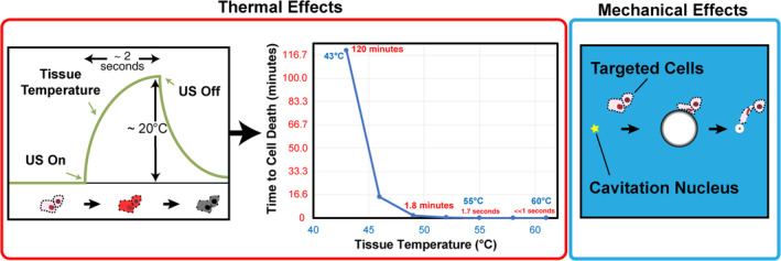
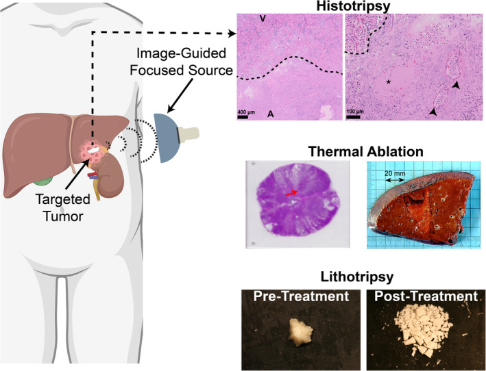
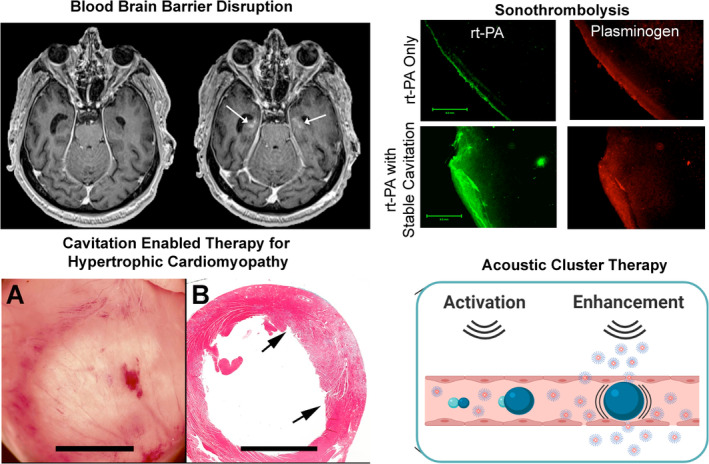
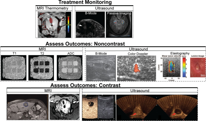
A 2012 review of therapeutic ultrasound was published to educate researchers and physicians on potential applications and concerns for unintended bioeffects (doi: 10.7863/jum.2012.31.4.623). This review serves as an update to the parent article, highlighting advances in therapeutic ultrasound over the past 12 years. In addition to general mechanisms for bioeffects produced by therapeutic ultrasound, current applications, and the pre-clinical and clinical stages are outlined. An overview is provided for image guidance methods to monitor and assess treatment progress. Finally, other topics relevant for the translation of therapeutic ultrasound are discussed, including computational modeling, tissue-mimicking phantoms, and quality assurance protocols.

Keywords: bioeffects; image‐guided therapy; therapeutic ultrasound.


Figures

Figure 1
Figure 1
(Left) Absorption of the ultrasound (US) energy heats tissue within the focal zone. Bioeffects that range from mild hyperthermia to thermal necrosis or boiling can be induced due to heating. (Middle) Schematic of temperature–time isodamage relationship leading to irreversible cell death as a thermal bioeffect. Cavitation and heating are not mutually exclusive effects. Cavitation can alter the rate of heating, and temperature elevation can reduce the cavitation threshold pressure. (Right) Schematic of bubble oscillations in response to ultrasound. Cavitation is capable of producing significant mechanical bioeffects, including cavitation, ablation, sonoporation, and streaming. Time increases from left to right as a cavitation nucleus under the influence of ultrasound grows into a microbubble that expands to a maximum diameter and then collapses. Damage can be imparted to cells surrounding the bubble during these oscillations.
Figure 2
Figure 2
A subset of therapeutic ultrasound applications without exogenous agents. Ultrasound is frequently applied transcutaneously to targets under image guidance, though intravascular sources are also under development. Histotripsy uses ultrasound pulses with sufficient tension to generate bubbles in situ that reduce ablated tissue (A) to acellular debris, as noted in the left image. At increased magnification there is a sharp boundary (dashed line) between viable (V) and tissue ablated mechanically (A). Treated regions exhibit viable vessels (arrowheads) and loss of cell nuclei (*) within the treatment zone (Reprinted from IEEE Trans Biomed Eng, vol. 71, no. 6, Development of Convolutional Neural Network to Segment Ultrasound Images of Histotripsy Ablation, pp. 1789–1797, Copyright 2024, under creative commons license CC BY‐NC‐ND 4.0). Thermal Ablation is achieved through absorption of ultrasound, and has been used to treat patients with pathological conditions in the prostate (left, Reprinted from Medical Physics, vol. 46, no. 2, MRI‐guided transurethral insonation of silica‐shell phase‐shift emulsions in the prostate with an advanced navigation platform, pp. 774–788, Copyright 2019, with permission from Wiley) and liver (right, Reprinted from PLoS ONE, vol. 10, no. 2, First Clinical Experience of Intra‐Operative High Intensity Focused Ultrasound in Patients with Colorectal Liver Metastases: A Phase I‐IIa Study, article number e0118212, Copyright 2015, reproduced under creative commons license CC BY 4.0). Several forms of Lithotripsy have been developed to break down mineralized tissue in the kidney and pancreas and vasculature. Burst wave lithotripsy applies tone bursts with broadly focused ultrasound that is able to comminute multiple types of kidney stones, including the struvite sample displayed above (Images provided courtesy Adam Maxwell). This figure was created in part in BioRender.com
Figure 3
Figure 3
A subset of therapeutic ultrasound applications that rely on exogenous agents to generate bubble activity. Blood–Brain Barrier Disruption uses transcranial or intracranial ultrasound in combination with microbubbles to enable large molecule drugs to pass from the vasculature into brain matter. An increase in brain permeability is apparent pre (left) and post (right) via contrast extravasation on T1‐weighted MRI (arrows, Reprinted from Brain, vol. 146, no. 3, Blood–brain barrier opening of the default mode network in Alzheimer's disease with magnetic resonance‐guided focused ultrasound, pp. 865–872, Copyright 2023, by permission of Oxford University Press). Sonothrombolysis enhances the penetration of a thrombolytic drug (eg, recombinant tissue plasminogen activator, rt‐PA) via microbubble activity into thrombus (left column) to promote fibrinolysis as indicated by plasminogen, a step in the fibrinolysis process highlighted in right column (Reprinted from Ultrasound in Medicine and Biology, vol. 34, no. 9, Ultrasound‐enhanced thrombolysis using Definity as a cavitation nucleation agent, pp. 1421–1433, Copyright 2008, by permission of Elsevier under creative commons license CC BY‐NC‐ND 4.0). Cavitation‐enabled therapy for hypertropic cardiomyopathy generates microlesions to reduce excessive heart muscle bulk. The treated region (light area in left image) indicated inflammation 2 days after bubble activity concurrent with swelling (arrows in right image) and inflammatory cells indicated via blue‐stained nuclei (Reprinted from Ultrasound in Medicine and Biology, vol. 44, no. 7, Ultrasound cavitation‐enabled treatment for therapy of hypertrophic cardiomyopathy: Proof of principle, pp. 1439–1450, Copyright 2018, by permission of Elsevier under creative commons license CC BY‐NC‐ND 4.0). Acoustic cluster therapy relies on a microbubble/droplet moiety to embolize vasculature to increase the penetration of therapeutics into the target (Reprinted from Journal of Controlled Release, vol. 10, no. 337, Acoustic Cluster Therapy (ACT®) enhances accumulation of polymeric micelles in the murine brain, pp. 285–295, Copyright 2021 under creative commons license CC BY 4.0).
Figure 4
Figure 4
Imaging for treatment monitoring and assessment of outcomes is critical for successful therapeutic ultrasound. Treatment monitoring: Temperature is tracked with MRI thermometry (Reprinted from Medical Physics, vol. 46, no. 2, MRI‐guided transurethral insonation of silica‐shell phase‐shift emulsions in the prostate with an advanced navigation platform, pp. 774–788, Copyright 2019, with permission from Wiley). Cavitation can be gauged by hyperechogenicity on B‐mode imaging or the detection of acoustic emissions (Reprinted from Brain, vol. 146, no. 3, Blood–brain barrier opening of the default mode network in Alzheimer's disease with magnetic resonance‐guided focused ultrasound, pp. 865–872, Copyright 2023, by permission of Oxford University Press). Noncontrast assessment of outcomes: Native MRI contrast (Reprinted from Physics in Medicine and Biology, vol. 62, no. 17, The response of MRI contrast parameters in in vitro tissues and tissue mimicking phantoms to fractionation by histotripsy, article number 7167, Copywrite 2017, with permission from IOP Publishing), standard ultrasound grayscale (Reprinted from Scientific Reports, vol. 9, Pilot in vivo studies on transcutaneous boiling histotripsy in porcine liver and kidney, article number 20176, Copyright 2019, with permission from Nature Publishing under creative commons license CC BY 4.0), bubble‐induced color Doppler (Reprinted from IEEE Transactions on Ultrasonics, Ferroelectrics, and Frequency Control, vol. 63, no. 6, Bubble‐induced color Doppler feedback for histotripsy tissue fractionation, pp. 408–419, Copyright 2016, with permission from IEEE), or elastography (Reprinted from IEEE Transactions on Ultrasonics, Ferroelectrics, and Frequency Control, vol. 59, no. 6, Imaging feedback of histotripsy treatments using ultrasound shear wave elastography, pp. 1167–1181, Copyright 2012, with permission from IEEE) can be used to gauge treatment outcomes. Contrast assessment of outcomes: Regions of nonperfusion on contrast MRI or ultrasound are primary methods used to assess ablation (Reprinted from International Journal of Hyperthermia, 39(1), First‐in‐man histotripsy of hepatic tumors: the THERESA trial, a feasibility study, pp. 1115–1123, Copyright 2022, with permission from Taylor and Francis Group under creative commons license CC BY 4.0; Reprinted from Medical Physics, vol. 46, no. 2, MRI‐guided transurethral insonation of silica‐shell phase‐shift emulsions in the prostate with an advanced navigation platform, pp. 774–788, Copyright 2019, with permission from Wiley; Reprinted from IEEE Transactions on Ultrasonics, Ferroelectrics, and Frequency Control, vol. 68, no. 9, Contrast‐Enhanced Ultrasound: A Useful Tool to Study and Monitor Hepatic Tumors Treated With Histotripsy, pp. 2853–2860, Copyright 2021, with permission from IEEE; Reprinted from European Journal of Radiology, vol. 81, no. 12, Clinical Utility of a Microbubble‐Enhancing Contrast (“SonoVue”) in Treatment of Uterine Fibroids with High Intensity Ultrasound: A Retrospective Study, pp. 3832–3838, Copyright 2012, with permission from Elsevier).
Figure 5
Figure 5
Harmonic beam profile plots for nonlinear pressure waves produced by a focused therapy transducer measured with a high‐resolution (100‐μm diameter geometrical sensitive element) fiber‐optic hydrophone. The harmonic number is denoted by n. Gaussian fits are shown in dotted lines. Error bars denote standard deviation in the measurement obtained from transverse scans obtained horizontally and vertically. The black vertical lines show the spatial extent of a medium‐resolution (400‐μm diameter geometrical sensitive element) low‐cost, robust needle hydrophone. The 400‐μm hydrophone can be used for accurate measurement of the acoustic field after spatial averaging correction is applied. Each panel is the same dataset but at differing extensions along the spatial dimension (Reprinted from IEEE Transactions on Ultrasonics, Ferroelectrics, and Frequency Control, vol. 66, no. 9, Correction for Spatial Averaging Artifacts in Hydrophone Measurements of High‐Intensity Therapeutic Ultrasound: An Inverse Filter Approach, pp. 1453–1464, Copyright 2019, with permission from IEEE). BBB, blood–brain barrier; FUS, focused ultrasound.

References

    1. American Institute of Ultrasound in Medicine . Conclusions Regarding Epidemiology for Obstetric Ultrasound. https://www.aium.org/resources/official‐statements/view/conclusions‐rega.... Accessed October 11, 2024.
    1. Fowlkes JB. American Institute of Ultrasound in medicine consensus report on potential bioeffects of diagnostic ultrasound. J Ultrasound Med 2008; 27:503–515. 10.7863/jum.2008.27.4.503. - DOI - PubMed
    1. International Electrotechnical Commission . Particular Requirements for the Basic Safety and Essential Performance of Ultrasonic Medical Diagnostic and Monitoring Equipment. 2.1st ed. IEC; 2015.
    1. American Institute of Ultrasound in Medicine . Recommended Maximum Scanning Times for Displayed Thermal Index (TI) Values. https://www.aium.org/resources/official‐statements/view/recommended‐maxi.... Accessed October 11, 2024. - PubMed
    1. Miller DL, Smith NB, Bailey MR, Czarnota GJ, Hynynen K, Makin IRS. Overview of therapeutic ultrasound applications and safety considerations. J Ultrasound Med 2012; 31:623–634. 10.7863/jum.2012.31.4.623. - DOI - PMC - PubMed