. 2024 Nov 1;17(11):dmm052002.
doi: 10.1242/dmm.052002. Epub 2024 Nov 11.

Modulation of SNARE-dependent exocytosis in astrocytes improves neuropathology in Huntington's disease

Affiliations

Modulation of SNARE-dependent exocytosis in astrocytes improves neuropathology in Huntington's disease

Annesha C King et al. Dis Model Mech. .

Abstract

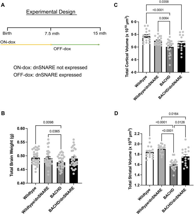
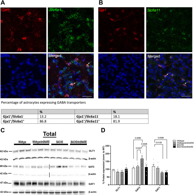
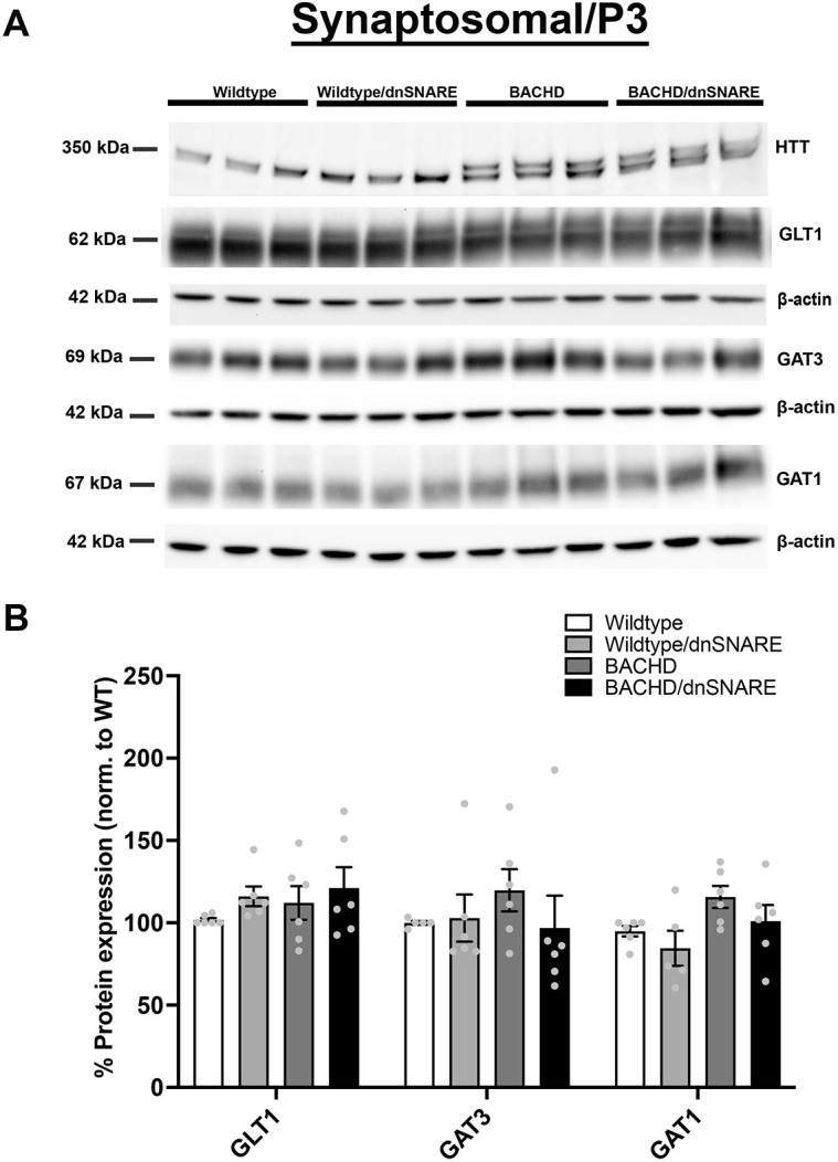
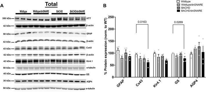
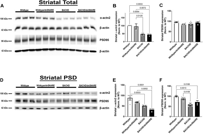
Huntington's disease (HD) is a fatal, progressive neurodegenerative disorder. Prior studies revealed an increase in extracellular glutamate levels after evoking astrocytic SNARE-dependent exocytosis from cultured primary astrocytes from mutant huntingtin (mHTT)-expressing BACHD mice compared to control astrocytes, suggesting alterations in astrocytic SNARE-dependent exocytosis in HD. We used BACHD and dominant-negative (dn)SNARE mice to decrease SNARE-dependent exocytosis from astrocytes to determine whether reducing SNARE-dependent exocytosis from astrocytes could rescue neuropathological changes in vivo. We observed significant protection against striatal atrophy and no significant rescue of cortical atrophy in BACHD/dnSNARE mice compared to BACHD mice. Amino acid transporters are important for modulating the levels of extracellular neurotransmitters. BACHD mice had no change in GLT1 expression, decreased striatal GAT1 expression and increased levels of GAT3. There was no change in GAT1 after reducing astrocytic SNARE-dependent exocytosis, and increased GAT3 expression in BACHD mice was normalized in BACHD/dnSNARE mice. Thus, modulation of astrocytic SNARE-dependent exocytosis in BACHD mice is protective against striatal atrophy and modulates GABA transporter expression.

Keywords: Astrocytes; BACHD; Huntington's disease; SNARE-dependent exocytosis; dnSNARE.


Conflict of interest statement

Competing interests The authors declare no competing or financial interests.

Figures

Fig. 1.
Fig. 1.
BACHD/dnSNARE mice have increased striatal volume. (A) Design of the doxycycline (dox) paradigm, showing the control of inducible astrocyte-specific dnSNARE expression. Mice were fed dox-containing chow for 7.5 months and removed from dox-containing food to allow transgene expression. (B) Total brain weight was assessed at 12-15 months of age (n=30/genotype). (C,D) Cortical (C) and striatal (D) volume was assessed at 12-15 months (n=16-22/genotype). Data are mean±s.e.m. Differences among the groups were assessed using Welch's ANOVA test followed by Dunnett T3 multiple comparison procedure. Non-significant P-values are not displayed on graphs. Refer to Table 1 for all P-values.
Fig. 2.
Fig. 2.
Expression of neurotransmitter transporters. (A,B) Representative RNAscope images of Slc6a1, Slc6a11and Gja1 in the striatum. Scale bars: 50 μm. The numbers of Gja1-positive cells co-labeled with Slc6a1 (encoding GAT1) or Slc6a11 (encoding GAT3) were counted. (C) Representative western blots of GLT1, GAT3 and GAT1 protein expression in the striatum of wild-type, wild-type/dnSNARE, BACHD and BACHD/dnSNARE mice (each lane represents a single sample; each sample is pooled from three mice of the same genotype). (D) Western blot quantification of GLT1, GAT3 and GAT1 (n=6/genotype). WT, wild type. The loading control β-actin shown for GAT1 in C is the same as the β-actin shown for Cx43 in Fig. 5A. The same membrane was probed for β-actin, GAT1 and Cx43. The loading control β-actin shown for GLT1 in C is the same as the β-actin used for GFAP in Fig. 5A. GLT1 and GFAP were probed on the same blot membrane. Data are mean±s.e.m. Differences among the groups were assessed by one-way ANOVA followed by Tukey's HSD multiple comparison procedure. Non-significant P-values are not displayed on graphs. Refer to Table 1 for all P-values.
Fig. 3.
Fig. 3.
Glutamate and GABA transporter levels in the synaptosomal fraction. (A) Representative western blots of glutamate and GABA transporter protein expression in wild-type, wild-type/dnSNARE, BACHD and BACHD/dnSNARE mice. (B) Western blot quantification of the transporters (shown are n=3 independent samples). HTT was probed to confirm the genotype of sample mice on the same blot probed for GAT1 (same β-actin loading control as shown for GAT1). No significant difference was observed in GLT1, GAT3 and GAT1 (n=5-6/genotype). Data are mean±s.e.m. Differences among the groups were assessed by one-way ANOVA followed by Tukey's HSD multiple comparison procedure. Non-significant P-values are not displayed on graphs. Refer to Table 1 for all P-values.
Fig. 4.
Fig. 4.
Number of S100β-, GFAP- and ALDH1L1-positive astrocytes in the striatum in wild-type, wild-type/dnSNARE, BACHD and BACHD/dnSNARE mice. (A) Immunofluorescence staining of S100β-, GFAP- and ALDH1L1-positive astrocytes (red) in wild-type, wild-type/dnSNARE, BACHD and BACHD/dnSNARE tissues. Scale bars: 50 μm. (B-D) No change in S100β-positive (B), GFAP-positive (C) or ALDH1L1-positive (D) astrocyte numbers was observed in these mice. Blue is DAPI staining (n=6-9/genotype). Data are mean±s.e.m. Differences among the groups were assessed by one-way ANOVA followed by Tukey's HSD multiple comparison procedure. Non-significant P-values are not displayed on graphs. Refer to Table 1 for all P-values.
Fig. 5.
Fig. 5.
Astrocyte-enriched protein expression in the striatal total cell homogenate. (A) Representative western blots of various astrocyte-enriched proteins in the striatal total cell homogenate of wild-type, wild-type/dnSNARE, BACHD and BACHD/dnSNARE mice. (B) Quantification of the western blot results (n=4/genotype). HTT was probed to confirm the genotype of our mouse models. The loading control β-actin shown for Cx43 in A is the same as the β-actin shown for GAT1 in Fig. 2C. The same membrane was probed for β-actin, GAT1 and Cx43. The loading control β-actin shown for GFAP in A is the same as the β-actin used for GLT1 in Fig. 2C. The same membrane was probed for β-actin, GLT1 and GFAP. Data are mean±s.e.m. Differences among the groups were assessed by one-way ANOVA followed by Tukey's HSD multiple comparison procedure. Non-significant P-values are not displayed on graphs. Refer to Table 1 for all P-values.
Fig. 6.
Fig. 6.
Reduction in SNARE-dependent exocytosis in BACHD mice had no effect on striatal ACTN2 or PSD95 expression. (A) Representative western blots of total homogenate for ACTN2 and PSD95 (n=4/genotype). (B,C) Quantitation of the western blots for ACTN2 (B) and PSD95 (C) in the total homogenate fraction (n=4/genotype). (D) Representative blots of ACTN2 and PSD95 in the postsynaptic density (PSD) fractions (n=4/genotype). (E,F) Quantitation of the western blots for ACTN2 (E) and PSD95 (F) in the PSD fractions (n=4/genotype). Data are mean±s.e.m. Differences among the groups were assessed by one-way ANOVA followed by Tukey's HSD multiple comparison procedure. Non-significant P-values are not displayed on graphs. Refer to Table 1 for all P-values.

References

    1. Anderson, C. M. and Swanson, R. A. (2000). Astrocyte glutamate transport: review of properties, regulation, and physiological functions. Glia 32, 1-14. 10.1002/1098-1136(200010)32:1<1::AID-GLIA10>3.0.CO;2-W - DOI - PubMed
    1. André, V. M., Cepeda, C. and Levine, M. S. (2010). Dopamine and glutamate in Huntington's disease: a balancing act. CNS Neurosci. Ther. 16, 163-178. 10.1111/j.1755-5949.2010.00134.x - DOI - PMC - PubMed
    1. André, V. M., Fisher, Y. E. and Levine, M. S. (2011). Altered balance of activity in the striatal direct and indirect pathways in mouse models of Huntington's disease. Front. Syst. Neurosci. 5, 46. 10.3389/fnsys.2011.00046 - DOI - PMC - PubMed
    1. Angulo, M. C., Kozlov, A. S., Charpak, S. and Audinat, E. (2004). Glutamate released from glial cells synchronizes neuronal activity in the hippocampus. J. Neurosci. 24, 6920. 10.1523/JNEUROSCI.0473-04.2004 - DOI - PMC - PubMed
    1. Araque, A., Sanzgiri, R. P., Parpura, V. and Haydon, P. G. (1999). Astrocyte-induced modulation of synaptic transmission. Can. J. Physiol. Pharmacol. 77, 699-706. 10.1139/y99-076 - DOI - PubMed

Substances