

FET PET to differentiate between post-treatment changes and recurrence in high-grade gliomas: a single center multidisciplinary clinic controlled study
- PMID: 39527264
- PMCID: PMC11893651
- DOI: 10.1007/s00234-024-03495-9
FET PET to differentiate between post-treatment changes and recurrence in high-grade gliomas: a single center multidisciplinary clinic controlled study
Abstract
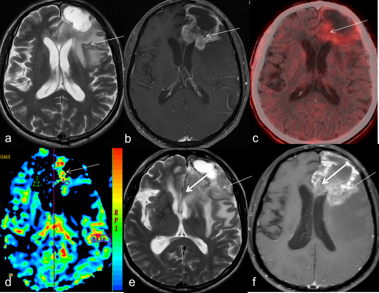
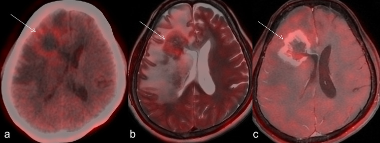
Purpose: The clinico-radiological dilemma in post-treatment high-grade gliomas, between disease recurrence (TR) and treatment-related changes (TRC), still persists. FET (Fluoro-ethyl-tyrosine) PET has been extensively used as problem-solving modality for cases where MR imaging is inconclusive. We incorporated a systematic imaging and clinical follow-up algorithm in a multi-disciplinary clinic (MDC) setting to analyse our cohort of FET PET in post-treatment gliomas.
Methods: We retrospectively analyzed 171 patients of post-treatment grade III and IV glioma with equivocal findings on MRI. 185-222 MBq of 18 F-FET was injected and dedicated static imaging of brain was performed at 20 min. TBR (Tumor to background ratio) was used as semi-quantitative parameter. Cutoff of 2.5 was used for image interpretation. Imaging findings were confirmed with histopathological diagnosis, wherever available or in a multidisciplinary joint clinic based on serial imaging.
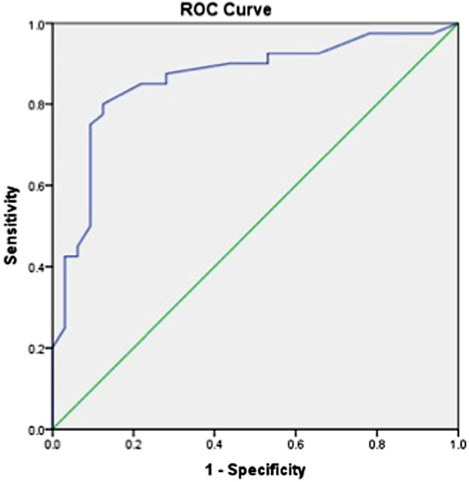
Results: 121 of 171 patients showed recurrent disease on FET PET, on follow up, 109 were confirmed with recurrence; 7 patients showed TRC, whereas 5 were treated with bevacizumab, with no further clinico-radiological deterioration, thus confirming TRC. 50 patients showed TRC on FET PET, on follow up on follow up, 40 were confirmed as true-negative. 10 patients who showed TBR less than 2.5 had confirmed TR on subsequent MR imaging. The overall sensitivity and specificity was 91.6 and 76.9% respectively, with a diagnostic accuracy of 87.13%.
Conclusion: There is potential for FET PET to be used along with MRI in the post treatment algorithm of high-grade glial tumors.
Keywords: FET PET; High grade glioma; MRI; Post-treatment changes; Recurrence.
© 2024. The Author(s).
Conflict of interest statement
Declarations. Ethical approval: The study was approved by Institutional Ethics Committee, Tata Memorial Hospital (Project No – 900717). Informed consent: Since this is a retrospective study, waiver of consent was given by Institutional Ethics Committee. However, as part of standard practice, written and informed consent was taken before undergoing FET PET and MRI procedure. Conflict of interest: There are no conflicts of interest. No competing interests or disclosures.
Figures
References
-
- Kumar AJ, Leeds NE, Fuller GN et al (2000) Malignant gliomas: MR imaging spectrum of radiation therapy- and chemotherapy-induced necrosis of the brain after treatment. Radiology 217:377–384. 10.1148/radiology.217.2.r00nv36377 - PubMed
-
- Wen PY, Macdonald DR, Reardon DA et al (2010) Updated response assessment criteria for high-grade gliomas: response assessment in neuro-oncology working group. J Clin Oncol off J Am Soc Clin Oncol 28:1963–1972. 10.1200/JCO.2009.26.3541 - PubMed
-
- Reardon DA, Weller M (2018) Pseudoprogression: fact or wishful thinking in neuro-oncology? Lancet Oncol 19:1561–1563. 10.1016/S1470-2045(18)30654-5 - PubMed
MeSH terms
Substances
LinkOut - more resources
Full Text Sources
Medical
