

Diagnostic accuracy of radiomics and artificial intelligence models in diagnosing lymph node metastasis in head and neck cancers: a systematic review and meta-analysis
- PMID: 39527265
- PMCID: PMC11893643
- DOI: 10.1007/s00234-024-03485-x
Diagnostic accuracy of radiomics and artificial intelligence models in diagnosing lymph node metastasis in head and neck cancers: a systematic review and meta-analysis
Abstract
Introduction: Head and neck cancers are the seventh most common globally, with lymph node metastasis (LNM) being a critical prognostic factor, significantly reducing survival rates. Traditional imaging methods have limitations in accurately diagnosing LNM. This meta-analysis aims to estimate the diagnostic accuracy of Artificial Intelligence (AI) models in detecting LNM in head and neck cancers.
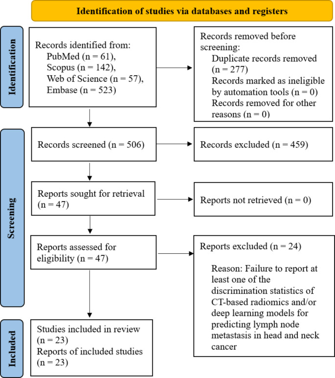
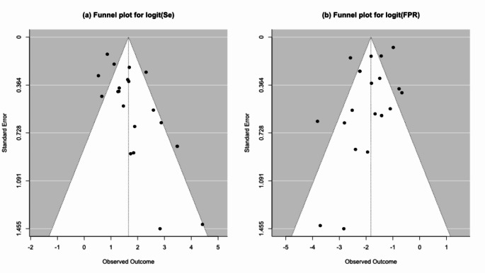
Methods: A systematic search was performed on four databases, looking for studies reporting the diagnostic accuracy of AI models in detecting LNM in head and neck cancers. Methodological quality was assessed using the METRICS tool and meta-analysis was performed using bivariate model in R environment.
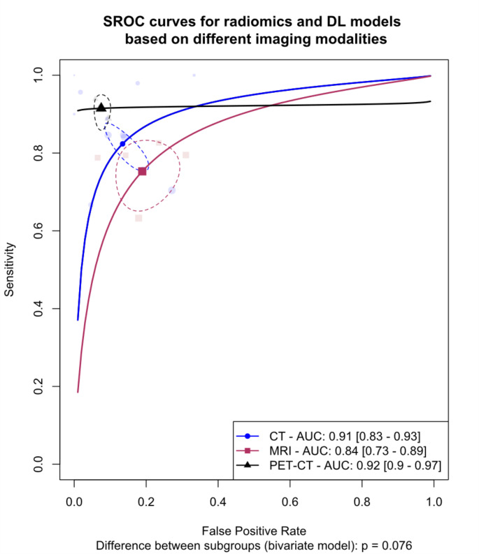
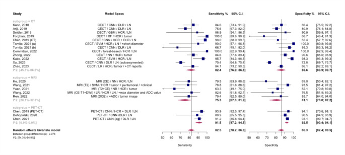
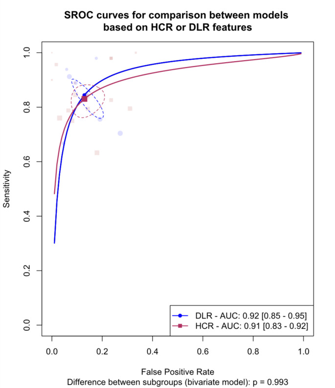
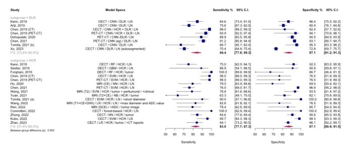
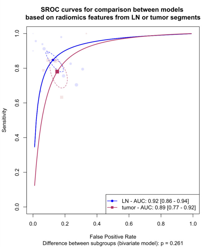
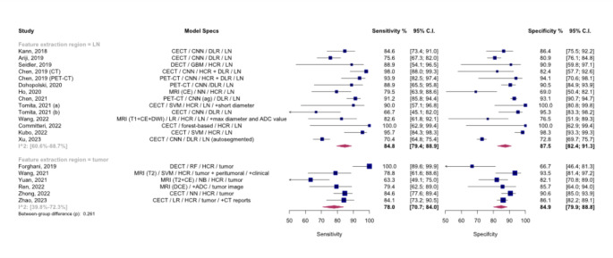
Results: 23 articles met the inclusion criteria. Due to the absence of external validation in most studies, all analyses were confined to internal validation sets. The meta-analysis revealed a pooled AUC of 91% for CT-based radiomics, 84% for MRI-based radiomics, and 92% for PET/CT-based radiomics. Sensitivity and specificity were highest for PET/CT-based models. The pooled AUC was 92% for deep learning models and 91% for hand-crafted radiomics models. Models based on lymph node features had a pooled AUC of 92%, while those based on primary tumor features had an AUC of 89%. No significant differences were found between deep learning and hand-crafted radiomics models or between lymph node and primary tumor feature-based models.
Conclusion: Radiomics and deep learning models exhibit promising accuracy in diagnosing LNM in head and neck cancers, particularly with PET/CT. Future research should prioritize multicenter studies with external validation to confirm these results and enhance clinical applicability.
Keywords: Deep learning; Head and neck cancer; Lymph node metastasis; PET/CT imaging; Radiomics.
© 2024. The Author(s).
Conflict of interest statement
Declarations. Conflict of interest: We declare that we have no conflict of interest. Conflict of interest: The authors have no relevant financial or non-financial interests to disclose. Declaration of generative AI and AI-assisted technologies in the writing process: We acknowledge ChatGPT, an OpenAI language model based on the GPT-4 architecture, for assisting with language corrections during the article’s editing. The model enhanced the readability and language quality of the publication. However, the authors retain full responsibility for the content, having reviewed and edited it as needed after using the tool. Ethical approval: This study, being a review and not involving patient data, did not require institutional ethical approval. Informed consent: As this study was a review and did not involve patient data, obtaining informed consent was not applicable.
Figures
Similar articles
-
Artificial Intelligence in CT for Predicting Cervical Lymph Node Metastasis in Papillary Thyroid Cancer Patients: A Meta-analysis.Acad Radiol. 2025 May;32(5):2554-2568. doi: 10.1016/j.acra.2025.02.007. Epub 2025 Feb 24. Acad Radiol. 2025. PMID: 40000328
-
Positron emission tomography (PET) and magnetic resonance imaging (MRI) for the assessment of axillary lymph node metastases in early breast cancer: systematic review and economic evaluation.Health Technol Assess. 2011 Jan;15(4):iii-iv, 1-134. doi: 10.3310/hta15040. Health Technol Assess. 2011. PMID: 21276372 Free PMC article.
-
MRI-Based Radiomics Methods for Predicting Ki-67 Expression in Breast Cancer: A Systematic Review and Meta-analysis.Acad Radiol. 2024 Mar;31(3):763-787. doi: 10.1016/j.acra.2023.10.010. Epub 2023 Nov 2. Acad Radiol. 2024. PMID: 37925343
-
Diagnostic accuracy of CT and PET/CT radiomics in predicting lymph node metastasis in non-small cell lung cancer.Eur Radiol. 2025 Apr;35(4):1966-1979. doi: 10.1007/s00330-024-11036-4. Epub 2024 Sep 2. Eur Radiol. 2025. PMID: 39223336
-
Artificial intelligence for diagnosing exudative age-related macular degeneration.Cochrane Database Syst Rev. 2024 Oct 17;10(10):CD015522. doi: 10.1002/14651858.CD015522.pub2. Cochrane Database Syst Rev. 2024. PMID: 39417312
Cited by
-
Fusobacterium nucleatum and IL-32 co-predict nodal metastasis preoperatively in head and neck cancer.Transl Cancer Res. 2025 Jun 30;14(6):3359-3372. doi: 10.21037/tcr-2024-2539. Epub 2025 Jun 19. Transl Cancer Res. 2025. PMID: 40687228 Free PMC article.
References
-
- Chow LQM (2020) Head and Neck Cancer. N Engl J Med 382:60–72. 10.1056/NEJMra1715715 - PubMed
-
- Oh LJ, Phan K, Kim SW et al (2020) Elective neck dissection versus observation for early-stage oral squamous cell carcinoma: systematic review and meta-analysis. Oral Oncol 105:104661. 10.1016/j.oraloncology.2020.104661 - PubMed
-
- Li B, Li D, Lau DH et al (2009) Clinical-dosimetric analysis of measures of dysphagia including gastrostomy-tube dependence among head and neck cancer patients treated definitively by intensity-modulated radiotherapy with concurrent chemotherapy. Radiat Oncol 4:52. 10.1186/1748-717X-4-52 - PMC - PubMed
Publication types
MeSH terms
LinkOut - more resources
Full Text Sources
Medical
Miscellaneous
