. 2024 Nov;40(11):1713-1731.
doi: 10.1007/s12264-024-01314-y. Epub 2024 Nov 11.

Glutamatergic Circuits in the Pedunculopontine Nucleus Modulate Multiple Motor Functions

Affiliations

Glutamatergic Circuits in the Pedunculopontine Nucleus Modulate Multiple Motor Functions

Yanwang Huang et al. Neurosci Bull. 2024 Nov.

Abstract

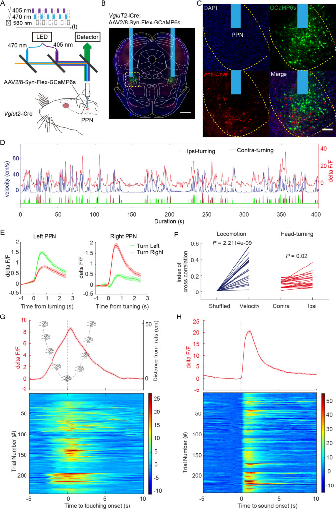
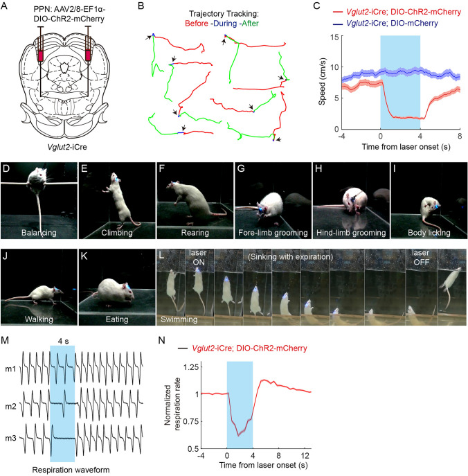
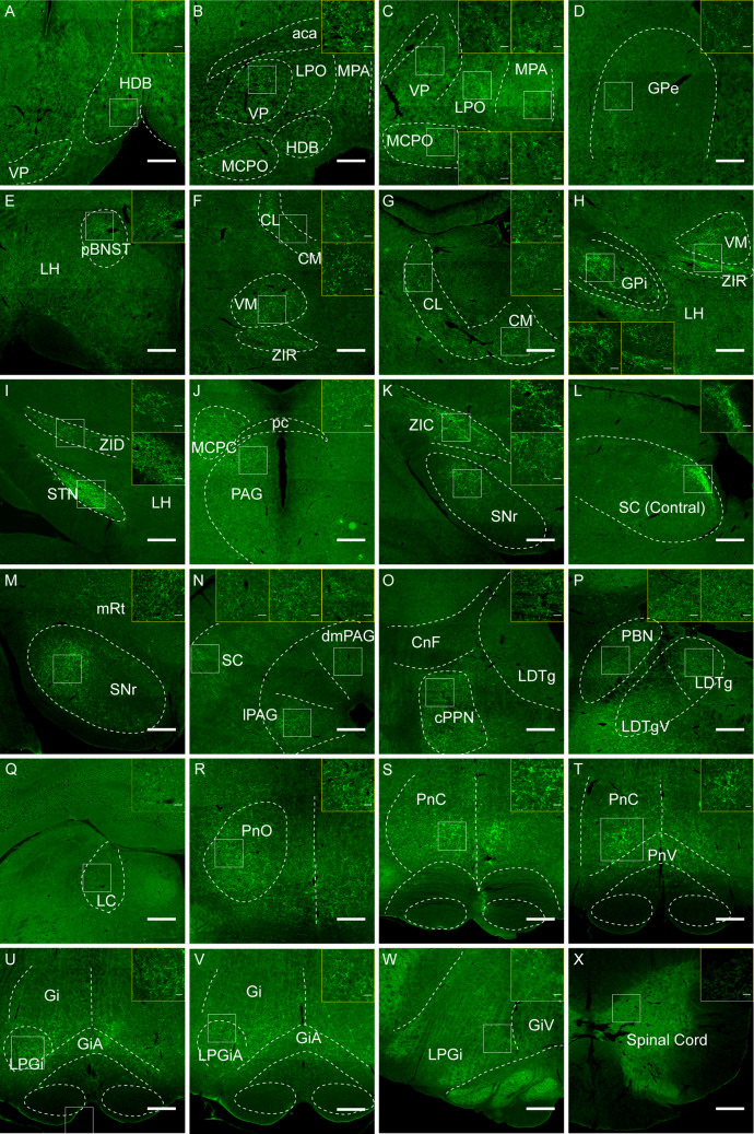
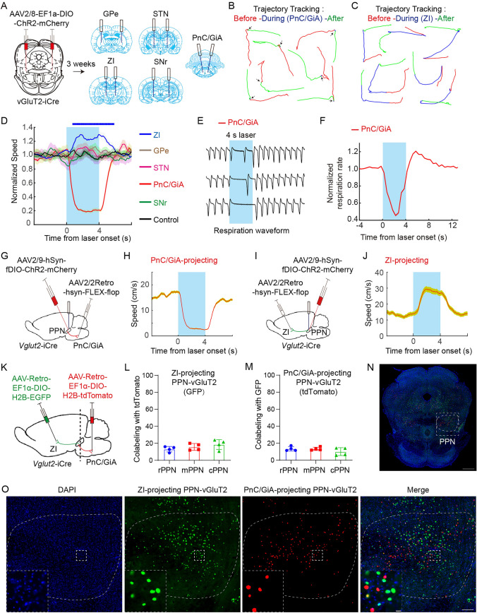
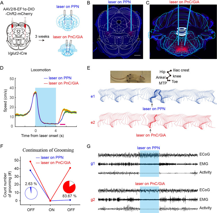
The functional role of glutamatergic (vGluT2) neurons in the pedunculopontine nucleus (PPN) in modulating motor activity remains controversial. Here, we demonstrated that the activity of vGluT2 neurons in the rostral PPN is correlated with locomotion and ipsilateral head-turning. Beyond these motor functions, we found that these rostral PPN-vGluT2 neurons remarkably respond to salient stimuli. Furthermore, we systematically traced the upstream and downstream projections of these neurons and identified two downstream projections from these neurons to the caudal pontine reticular nucleus/anterior gigantocellular reticular nucleus (PnC/GiA) and the zona incerta (ZI). Our findings indicate that the projections to the PnC/GiA inhibit movement, consistent with 'pause-and-play' behavior, whereas those to the ZI promote locomotion, and others respond to a new 'pause-switch-play' pattern. Collectively, these findings elucidate the multifaceted influence of the PPN on motor functions and provide a robust theoretical framework for understanding its physiological and potential therapeutic implications.

Keywords: Anterior gigantocellular reticular nucleus; Caudal pontine reticular nucleus; Glutamatergic neuron; Pause-and-play; Pedunculopontine nucleus; Zona incerta.


Conflict of interest statement

Conflict of interest: The authors declare no competing financial interests.

Figures

Fig. 1
Fig. 1
Fiber photometry recording of rPPN-vGluT2 neuronal activity during locomotion and ipsilateral head-turning, and in response to salient stimuli. A Schematic of fiber photometric recording of PPN vGluT2 neuronal activity. B Representative histology confirming the fiber placement and specificity of GCaMP6s expression in the PPN. Scale bar, 1000 μm. C Representative histology showing co-localization of GCaMP6s and ChAT in the PPN. Green, GCaMP6s; red, anti-ChAT. Scale bar, 200 μm. D Concurrent recording of locomotor velocity, directional head-turning, and PPN vGluT2 neuronal Ca2+ signals in freely-moving rats. Blue line, velocity of rat; red line, Ca2+ signals; green, ipsilateral head-turning; carmine, contralateral head-turning. E Averaged PPN-vGluT2 neuronal Ca2+ signal in response to head-turning behavior. Left panel, activity of the left PPN; right panel, activity of the right PPN, during left (red) and right (green) head-turning. Data are presented as the mean ± SEM. F Cross-correlation analysis between locomotor velocity and Ca2+ signals (left, n = 24, paired t-test, P = 2.2−9), and between head-turning and Ca2+ signals (right, n = 24 paired t-test, P = 0.02). G Activation of PPN-vGluT2 neurons during hand-touching stimulation. The red line is the averaged PPN-vGluT2 neuronal Ca2+ signal in response to hand-touching. Grey line, diagram of the distance of the hand from the rat during hand-touching, with the onset points as touching time points. Lower panel, heatmap of the PPN-vGluT2 neuronal Ca2+ signal in response to the onset of hand-touching. H Activation of PPN-vGluT2 neurons during salient sound stimulation. Red line, averaged PPN-vGluT2 neuronal Ca2+ signal in response to salient sound stimulation. Lower panel, heatmap of the PPN-vGluT2 neuronal Ca2+ signal in response to the onset of salient sound stimulation.
Fig. 2
Fig. 2
Inhibition of voluntary and involuntary movements by activation of rPPN-vGluT2 neurons. A Schematic of viral injection sites and fiber placement in the PPN. B Representative traces of 4-s locomotor trajectories before (red), during (blue), and after (green) optogenetic stimulation of PPN-vGluT2 neurons. C Averaged velocity shows that activation of PPN-vGluT2 neurons halts locomotion. Red line, experiment group with ChR2 expression, n = 6. Blue line, control group with mCherry expression, n = 6. Data are presented as the mean ± SEM. D-L Stationary postures are maintained during optogenetic activation of PPN-vGluT2 neurons. Rats are balancing (D), climbing (E), rearing (F), forelimb grooming (G), hindlimb grooming (H), body licking (I), walking (J), and eating (K) during optogenetic stimulation. Moreover, rats stop swimming and sink with a stationary posture when PPN-vGluT2 neurons are activated, and resume swimming after stimulation offset (L). M, N Example traces of involuntary respiration of anesthetized rats before, during, and after optogenetic activation of PPN-vGluT2 neurons (M) and normalized respiration rates (N) (n = 9). Data are presented as the mean ± SEM.
Fig. 3
Fig. 3
Downstream targets of rPPN-vGluT2 neurons. A-X Representative histology of downstream regions of PPN-vGluT2 neurons, including the horizontal limb of the diagonal band of Broca (HDB), ventral pallidum (VP), lateral preoptic area (LPO), medial preoptic area (MPA), magnocellular preoptic nucleus (MCPO), globus pallidus externus (GPe), posterior bed nucleus of stria terminalis (pBNST), centromedian nucleus (CM), centrolateral nucleus (CL), ventromedial nucleus (VM), ZI, rostral ZI (ZIR), dorsal ZI (ZID), caudal ZI (ZIC), globus pallidus internus (GPi), lateral hypothalamus (LH), subthalamic nucleus (STN), magnocellular nucleus of the posterior commissure (MCPC), superior colliculus (SC), mesencephalic reticular formation (mRt), periaqueductal gray (PAG), dorsomedial periaqueductal gray (dmPAG), lateral periaqueductal gray (lPAG), cuneiform nucleus (CnF), laterodorsal tegmental nucleus (LDT), parabrachial nucleus (PBN), locus coeruleus (LC), oral pontine reticular nucleus (PnO), PnC, ventral pontine reticular nucleus (PnV), GiA, Gi, lateral paragigantocellular nucleus (LPGi), gigantocellular reticular nucleus, ventral part (GiV), and the spinal cord. Scale bars, 1000 μm. The region of interest is windowed and enlarged in each panel. Scale bars, 60 μm.
Fig. 4
Fig. 4
The dual function of locomotor modulation is controlled by different neural circuits. A Schematic of viral labeling (left) and fiber placement (right). B, C Representative traces of 4-s locomotor trajectories before (red), during (blue), and after (green) optogenetic stimulation of PPN-vGluT2 neuron terminals in the PnC/GiA (B) or ZI (C). D Changes in the averaged velocity resulting from optogenetic activation of PPN-vGluT2 neuron terminals in the PnC/GiA (red, n = 7), GPe (brown, n = 6), STN (pink, n = 6), SNr (green, n = 6), and ZI (blue, n = 8). The control group (n = 6) received laser stimulation to the ZI with mCherry-expression in PPN-vGluT2 neurons. Data are presented as the mean ± SEM. E, F Example traces of respiration of anesthetized rats before, during, and after optogenetic activation of PPN-vGluT2 neuron terminals in PnC/GiA (E) and normalized respiration rates (F) (n = 7). Data are presented as the mean ± SEM. G Schematic of optogenetic activation of PnC/GiA-projecting PPN-vGluT2 neurons. H Changes in averaged velocity during locomotion induced by optogenetic activation of PnC/GiA-projecting PPN-vGluT2 neurons (n = 6). Data are presented as the mean ± SEM. I Schematic of optogenetic activation of ZI-projecting PPN-vGluT2 neurons. J Changes in averaged velocity during locomotion induced by optogenetic activation of ZI-projecting PPN-vGluT2 neurons (n = 4). Data are presented as the mean ± SEM. K Schematic of labeling neuronal nuclei of PnC/GiA-projecting and ZI-projecting PPN-vGluT2 neurons. L, M Quantitation of the overlapped neurons among PnC/GiA-projecting and ZI-projecting PPN-vGluT2 neurons. L, the percentage of ZI-projecting PPN-vGluT2 neurons (GFP) co-labeled with PnC/GiA-projecting PPN-vGluT2 neurons (tdTomato) in the rPPN, mPPN, and cPPN; M, the percentage of PnC/GiA-projecting PPN-vGluT2 neurons (tdTomato) co-labeled with ZI-projecting PPN-vGluT2 neurons (GFP) in the rPPN, mPPN, and cPPN. N, O Representative histology showing the different distribution of PnC/GiA-projecting (red, tdTomato) and ZI-projecting (green, GFP) PPN-vGluT2 neurons. N, Representative histology in the rPPN. Scale bar, 1000 μm; O, Window from N. Scale bar, 200 μm. The left-lower panel, enlarged window. Scale bar, 20 μm.
Fig. 5
Fig. 5
PPN-vGluT2 neurons projecting to the PnC/GiA are responsible for pause-and-play pattern arrest behavior. A Schematic of virus injection and fiber placement. B Representative histology of a coronal section showing ChR2-mCherry expression (red) in somata of PPN-vGluT2 neurons. Blue rectangle, fiber position. Scale bar, 1000 μm. C Representative histology of a coronal section showing PPN-vGluT2 neuron terminals in the PnC/GiA with ChR2-mCherry expression (red). Blue rectangle, fiber position. D Changes in the averaged velocity resulting from optogenetic activation of PPN-vGluT2 neuron somata (blue line) or their terminals in the PnC/GiA (red line) (n = 7). Data are presented as the mean ± SEM. Pink bar, time window with a significant change at the P<0.05 level. E Changes in the pause-and-play pattern of hindlimb dynamics resulting from optogenetic activation of PPN-vGluT2 neuron somata (blue line) or their dendrites in the PnC/GiA (red line) during locomotion in a linear corridor. The arrow indicates the period of pause. F Changes in the pause-and-play pattern of grooming resulting from optogenetic activation of PPN-vGluT2 neuron somata (blue line, 1/38) or their dendrites in the PnC/GiA (red line, 41/49). G Changes in representative ECoG, EMG, and activity signals of ongoing grooming resulting from optogenetic activation of PPN-vGluT2 neuron somata (blue line) or their dendrites in the PnC/GiA (red line).
Fig. 6
Fig. 6
Inputs of rPPN-vGluT2 neurons. A Schematic of injections of helper and rabies viruses into the PPN. B Representative histology showing the viral injection site. B1, green is a signal of rabies labeling; b2, PPN-vGluT2 neurons with helper virus labeling; b3, yellow cells are the start neurons for retrograde trans-synaptic tracing. C-N Representative histology of areas upstream of PPN-vGluT2 neurons, including secondary motor cortex (M2), anterior cingulate cortex (ACC), prelimbic cortex (Prl), nucleus accumbens (NAc), MPA, LPO, GPi, central medial amygdala (CeM), paraventricular nucleus (PVN), LH, ZI, SNr, pararubral nucleus (PaR), SC, PAG, dorsal raphe nucleus (DRN), lateral parabrachial nucleus (LPB), medial parabrachial nucleus (MPB), LDT, and medial cerebellum (Med).
Fig. 7
Fig. 7
Supraspinal circuits via rPPN glutamatergic neurons modulate multiple motor actions. A Connectome of PPN-vGluT2 neurons in the whole brain. Arrows represent the downstream outputs of PPN vGluT2 neurons; yellow dots represent the upstream inputs. B Schematic of different motor modulation underlying different circuits of PPN-vGluT2 neurons. The projection from PPN-vGluT2 to ZI promotes locomotion, and the projection to PnC/GiA plays an important role in pause-and-play pattern arrest behavior.

Similar articles

Cited by

References

    1. Parkinson J. An essay on the shaking palsy. J Neuropsychiatry Clin Neurosci 2002, 14: 223–236. - PubMed
    1. Shik ML, Severin FV, Orlovskiĭ GN. Control of walking and running by means of electric stimulation of the midbrain. Biofizika 1966, 11: 659–666. - PubMed
    1. Ryczko D, Dubuc R. The multifunctional mesencephalic locomotor region. Curr Pharm Des 2013, 19: 4448–4470. - PubMed
    1. Fasano A, Aquino CC, Krauss JK, Honey CR, Bloem BR. Axial disability and deep brain stimulation in patients with Parkinson disease. Nat Rev Neurol 2015, 11: 98–110. - PubMed
    1. Thevathasan W, Debu B, Aziz T, Bloem BR, Blahak C, Butson C. Pedunculopontine nucleus deep brain stimulation in Parkinson’s disease: A clinical review. Mov Disord 2018, 33: 10–20. - PubMed

Substances

LinkOut - more resources