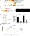
. 2024 Nov 11;20(11):e1012294.
doi: 10.1371/journal.ppat.1012294. eCollection 2024 Nov.

Critical role of Babesia bovis spherical body protein 3 in ridge formation on infected red blood cells

Affiliations

Critical role of Babesia bovis spherical body protein 3 in ridge formation on infected red blood cells

Atefeh Fathi et al. PLoS Pathog. .

Abstract

Babesia bovis, an apicomplexan intraerythrocytic protozoan parasite, causes serious economic loss to cattle industries around the world. Infection with this parasite leads to accumulation of infected red blood cells (iRBCs) in the brain microvasculature that results in severe clinical complications known as cerebral babesiosis. Throughout its growth within iRBCs, the parasite exports various proteins to the iRBCs that lead to the formation of protrusions known as "ridges" on the surface of iRBCs, which serve as sites for cytoadhesion to endothelial cells. Spherical body proteins (SBPs; proteins secreted from spherical bodies, which are organelles specific to Piroplasmida) are exported into iRBCs, and four proteins (SBP1-4) have been reported to date. In this study, we elucidated the function of SBP3 using an inducible gene knockdown (KD) system. Localization of SBP3 was assessed by immunofluorescence assay, and only partial colocalization was detected between SBP3 and SBP4 inside the iRBCs. In contrast, colocalization was observed with VESA-1, which is a major parasite ligand responsible for the cytoadhesion. Immunoelectron microscopy confirmed localization of SBP3 at the ridges. SBP3 KD was performed using the glmS system, and effective KD was confirmed by Western blotting, immunofluorescence assay, and RNA-seq analysis. The SBP3 KD parasites showed severe growth defect suggesting its importance for parasite survival in the iRBCs. VESA-1 on the surface of iRBCs was scarcely detected in SBP3 KD parasites, whereas SBP4 was still detected in the iRBCs. Moreover, abolition of ridges on the iRBCs and reduction of iRBCs cytoadhesion to the bovine brain endothelial cells were observed in SBP3 KD parasites. Immunoprecipitation followed by mass spectrometry analysis detected the host Band 3 multiprotein complex, suggesting an association of SBP3 with iRBC cytoskeleton proteins. Taken together, this study revealed the vital role of SBP3 in ridge formation and its significance in the pathogenesis of cerebral babesiosis.


Conflict of interest statement

The authors have declared that no competing interests exist.

Figures
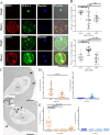
Fig 1
Fig 1. Localization analysis of SBP3 by indirect immunofluorescence assay (IFA) and immunoelectron microscopy (IEM).
(A) IFA images of B. bovis SBP3-Myc parasites. The parasites were stained with anti-Myc antibody (SBP3, red), anti-VESA-1 antibody (green), and anti-SBP4 antibody (green). Nuclei (Nuc) were stained with Hoechst 33342 (Hoechst, blue). Overlay shows bright field, anti-Myc antibody (SBP3, red) and anti-VESA-1 antibody (green) or SBP4 antibody (green). Scale bar = 5 μm. (B) Pearson’s correlation coefficient (PCC) values of the fluorescence images. Upper row: SBP3 and VESA-1, lower row: SBP3 and SBP4. Images were analyzed nearby the surface of iRBC, inside the parasites, and iRBC cytosol (n = 20, ns, non-significant; *** p < 0.001; **** p < 0.0001; determined by multiple comparisons one-way ANOVA test). (C) IEM images of B. bovis SBP3-Myc parasites. The parasites were stained with anti-Myc antibody. Gold particles (SBP3) were detected in spherical bodies (SB; arrowheads) and on the iRBC surface ridges (arrows). Scale bar = 500 nm. (D) Quantification of the number of gold particles nearby the ridges (Ridges) and other part of the iRBC surface (Surface of iRBC). Anti-Myc: images reacted with anti-Myc antibody. Control: negative control images reacted with rabbit IgG. (n = 20, n: the number of iRBCs used for counting the gold particles, **** p < 0.0001; determined by Mann-Whitney U test) (E) Quantification of the number of gold particles inside the parasites. Gold particles were counted on spherical bodies, nucleus, apical organelles and cytosol of the parasites. (n = 20, **** p < 0.0001; determined one-way ANOVA test).
Fig 2
Fig 2. Generation of B. bovis SBP3-inducible knockdown lines using glmS system.
(A) Schematic of plasmid to insert Myc-glmS sequences at the 3′ end of the sbp3 ORF showing the two Myc-tag, glmS, thioredoxin peroxidase-1 (tpx-1) 3′ noncoding region (NR), human dihydrofolate reductase (hDHFR) expression cassette, and homologous regions (HR 1 and 2). (B) Agarose gel electrophoresis image of diagnostic PCR to confirm integration of the Myc-glmS sequence. Tg1 and Tg2 indicate 2 independent clones. WT: parental strain parasite. (C) Western blot analysis of SBP3-glmS clones with (+) or without (-) glucosamine (GlcN). Anti-MSA2a antibody was used to detect MSA2a protein as a loading control. The image is representative of the experiments (other images are shown in S5 Fig). (D) Densitometry of SBP3 protein levels with (+) or without (-) GlcN. The data show average ± SE results of three independent experiments (* p ≤ 0.05; determined by paired Student t-test). (E) Growth of WT and SBP3-glmS lines with (+) or without (-) GlcN. Initial parasitemia was 0.1%, and parasitemia was monitored for 4 days with daily culture medium replacement. The data are shown as mean ± SE for three independent experiments. (** p < 0.01; **** p < 0.0001; determined by multiple t-test).
Fig 3
Fig 3. SBP3 KD decreases the surface localization of VESA-1.
(A) IFA images of SBP3-glmS parasite with (+) or without (-) GlcN. Parasites stained by anti-Myc antibody (SBP3, red), anti-VESA-1 antibody (green), anti-SBP4 antibody (green), and nuclei stained with Hoechst 33342 (Hoechst, blue) are shown. Overlay shows bright field and fluorescent images. Scale bar = 5 μm. (B) Fluorescence intensities of SBP3, VESA-1 and SBP4 near the surface of iRBCs before (-) and after (+) SBP3 KD (n = 10, ns; non-significant; **** p < 0.0001; determined by multiple t-test) (C) Western blot analysis of SBP3-glmS clones with (+) or without (-) of glucosamine (GlcN). Anti-VESA-1 and Anti-SBP4 antibodies were used to detect the proteins. Anti-MSA2a antibody was used to detect MSA2a protein as a loading control. The image is representative of three independent experiments. (D) Densitometry of SBP4 and VESA-1 protein levels with (+) or without (-) GlcN. The data show average±SE of three independent experiments (ns, non-significant; determined by paired Student t-test).
Fig 4
Fig 4. Severe decrease in ridge numbers on the surface of iRBCs.
(A) Transmission electron microscopy (TEM) images of SBP3-glmS parasite iRBCs with (+) or without (-) GlcN. Arrows indicate ridges and arrowheads indicate spherical bodies. Scale bar = 1 μm. (B) Quantification of ridge numbers on the surface of iRBCs of SBP3-glmS parasites in the presence or absence of GlcN. Ridge numbers were counted for 25 iRBCs (**** p < 0.0001 as determined by Mann-Whitney U test).
Fig 5
Fig 5. SBP3 KD decreases cytoadhesion of iRBCs to bovine brain endothelial cells (BBECs).
(A) Cytoadhesion assay of SBP3-glmS parasites with (+) or without (-) GlcN. Arrow indicate the BBECs and arrowheads indicate iRBCs. (B) Quantification of cytoadhered SBP3-glmS and WT parasite iRBCs to the 100 BBECs. The data are shown as the mean ± SE of a triplicate assay (ns, non-significant; ** p < 0.01 as determined by paired Student t-test).
Fig 6
Fig 6. Schematic diagram of ridge-related proteins in Babesia iRBC.
(A) Effect of SBP3 knockdown on ridge formation and localization of SBP3, VESA-1, and SBP4. When SBP3 was knocked down, VESA-1 was not exported to the surface of iRBCs, while there was no change in the expression and localization of SBP4. (B) B. bovis exportome localization and interaction of ridge-related proteins with iRBC proteins. SBP3 localizes at the ridge and interacts with iRBC cytoskeleton proteins (Band 3, Band 4.2, ankyrin 1 and spectrin filaments). MTM and VESA-1 are localized on the surface of ridges. VEAP and SBP4 are located in the iRBC cytoplasm. The schematic diagram was created with BioRender.com software.

References

    1. Bock R, Jackson L, De Vos A, Jorgensen W. Babesiosis of cattle. Parasitology. 2004;129(S1):S247–69. doi: 10.1017/s0031182004005190 - DOI - PubMed
    1. Florin-Christensen M, Suarez CE, Rodriguez AE, Flores DA, Schnittger L. Vaccines against bovine babesiosis: where we are now and possible roads ahead. Parasitology. 2014;141(12):1563–92. doi: 10.1017/S0031182014000961 - DOI - PubMed
    1. Vudriko P, Okwee-Acai J, Byaruhanga J, Tayebwa DS, Omara R, Muhindo JB, et al.. Evidence-based tick acaricide resistance intervention strategy in Uganda: concept and feedback of farmers and stakeholders. Ticks Tick Borne Dis. 2018;9(2):254–65. doi: 10.1016/j.ttbdis.2017.09.011 - DOI - PubMed
    1. Gohil S, Kats LM, Sturm A, Cooke BM. Recent insights into alteration of red blood cells by Babesia bovis: moovin’ forward. Trends Parasitol. 2010;26(12):591–9. - PubMed
    1. Aikawa M, Pongponratn E, Tegoshi T, Nakamura K, Nagatake T, Cochrane A, et al.. A study on the pathogenesis of human cerebral malaria and cerebral babesiosis. Mem Inst Oswaldo Cruz. 1992;87 Suppl 3:297–301. - PubMed

Substances

LinkOut - more resources