. 2024 Nov 11;20(11):e1012644.
doi: 10.1371/journal.ppat.1012644. eCollection 2024 Nov.

A model of lymphocryptovirus-associated post-transplant lymphoproliferative disorder in immunosuppressed Mauritian cynomolgus macaques

Affiliations

A model of lymphocryptovirus-associated post-transplant lymphoproliferative disorder in immunosuppressed Mauritian cynomolgus macaques

Helen L Wu et al. PLoS Pathog. .

Abstract

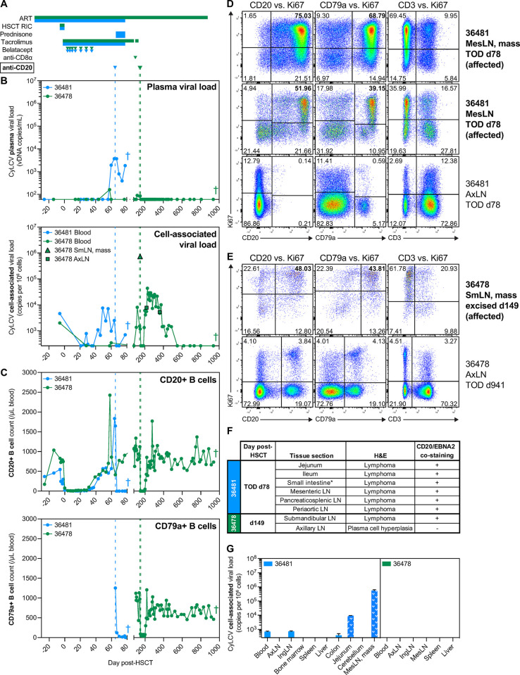
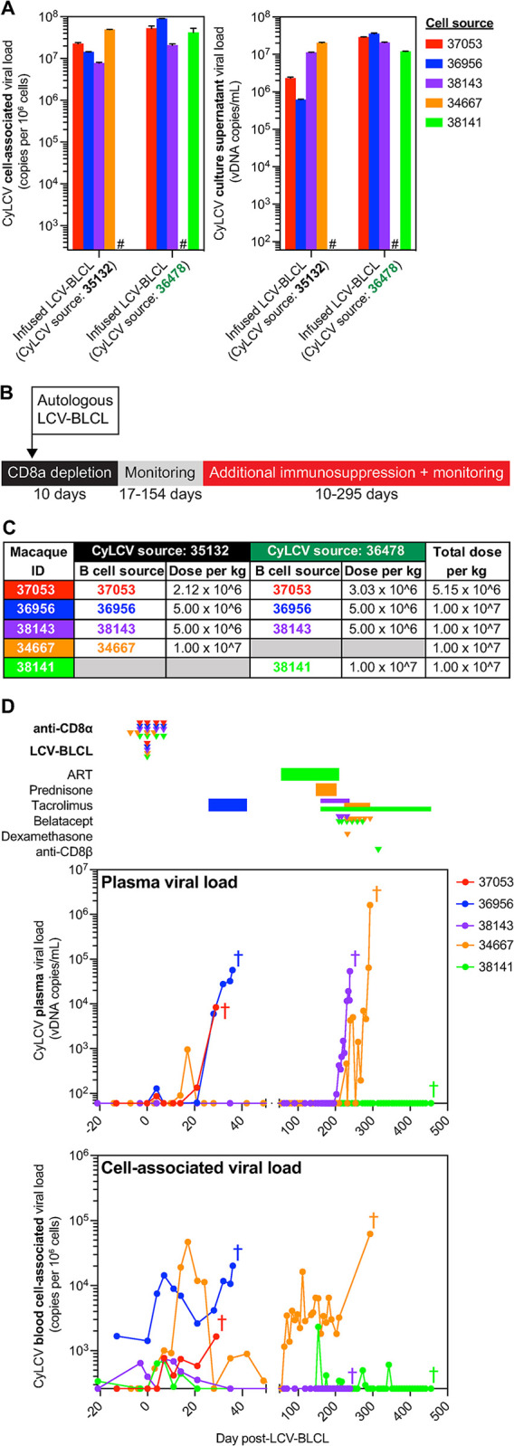
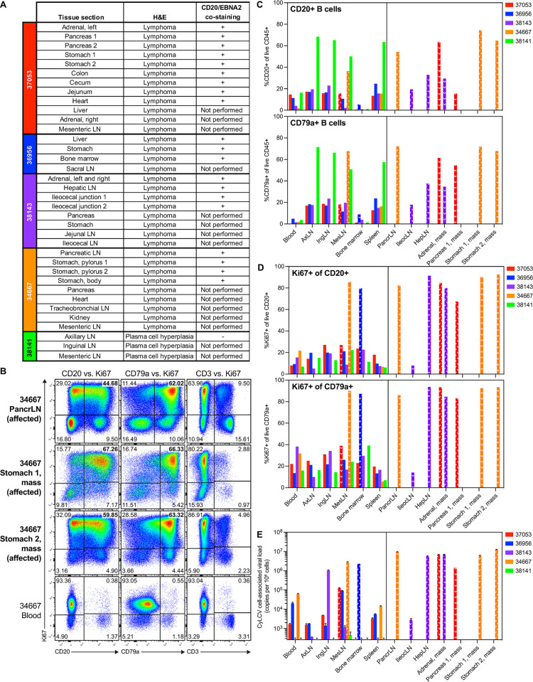
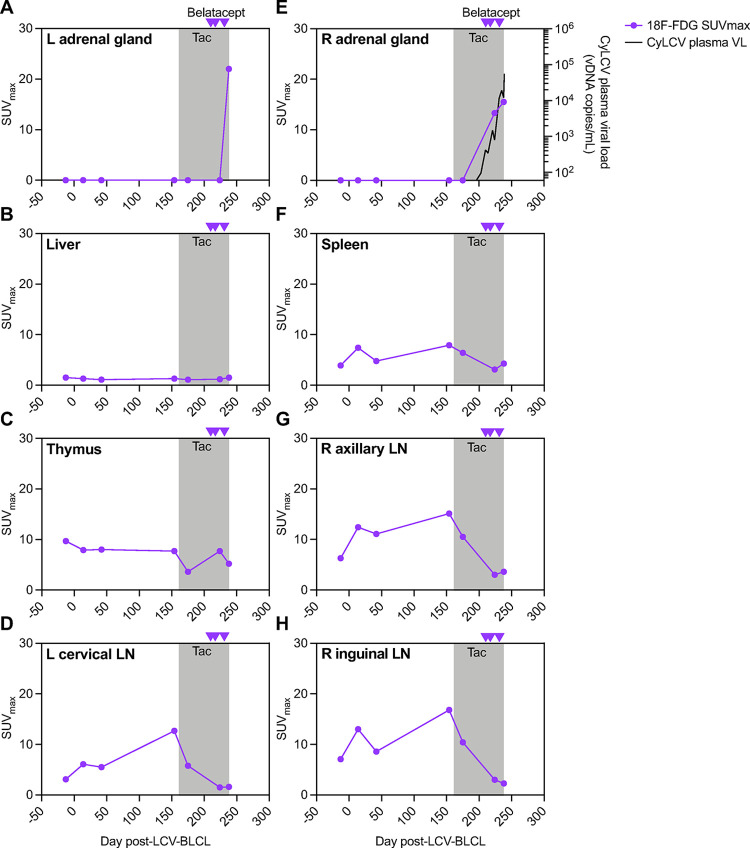
Immunocompromised individuals are at risk for developing lymphocryptovirus-associated lymphoproliferative diseases, such as Epstein Barr virus (EBV)-associated B cell lymphomas and post-transplant lymphoproliferative disorder (PTLD). We previously reported development of cynomolgus lymphocryptovirus (CyLCV)-associated PTLD in Mauritian cynomolgus macaques (MCMs) undergoing hematopoietic stem cell transplantation (HSCT), which mirrored EBV-PTLD in transplant patients. Here, we sought to develop a MCM model of lymphocryptovirus-associated lymphoproliferative disease in immunosuppressed MCMs without HSCT. Five simian immunodeficiency virus (SIV)-infected, CD8α+ cell-depleted MCMs received an infusion of autologous B-lymphoblastoid cells transformed with CyLCV, followed by varying degrees of immunosuppression. Four of five infused macaques developed masses coincident with increasing CyLCV plasma viremia, and necropsies confirmed the presence of multicentric lymphomas, which most commonly manifested in lymph nodes, gastrointestinal tract, adrenal glands, and pancreas. Affected tissues harbored neoplastic lymphocytes double-positive for CD20 and CyLCV EBNA2 antigen, large frequencies of proliferating B cells, and high levels of cell-associated CyLCV DNA. In addition, longitudinal 18F-fluorodeoxyglucose positron-emission tomography (18F-FDG PET) of one MCM successfully detected lymphoproliferative disease in the adrenal glands prior to clinical signs of disease. These data demonstrate successful induction of lymphocryptovirus-associated PTLD-like disease in 4 of 5 MCMs, and thus support the use of MCMs as a preclinical NHP model of EBV-associated lymphoproliferative disease that could be employed to test novel diagnostic and therapeutic modalities.


Conflict of interest statement

The authors have declared that no competing interests exist.

Figures

Fig 1
Fig 1. Treatment of CyLCV-associated post-transplant lymphoproliferative disease in HSCT recipient MCMs is challenging.
(A) Drug regimens with timepoints aligned to graphs in B, C, with colors corresponding to each MCM shown in graphs. (B) Longitudinal plasma (top) and cell-associated (bottom) CyLCV DNA viral loads. Limit of quantification (LOQ) = 60 copies/mL for plasma, 260 copies/million cells for cell-associated. Undetectable and below LOQ measurements graphed at the LOQ. Colored vertical dashed lines represent timepoints of anti-CD20 monoclonal antibody administration. Graphs show mean viral loads of two qPCR replicates. (C) Longitudinal CD20+ B cell (top) and CD79a+ B cell (bottom) absolute counts in blood. (D, E) Representative flow cytometry plots showing intracellular Ki67 staining of B cells and T cells in MCM 36481 (D) and 36478 (E) tissues. Plots are gated on live, CD45+ singlets. Numbers indicate cell frequencies within each quadrant. Bolded tissues represent affected tissues positive for lymphoma. (F) Table summarizes tissue sections with signs of lymphoproliferative disease assessed by H&E and presence of cells double-positive for CD20 and EBNA2 assessed by dual immunohistochemistry. *section not specified. (G) Cell-associated CyLCV DNA viral loads of necropsy tissues. White patterned bars denote tissues positive for lymphoma. Bars show mean ±SD of two qPCR replicates. Limit of quantification (LOQ) = 260 copies/million cells for cell-associated. Undetectable and below LOQ measurements graphed at the LOQ. Colored crosses next to datapoints indicate time of euthanasia/death for each MCM. ART = antiretroviral therapy, RIC = reduced intensity conditioning, SmLN = submandibular lymph node, AxLN = axillary lymph node, IngLN = inguinal lymph node, MesLN = mesenteric lymph node, TOD = time of death.
Fig 2
Fig 2. Generation and infusion of CyLCV-BLCLs into immunosuppressed LCV model MCMs.
(A) Cell-associated (left) and supernatant (right) CyLCV DNA viral loads in ex vivo-generated CyLCV-BLCL cultures using CyLCV from PTLD-experiencing HSCT recipient MCM. Bars show mean ±SD of two qPCR replicates. Hashtags (#) indicate CyLCV-BLCL lines not generated. (B) Study outline for LCV model MCMs. (C) Table summarizing dosing of autologous CyLCV-BLCL infusions into LCV model MCMs. (D) Longitudinal plasma (top) and blood cell-associated (bottom) CyLCV DNA viral loads. LOQ = 60 copies/mL for plasma, 260 copies/million cells for blood cells. Graphs show mean viral loads of two qPCR replicates. Drug regimens and CyLCV-BLCL infusion timepoints indicated above graphs with colors corresponding to each MCM shown in graphs. Colored crosses next to datapoints indicate time of euthanasia/death for each MCM.
Fig 3
Fig 3. Majority of CyLCV-BLCL-infused, immunosuppressed LCV model MCMs develop CyLCV-associated lymphoproliferative disease.
(A) Table summarizes tissue sections with signs of lymphoproliferative disease assessed by H&E staining and presence of cells double-positive for CD20 and EBNA2 assessed by dual immunohistochemistry. (B) Representative flow cytometry plots showing intracellular Ki67 staining of B cells and T cells in MCM 34667 necropsy tissues. Plots are gated on live, CD45+ singlets. Numbers indicate cell frequencies within each quadrant. Bolded tissues represent affected tissues positive for lymphoma. (C, D) Summary graphs of flow cytometry staining of LCV model MCM necropsy tissues. Frequencies of CD20+ B cells and CD79a+ B cells among live CD45+ cells (C). Frequencies of Ki67+ cells among live CD20+ B cells and CD79a+ B cells (D). White patterned bars denote affected tissues positive for lymphoma. Bars not shown indicate samples not assayed. (E) Cell-associated CyLCV DNA viral loads of necropsy tissues. White patterned bars denote affected tissues positive for lymphoma. Bars show mean ±SD of two qPCR replicates. LOQ = 260 copies/million cells. Asterisks (*) indicate samples that measured below LOQ. Other bars not shown indicate samples not assayed. PancrLN = pancreatic lymph node, AxLN = axillary lymph node, IngLN = inguinal lymph node, MesLN = mesenteric lymph node, IleocLN = ileocecal lymph node, HepLN = hepatic LN.
Fig 4
Fig 4. 18F-FDG PET of model MCM 38143.
(A-H) Longitudinal maximum standardized uptake values (SUVmax) for 18F-FDG in various tissues from scans of model MCM 38143. Gray boxes denote the period of daily tacrolimus (tac) treatment. Purple arrowheads denote timepoints of belatacept administration. Longitudinal 38143 plasma CyLCV DNA viral loads are overlayed on right adrenal gland graph (E, right axis), line shows mean viral load of two qPCR replicates. LOQ = 60 copies/mL plasma. Undetectable and below LOQ measurements graphed at the LOQ. L = left, R = right, LN = lymph node.

References

    1. Hanto DW, Frizzera G, Gajl-Peczalska KJ, Simmons RL. Epstein-Barr virus, immunodeficiency, and B cell lymphoproliferation. Transplantation. 1985;39: 461–472. doi: 10.1097/00007890-198505000-00001 - DOI - PubMed
    1. Young LS, Yap LF, Murray PG. Epstein–Barr virus: more than 50 years old and still providing surprises. Nat Rev Cancer. 2016;16: 789–802. doi: 10.1038/nrc.2016.92 - DOI - PubMed
    1. Snow AL, Martinez OM. Epstein-Barr Virus: Evasive Maneuvers in the Development of PTLD. Am J Transplant. 2007;7: 271–277. doi: 10.1111/j.1600-6143.2006.01650.x - DOI - PubMed
    1. Allen U, Alfieri C, Preiksaitis J, Humar A, Moore D, Tapiero B, et al.. Epstein-Barr virus infection in transplant recipients: Summary of a workshop on surveillance, prevention and treatment. Can J Infect Dis J Can des Mal Infect. 2001;13: 89–99. doi: 10.1155/2002/634318 - DOI - PMC - PubMed
    1. Martinez OM, Gruijl FRD. Molecular and Immunologic Mechanisms of Cancer Pathogenesis in Solid Organ Transplant Recipients. Am J Transplant. 2008;8: 2205–2211. doi: 10.1111/j.1600-6143.2008.02368.x - DOI - PubMed

MeSH terms

LinkOut - more resources