. 2024 Nov 19;121(47):e2409790121.
doi: 10.1073/pnas.2409790121. Epub 2024 Nov 11.

Subchronic elevation in ambient temperature drives alterations to the sperm epigenome and accelerates early embryonic development in mice

Affiliations

Subchronic elevation in ambient temperature drives alterations to the sperm epigenome and accelerates early embryonic development in mice

Natalie Trigg et al. Proc Natl Acad Sci U S A. .

Abstract

Forecasted increases in the prevalence and severity of extreme weather events accompanying changes in climatic behavior pose potential risk to the reproductive capacity of humans and animals of ecological and agricultural significance. While several studies have revealed that heat stress induced by challenges such as testicular insulation can elicit a marked negative effect on the male reproductive system, and particularly the production of spermatozoa, less is known about the immediate impact on male reproductive function following subchronic whole-body exposure to elevated ambient temperature. To address this knowledge gap, we exposed unrestrained male mice to heat stress conditions that emulate a heat wave (daily cycle of 8 h at 35 °C followed by 16 h at 25 °C) for a period of 7 d. Neither the testes or epididymides of heat-exposed male mice exhibited evidence of gross histological change, and similarly, spermatozoa of exposed males retained their functionality and ability to support embryonic development. However, the embryos generated from heat-exposed spermatozoa experienced pronounced changes in gene expression linked to acceleration of early embryo development, aberrant blastocyst hatching, and increased fetal:placental weight ratio. Such changes were causally associated with an altered sperm small noncoding RNA (sncRNA) profile, such that these developmental phenotypes were recapitulated by microinjection of wild-type embryos sired by control spermatozoa with RNAs extracted from heat-exposed spermatozoa. Such data highlight that even relatively modest excursions in ambient temperature can affect male reproductive function and identify the sperm sncRNA profile as a particular point of vulnerability to this imposed environmental stress.

Keywords: embryo development; epididymis; heat; small non-protein-coding RNA; sperm.


Conflict of interest statement

Competing interests statement:The authors declare no competing interest.

Figures

Fig. 1.
Fig. 1.
Effect of paternal heat stress on the quality of mouse spermatozoa. At necropsy, cauda epididymal spermatozoa were isolated from heat-treated or control mice and prepared for assessment of (AF) motility parameters. Several sperm motility and velocity parameters were objectively assessed via CASA including (A) total motility, (B) progressive motility, (C) linearity (LIN, %), (D) average path velocity (VAP, μm/s), (E) straight line velocity (VSL, μm/s), and (F) amplitude of lateral head displacement (ALH, μm). (G) Spermatozoa were also induced to undergo in vitro capacitation before assessment of tyrosine phosphorylation status via immunoblotting of cell lysates and immunostaining of fixed cells with anti-phosphotyrosine antibodies. Additional measures of sperm quality including (H) MSR, (I) sperm membrane fluidity (MC540), (J) area of Halo, and (K) intensity of 8-OHdG labeling within the sperm Halo were also recorded. Data are presented as mean ± SEM having been calculated based on the assessment of spermatozoa from n = 4 to 12 mice/group for each assay; circle symbols depict values obtained from populations of spermatozoa from individual mice. Differences between groups were assessed by unpaired Student’s t test for normally distributed data or unpaired Mann–Whitney test for data not normally distributed.
Fig. 2.
Fig. 2.
Impact of paternal heat stress on IVF and embryo development. Cauda epididymal spermatozoa from control and heat-exposed mice were isolated and added into droplets containing oocytes from untreated females to permit IVF. Note that all data are presented as arcsine transformed values and each circle symbol represents an individual IVF experiment (i.e., replicate) featuring the spermatozoa from one male and oocytes from six female mice. (A) The number of fertilized oocytes after 4 h of gamete coincubation was recorded, and data are presented as the transformed total 2-cell embryos at 24 h. (B) Zygotes were cultured for 96 h during which development was tracked. The development rate for each group is represented as transformed data based on the proportion of 2-cell embryos that developed to blastocyst. The developmental stage of embryos sired from control and heat-exposed spermatozoa at (C) 24, (D) 48, (E) 72, and (F) 96 h is depicted. (G) Following 96 h of culture, the number of embryos displaying multiple hatching sites was recorded and presented as transformed data based on the percentage of hatching blastocysts. (H) Representative phase images of embryos cultured for 96 h are presented to illustrate the phenomenon of embryos with multiple hatching sites (black arrowheads). (I) Blastocysts were stained for polymeric actin (phalloidin-fluorescein isothiocyanate; green) and alpha tubulin (red) prior to counterstaining with the nuclear marker, DAPI (blue). Representative phase and merged images captured by confocal microscopy are presented. (J) Alternatively, blastocysts were dual stained for γH2AX (green), a molecular marker of DNA damage (specifically, DNA double-strand breaks), and DAPI (blue). (K) Quantitation of γH2AX foci was performed using corrected total cell fluorescence (CTCF) protocols and graphical data are presented as mean ± SEM wherein circle symbols depict the CTCF values recorded in individual embryos (n = 36 or n = 40 embryos fertilized with the spermatozoa of control or heat-treated males, respectively). Embryo development data were arcsine transformed prior to statistical analysis. Differences between groups were assessed by (AG) paired Student’s t test for normally distributed data, or paired Wilcoxon matched-pairs signed-rank test for data not normally distributed, or (K) unpaired Mann–Whitney test for data not normally distributed. * indicates P ≤ 0.05, # indicates P ≤ 0.1. (HJ) (Scale bars, 40 μm.).
Fig. 3.
Fig. 3.
Effect of paternal heat stress on pregnancy outcomes. At the completion of their treatment regimen, heat-exposed and control male mice (n = 14 per treatment) were mated with untreated females (n = 2 per breeding pair). Thereafter, females were killed at 17.5 d postcoitus, and the following parameters were assessed: (A) Plug:pregnancy ratio (defined as the number of mated females with at least one viable implantation site), (B) total number of implantation sites per pregnant mouse, (C) total number of viable implantation sites per pregnant mouse, and (D) the proportion of implantation sites per pregnant female undergoing resorption. (E) Genomic DNA from fetal tail clippings was used to determine fetal sex. (F) Representative gel image of Sly/Xlr gene products amplified by PCR. This approach yields 685-bp, 660-bp, and 480-bp products for female gDNA (i.e., white arrow; F) or alternatively, a 280-bp amplicon and often faint “X chromosome” bands for male gDNA (i.e., blue arrow; M). Lane 1 contains a 100 bp DNA ladder; lanes 2 to 14 contain fetal gDNA; lane 15 contains a no template control (NTC) sample. (G) Fetal weight, (H) placental weight, and (I) fetal:placental weight ratio (surrogate marker for placental efficiency). Assessment of the placenta was performed on histological sections stained with Masson’s trichrome, and the (J) total area, as well as the area of the (K) labyrinth, (L) junctional, and (M) decidual zones, was recorded. Representative images of fetal placentae from pups sired by either (N) control or (O) heat-exposed males are depicted, with dashed lines demarcating the boundaries of the different zones assessed in this study. (Scale bars, 1,000 μm.). Graphical data are presented as (BD and JM) mean ± SEM values with circle symbols depicting values from individual mice or (GI) estimated marginal mean ± SEM. The effect of heat stress was assessed from n = 16 to 19 litters by (A and E) chi-square analysis, (BE and JM) unpaired Student’s t test for normally distributed data or unpaired Mann–Whitney test for data not normally distributed, and (GI) Generalized Linear Mixed Model and post hoc least significant difference test, with the subject as father (mother nested) and viable pups as a covariate. * indicates P ≤ 0.05; # indicates P ≤ 0.1.
Fig. 4.
Fig. 4.
The influence of heat stress on the sncRNA payload of cauda epididymal spermatozoa. At the cessation of heat exposure, spermatozoa were isolated from the cauda epididymides of control and heat-treated mice and prepared for sncRNA isolation and sequencing (n = 3). (A) Proportional contribution of each sncRNA subclass to the global sncRNA payload of control and heat-exposed spermatozoa. (B) Size distribution of tRFs, piRNA, and miRNA mapping sncRNA transcripts. Data represent average of three replicates for each treatment group. (C) Volcano plots depicting differentially accumulated sperm sncRNAs from heat-exposed compared to control males. Blue and red circle symbols denote individual sncRNAs that were either significantly reduced or increased in abundance in heat-exposed compared to control spermatozoa, respectively. Graphical data are presented as mean values of three biological replicates. The effect of heat stress was assessed by (B) an unpaired t test or (C) DESeq2, represented by volcano plots of tRF, piRNA, and miRNA expression values with a significance threshold of fold change ± 1.5 and P-value ≤ 0.05.
Fig. 5.
Fig. 5.
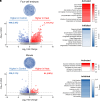
The legacy of paternal heat stress on the preimplantation embryo transcriptome. Following IVF, embryos were cultured for either (AC) 46 h (i.e., 4-cell embryos) or (DF) 72 h (i.e., morula embryos) before being prepared for single embryo mRNA-Seq (n = 16 to 22 single embryos per group). (A and D) Volcano plots depicting fold change (x-axis, log2) and P-value (y-axis, −log10) of identified mRNA transcripts in 4-cell and morula stage embryos generated with spermatozoa from heat-exposed compared to control sires. Blue and red circle symbols indicate individual genes deemed to be significantly down- and up-regulated (P-value ≤0.05 and fold change ±1.5) in embryos sired by the spermatozoa of heat-exposed males, respectively. (B, C, E, and F) Highest ranked disease and functions identified by Ingenuity Pathway Analysis (IPA) software as being either (B and E) activated or (C and F) inhibited in embryos fertilized with the spermatozoa of heat-exposed compared to control males.
Fig. 6.
Fig. 6.
Representative network analysis linking heat-sensitive sperm miRNAs to dysregulation of embryo gene expression. The MicroRNA Target Prediction Database software was used to identify the predicted gene targets of miRNAs that were differentially accumulated in the spermatozoa of heat-exposed males and that were themselves dysregulated in the 4-cell embryos generated from this population of spermatozoa. These data were interrogated using IPA to determine embryonic cellular pathways that may be susceptible to dysregulation by heat-associated changes in the sperm miRNA landscape. Among the dysregulated pathways identified by this strategy, “size of body” is presented to illustrate the reciprocal relationship that exists between the expression profile of heat-sensitive sperm miRNAs and that of their predicted target genes in 4-cell embryos. Specifically, red = up-regulated miRNAs/genes, blue = down-regulated miRNAs/genes, and connecting lines indicate the interaction network between sperm miRNAs and the embryonic genes they are predicted to regulate. The network schematic was initially generated by IPA before being redrawn using BioRender software (BioRender).
Fig. 7.
Fig. 7.
The impact of heat-exposed sperm RNA on embryo development. (A) Zygotes generated by conventional IVF were microinjected with total RNA isolated from the spermatozoa of either control male mice or those subjected to heat stress. Note that all data are presented as arcsine transformed values and each circle symbol represents an individual IVF/microinjection experiment (i.e., replicate) in which naïve control mice were used as initial oocyte (3 to 4 females) and sperm (1 male) donors. (B) Development of embryos at 24 h postfertilization that were injected with control or heat-exposed sperm RNA. Embryos were cultured until 96 h, and development was recorded at intervals of (C) 48 h, (D) 72 h, and (E) 96 h. Graphical data depict the arcsine transformed value calculated from the percentage of 2-cell embryos at each developmental milestone and are presented as mean ± SEM. Differences between groups were assessed by paired Student’s t test for normally distributed data or paired Wilcoxon matched-pairs signed-rank test for data not normally distributed. * indicates P ≤ 0.05; # indicates P < 0.1.

References

    1. Climate Change 2014: Synthesis Report, Contribution of Working Groups I, II and III to the Fifth Assessment Report of the Intergovernmental Panel on Climate Change, Pachauri R. K., Meyer L. A., Eds. (Intergovernmental Panel on Climate Change, Geneva, Switzerland, 2014), p. 151.
    1. Robinson P. J., On the definition of a heat wave. J. Appl. Meteorol. 40, 762–775 (2001).
    1. Nienaber J. A., Hahn G. L., Brown-Brandl T. M., Eigenberg R. A., “Summer heat waves–Extreme years” in 2007 ASAE Annual Meeting (American Society of Agricultural and Biological Engineers, 2007).
    1. Nidumolu U., et al. , Spatio-temporal modelling of heat stress and climate change implications for the Murray dairy region, Australia. Int. J. Biometeorol. 58, 1095–1108 (2014). - PubMed
    1. Thompson V., et al. , The 2021 western North America heat wave among the most extreme events ever recorded globally. Sci. Adv. 8, eabm6860 (2022). - PMC - PubMed