. 2024 Nov 11;15(1):9746.
doi: 10.1038/s41467-024-53554-1.

A cyclic peptide toolkit reveals mechanistic principles of peptidylarginine deiminase IV regulation

Affiliations

A cyclic peptide toolkit reveals mechanistic principles of peptidylarginine deiminase IV regulation

M Teresa Bertran et al. Nat Commun. .

Abstract

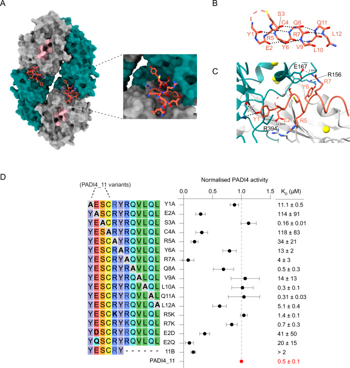
Peptidylarginine deiminase IV (PADI4, PAD4) deregulation promotes the development of autoimmunity, cancer, atherosclerosis and age-related tissue fibrosis. PADI4 additionally mediates immune responses and cellular reprogramming, although the full extent of its physiological roles is unexplored. Despite detailed molecular knowledge of PADI4 activation in vitro, we lack understanding of its regulation within cells, largely due to a lack of appropriate systems and tools. Here, we develop and apply a set of potent and selective PADI4 modulators. Using the mRNA-display-based RaPID system, we screen >1012 cyclic peptides for high-affinity, conformation-selective binders. We report PADI4_3, a cell-active inhibitor specific for the active conformation of PADI4; PADI4_7, an inert binder, which we functionalise for the isolation and study of cellular PADI4; and PADI4_11, a cell-active PADI4 activator. Structural studies with PADI4_11 reveal an allosteric binding mode that may reflect the mechanism that promotes cellular PADI4 activation. This work contributes to our understanding of PADI4 regulation and provides a toolkit for the study and modulation of PADI4 across (patho)physiological contexts.


Conflict of interest statement

Competing interests M.T.B., R.W., T.C., M.A.C. and L.J.W. are inventors on UK Patent application No. 230979.0, which has been filed with respect to methods that modulate cellular reprogramming through enhancing the activity of the PADI4 enzyme. The remaining authors declare no competing interests.

Figures

Fig. 1
Fig. 1. Schematic of the RaPID selection strategies used to identify PADI4-binding cyclic peptides.
RaPID selections were carried out against PADI4 as bait under three different conditions to target different conformations of PADI4.
Fig. 2
Fig. 2. PADI4_3 is a potent inhibitor of calcium-bound PADI4.
A Inhibition of PADI4 by cyclic peptides identified in the RaPID selections. COLDER assays were performed at different peptide concentrations (10–0.0003 µM) in the presence of 10 mM CaCl2. Data were normalised to activity of PADI4 in the presence of 0.1% DMSO. Data shows mean ± SEM of three independent replicates. Each replicate was done in triplicate. B Overall cryoEM structure of PADI4 homodimer (N-terminal immunoglobulin-like domains shown as grey surface, C-terminal catalytic domain as green surface) bound to PADI4_3 (orange sticks). Inset shows a zoomed-in view of the peptide binding site. C View from a cryoEM structure of PADI4 homodimer (grey cartoon) bound to PADI4_3 (orange sticks) highlighting a network of inter- and intramolecular interactions formed by PADI4_3. Intramolecular hydrogen bonds are shown as orange dashed lines, and intermolecular bonds as black dashed lines. D View from a cryoEM structure of PADI4 homodimer (grey cartoon) bound to PADI4_3 (orange sticks) highlighting the core catalytic residues of PADI4. His4 of PADI4_3 occupies the site bound by arginine residues in substrate peptides. E. Mutational scanning reveals which regions of PADI4_3 appear tolerant to amino acid substitutions. Columns show each position in the PADI4_3 sequence, and rows show individual amino acid substitutions. Data were plotted as the mean enrichment score from three independent selections where red represents amino acids with high binding affinity and blue represents amino acids with low binding affinity. Black squares highlight the parent amino acids. F. Sequence of the PADI4_3 analogues synthesised by SPPS (left) and their calculated IC50 and Kd values (right). Activity data was measured as in A and Kd values determined by SPR. Both IC50 and Kd values show mean ± SEM from three independent replicates.
Fig. 3
Fig. 3. PADI4_3 inhibits PADI4 in mES cells and human neutrophils.
A Representative high-content immunofluorescence images of hPADI4-stable mES cells stimulated with KSR/2i in the presence of increasing concentrations of PADI4_3 for 3 h. Immunostaining of citrullinated histone H3 (H3Cit) is shown in green, and DAPI in blue. Scale bar = 50 µm. B High content imaging-based quantification of mean H3Cit immunofluorescence intensity in hPADI4-stable mES cells stimulated with KSR/2i in the presence of increasing concentrations of PADI4_3 or PADI4_3i for 3 h. Data represent mean ± SD from at least 500 cells from a representative experiment. C High content imaging-based quantification of mean H3Cit immunofluorescence intensity in hPADI4-stable mES cells treated with increasing concentrations of PADI4_3 or PADI4_3i for 3 h. Each data point represents the average H3Cit intensity per condition of one experiment. D Immunoblots showing citrullinated histone H3, total histone H3 and MPO levels from NET preparations following stimulation with 1 mg/ml cholesterol crystals and either DMSO (0.5%), 50 μM PADI4_3 or 50 μM PADI4_3i control. Data show a representative experiment after two repeats. E (Left) representative immunofluorescence confocal micrographs of human neutrophils pre-incubated with vehicle, PADI4_3 or PADI4_3i peptide for 60 minutes and stimulated with Ca-Ionophore (Time stamp = 12 h). Citrullinated H3 is shown in cyan and DAPI in blue. Scale bar = 40 µm. (right) Quantification of the immunofluorescence micrographs of the percentage of citrullinated H3-positive cells over the total number of cells. Data represent mean ± SD from six technical replicates. Kruskal–Wallis test was used to determine if the differences among means were significantly different from each other. p-values were adjusted using Dunn’s correction for multiple comparisons. Data show a representative experiment after two repeats. F Cell lysates from calcium ionophore-stimulated and unstimulated human neutrophils pre-incubated with vehicle or PADI4_3 peptide (50 µM). Cells were collected at 10, 30 and 90 min post-stimulation and immunoblotted for citrullinated H3 and MPO. G (Left) Representative bright field micrographs of human neutrophils pre-incubated with vehicle, PADI4_3 or PADI4_3i peptide, imaged 12 h post-stimulation with Ca-ionophore. (Right) Quantification of the percentage of NETotic cells over total cells imaged in the micrographs. Quantification was done as in (E).
Fig. 4
Fig. 4. PADI4_11 is a PADI4 activator.
A PADI4_11 and PADI4_12 are PADI4 activators in vitro. Activation COLDER assays with PADI4 and different PADI4-binding peptides. Assays were performed with different concentrations of CaCl2 (10–0.003 mM) and 30 µM peptide. K50Ca2+ is the concentration of CaCl2 that yields half maximum PADI4 activity. Data were normalised against the activity of PADI4 in the presence of 0.1% DMSO and 10 mM CaCl2 and represents mean ± SEM of three independent replicates. Each replicate was done in triplicate. B PADI4_11 and PADI4_12 activate PADI4 at low calcium concentrations. COLDER assays were performed at different concentrations of peptides (0.003–100 µM) and 0.1 mM CaCl2. Data represents mean ± SEM of three independent replicates done in triplicate. C Representative high-content immunofluorescence images of hPADI4-stable mES cells in the presence of increasing concentrations of PADI4_11 for 1 h. Citrullinated histone H3 (H3Cit) is shown in green, and DAPI in blue. Scale bar = 50 µm. D High content imaging-based quantification of mean H3Cit immunofluorescence intensity in hPADI4-stable mES cells treated with increasing concentrations of PADI4_11 or PADI4_11i for 1 h. Each data point represents the mean H3Cit intensity per cell. All cells from three technical replicates and three individual experiments are included. E High content imaging-based quantification of mean H3Cit immunofluorescence intensity in hPADI4-stable mES cells treated with increasing concentrations of PADI4_11 or PADI4_11i for 1 h. Each data point represents the average H3Cit intensity per condition from three technical replicates and three individual experiments. F High content imaging-based quantification of mean H3Cit immunofluorescence intensity in control mES cells treated with increasing concentrations of PADI4_11 or PADI4_11i for 1 h. Each data point represents the mean ± SD H3Cit intensity of at least 3000 cells per condition from a representative experiment. G Calcium influx into cells, as measured by Calbryte intensity, after treatment with 25 μM PADI4_11 or PADI4_11i. Calcium ionophore A23187 (10 μM) was used as a positive control for calcium influx. Data represent mean ± SD of two biological replicates. H Flow cytometry-based quantification of propidium iodide (PI) incorporation into cells, as a measure of cell toxicity and membrane permeability, after treatment with increasing concentrations of PADI4_11 or PADI4_11i. Hydrogen peroxide (H2O2) is used as a positive control for cell toxicity. Data represent mean ± SD of three biological replicates.
Fig. 5
Fig. 5. PADI4_11 activates PADI4 by allosteric binding.
A Views from a cryoEM structure of PADI4 homodimer (N-terminal immunoglobulin-like domains in grey surface, C-terminal catalytic domain in green surface and active site in pink) bound to PADI4_11 (coral sticks). The inset shows a zoomed-in view of the peptide binding site. B View of PADI4_11 forming an alpha-helical structure. Intramolecular backbone interactions are shown as black dashed lines. C View from PADI4 homodimer (grey and cyan) bound to PADI4_11 (coral) showing a network of intramolecular interactions between PADI4_11 and PADI4. Interactions are shown as black dashed lines. D (Left) Sequence of the different PADI4_11 analogues synthesised by SPPS and in vitro activity measured by COLDER assay (centre). Assays were performed with 50 nM PADI4 in the presence of 0.1 mM CaCl2 and 100 µM peptide. Data were normalised against the activity of PADI4 with 0.1 mM CaCl2 and 100 µM of PADI4_11. KD values were measured by SPR (right). Data represents mean ± SEM of three independent repeats. Each COLDER experiment was performed in technical triplicate.
Fig. 6
Fig. 6. Pull-downs with bio-PADI4_7.
A Immunoblot analysis for hPADI4. Pull-down assays were performed from hPADI4-stable or Control mES cells using bio-PADI4_7 as bait, or biotin-coated beads as negative control. Data show a representative experiment after two repeats. B Volcano plot representing the proteins enriched in bio-PADI4_7 versus bio-PADI4_7scr pull-downs from hPADI4-stable mES cells cultured in Serum conditions. Proteins highlighted in red were identified in all three replicates, with two sample Student’s t-tests S0 = 0, both sides and p value ≤ 0.05 and Log2FC ≥2 for enrichment with the bio-PADI4_7 against bio-PADI4_7scr. C Volcano plot representing the proteins enriched in bio-PADI4_7 versus bio-PADI4_7scr pull-downs from hPADI4-stable mES cells cultured in KSR medium for 3 h. Proteins highlighted in red were identified in all three replicates, with two sample Student’s t-tests S0 = 0, both sides and p value ≤ 0.05 and Log2FC ≥2 for enrichment with the bio-PADI4_7 against bio-PADI4_7scr. D Volcano plot representing the proteins enriched in bio-PADI4_7 pull-downs from hPADI4-stable mES cells stimulated with KSR- versus serum-containing medium for 3 h. Two sample Student’s t-tests (S0 = 0, both sides and p value, FDR at 0.05; Log2FC ≥1) were used on proteins found in all three replicates in each condition. Proteins highlighted in red (for enrichment in KSR) or blue (for enrichment in Serum) are also enriched in bio-PADI4_7 over PADI4_7scr pulldowns with Log2FC ≥2.

References

    1. Vossenaar, E. R., Zendman, A. J. W., van Venrooij, W. J. & Pruijn, G. J. M. PAD, a growing family of citrullinating enzymes: genes, features and involvement in disease. BioEssays25, 1106–1118 (2003). - PubMed
    1. Christophorou, M. A. The virtues and vices of protein citrullination. R. Soc. Open Sci.9, 220125 (2022). - PMC - PubMed
    1. Cuthbert, G. L. et al. Histone deimination antagonizes arginine methylation. Cell118, 545–553 (2004). - PubMed
    1. Wang, Y. et al. Human PAD4 regulates histone arginine methylation levels via demethylimination. Science306, 279–283 (2004). - PubMed
    1. Hagiwara, T., Nakashima, K., Hirano, H., Senshu, T. & Yamada, M. Deimination of arginine residues in nucleophosmin/B23 and histones in HL-60 granulocytes. Biochem. Biophys. Res. Commun.290, 979–983 (2002). - PubMed

Publication types

MeSH terms

LinkOut - more resources