. 2024 Nov 11;15(1):9751.
doi: 10.1038/s41467-024-54150-z.

Split-design approach enhances the therapeutic efficacy of ligand-based CAR-T cells against multiple B-cell malignancies

Affiliations

Split-design approach enhances the therapeutic efficacy of ligand-based CAR-T cells against multiple B-cell malignancies

Shuhong Li et al. Nat Commun. .

Abstract

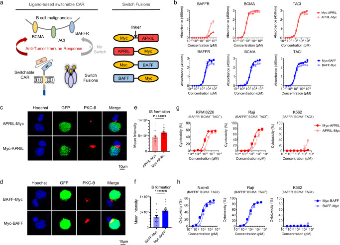
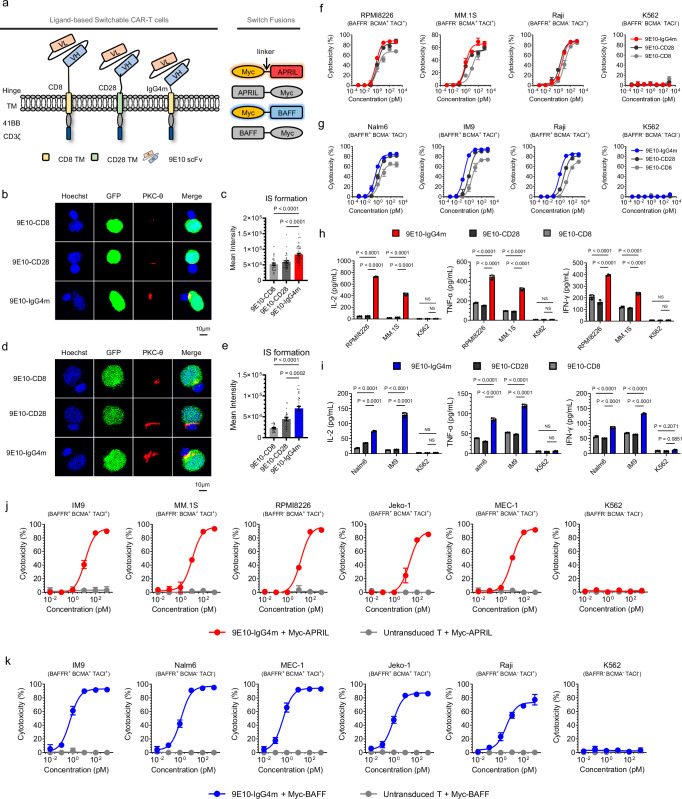
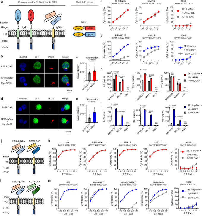
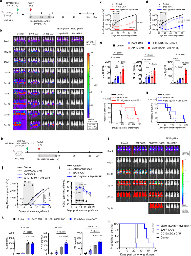
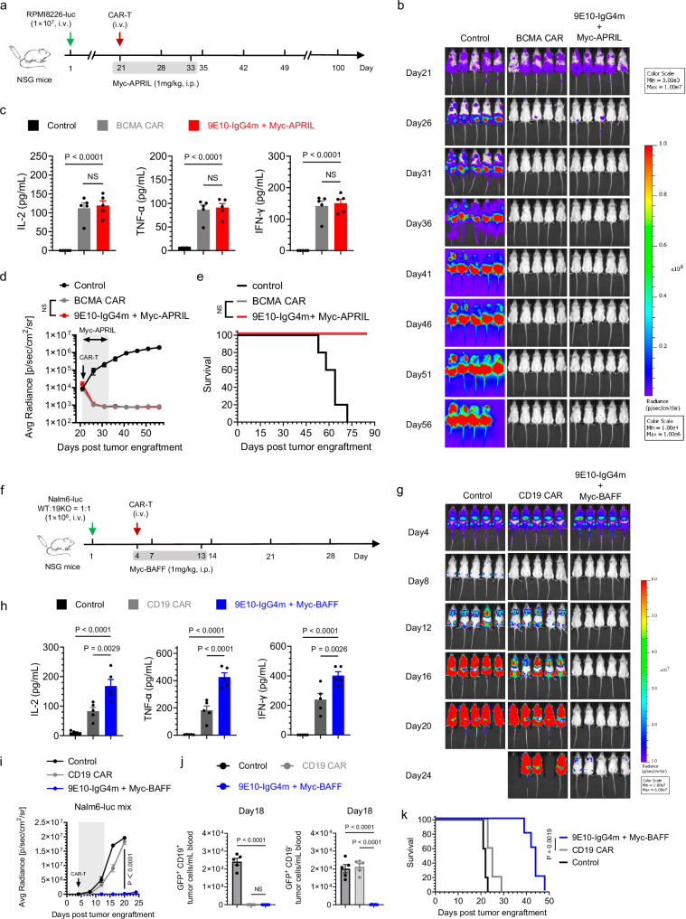
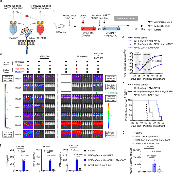
To address immune escape, multi-specific CAR-T-cell strategies use natural ligands that specifically bind multiple receptors on malignant cells. In this context, we propose a split CAR design comprising a universal receptor expressed on T cells and ligand-based switch molecules, which preserves the natural trimeric structure of ligands like APRIL and BAFF. Following optimization of the hinges and switch labeling sites, the split-design CAR-T cells ensure the native conformation of ligands, facilitating the optimal formation of immune synapses between target cancer cells and CAR-T cells. Our CAR-T-cell strategy demonstrates antitumor activities against various B-cell malignancy models in female mice, potentially preventing immune escape following conventional CAR-T-cell therapies in the case of antigen loss or switching. This ligand-based split CAR design introduces an idea for optimizing CAR recognition, enhancing efficacy and potentially improving safety in clinical translation, and may be broadly applicable to cellular therapies based on natural receptors or ligands.


Conflict of interest statement

Competing interests The Chinese patent “A method to prepare natural ligand-mediated switchable gene-modified immune cells against multiple targets” with the application number CN202010073487.1 is related to the present work. The applicant for the patent is Peking University Shenzhen Graduate School. The authors declare that they have no competing interests.

Figures

Fig. 1
Fig. 1. Design and comparison of BAFF- and APRIL-based switches.
a Schematic representation of the ligand-based CAR-T-cell system using a split-design approach. The system comprises Myc-targeted sCAR-T cells and ligand-Myc-based switch fusions, allowing redirection to the B-cell malignant tumor antigens BCMA, TACI and BAFFR. The Myc tag was fused to the N- or C-terminus of the extracellular domain of APRIL or BAFF ligand with a flexible GGGGS linker. Consequently, four switches were generated and referred to as Myc-APRIL, APRIL-Myc, Myc-BAFF and BAFF-Myc. b Binding profiles of APRIL- or BAFF-based switches to antigens BAFFR, BCMA, and TACI were determined by ELISA in triplicate. c, d Representative confocal images of synapses. sCAR-T cells were co-cultured with RPMI8226-GFP cells in the presence of ARPIL-based switches (c) or co-cultured with Nalm6-GFP cells in the presence of BAFF-based switches (d) for 1 h, and cell‒cell conjugates were imaged at 100× oil objective magnification using a laser scanning confocal microscope (Nikon, A1R). Hoechst (blue), anti-PKC-θ (red), GFP (green) and merged images of all the stains are shown. Scale bar=10 μm. e, f Statistical analysis of mean fluorescence intensity of PKC-θ at the IS in the panels (c) and (d), respectively. Sample sizes: APRIL-Myc/Myc-APRIL, n = 39; BAFF-Myc, n = 31; Myc-BAFF, n = 48. All n values represent individual cells. P values determined by paired two-tailed t-tests. g, h Cytotoxicity assays of different APRIL- (g) or BAFF-based switches (h) were performed with sCAR-T cells against the indicated target cells at an E:T ratio of 10:1 for 24 h in triplicate. Data in this figure are representative of three independent experiments. Error bars represent mean ± SD. NS indicates not significant. Source data are provided in the Source Data file.
Fig. 2
Fig. 2. Characterization of sCAR-T hinge designs.
a Schematic representations of different ligand-based sCAR structures. Second-generation sCARs were engineered comprising a Myc-targeted scFv (clone 9E10), a hinge-transmembrane domain (CD8-CD8, CD28-CD28 or IgG4m-CD8) and an intracellular domain (41BB-CD3ζ), designated as 9E10-CD8, 9E10-CD28 and 9E10-IgG4m, respectively. b, d Representative images of cell‒cell conjugates were acquired at 100 × oil objective magnification under a laser scanning confocal microscope (Nikon, A1R). sCAR-T cells, as indicated, were pre-incubated with Myc-APRIL or Myc-BAFF and co-cultured with their respective target cells for 1 h. Fluorescent labeling included Hoechst (blue), anti-PKC-θ (red), and GFP (green) and a merged view of all stains. Scale bar = 10 μm. c, e Statistical analysis of the mean fluorescence intensity of PKC-θ at the IS in panels b and d, respectively. In panel c, samples sizes: 9E10-CD8, n = 28; 9E10-CD28, n = 38; 9E10-IgG4m, n = 43. In panel e, 9E10-CD8, n = 23; 9E10-CD28, n = 22; 9E10-IgG4m, n = 32. All n values represent individual cells. P values were determined by paired two-tailed t-tests. f, g Cytotoxicity assays of different sCAR-T cells against the indicated target cells with Myc-APRIL (f) or Myc-BAFF (g) at an E:T ratio of 10:1 for 24 h in triplicate. h, i Inflammatory cytokine release assay. sCAR-T cells were co-cultured with the specified target cells in the presence of 100 pM Myc-APRIL (h) or Myc-BAFF (i) for 24 h at an E:T ratio of 1:1 in triplicate. Two-way ANOVA multiple comparisons in Dunnett correction were used to assess significance. j, k Cytotoxicity assays of 9E10-IgG4m CAR-T cells against the indicated target cells with Myc-APRIL (j) or Myc-BAFF (k) at an E:T ratio of 1:1 for 24 h in triplicate. Data in this figure are representative of three independent experiments. Error bars represent mean ± SD. NS indicates not significant. Source data are provided in the Source Data file.
Fig. 3
Fig. 3. In vitro comparison of the split-design CAR approach with conventional CAR.
a Schematic representations of ligand-based conventional and split-design CAR approaches. The extracellular domains of BAFF and APRIL were used as target moieties to generate conventional CAR-T cells, referred to as APRIL CAR and BAFF CAR, respectively. b, d Representative images of cell‒cell conjugates captured at 100× oil objective magnification using a laser scanning confocal microscope (Nikon, A1R). APRIL or 9E10-IgG4m (pre-incubated with Myc-APRIL) CAR-T cells were co-cultured with RPMI8226-GFP cells (b), while BAFF or 9E10-IgG4m (pre-incubated with Myc-BAFF) CAR-T cells were co-cultured with IM9-GFP cells (d). Fluorescent labels included Hoechst (blue), anti-PKC-θ (red), and GFP (green) and a merged view of all stains. Scale bar = 10 μm. c, e Statistical analysis of the mean fluorescence intensity of PKC-θ at the IS in panels b and d, respectively. In panel c, sample sizes: APRIL CAR, n = 37; 9E10-IgG4m, n = 39. In panel e, BAFF CAR, n = 34; 9E10-IgG4m, n = 44. All n values represent individual cells. P values were determined by paired two-tailed t-tests. f, g Cytotoxicity assays of conventional and split-design CAR-T cells against the indicated target cells at various E:T ratios for 24 h in triplicate. h, i Inflammatory cytokine release assay. Conventional CAR-T cells or sCAR-T cells along with 1 nM corresponding switches were co-cultured with the specific target cells for 24 h at an E:T ratio of 1:1 in triplicate. Two-way ANOVA multiple comparisons in Dunnett correction were used to assess significance. j, l Schematic representations of ligand-based split-design CAR and FDA-approved CAR, referred to as BCMA CAR (j) and CD19 CAR (l), respectively. k, m Cytotoxicity assays of FDA-approved CAR-T cells and split-design CAR-T cells against the indicated target cells at various E:T ratios for 24 h in triplicate. Data in this figure are representative of three independent experiments. Error bars represent mean ± SD. NS indicates not significant. Source data are provided in the Source Data file.
Fig. 4
Fig. 4. In vivo comparison of the split-design CAR approach with ligand-based conventional CARs.
a Timeline of in vivo experiments. Consistent results were obtained in two independent experiments (n = 5 mice). b Representative bioluminescence images of mice subjected to different treatments. Colors represent the luminescence intensity (red, highest; blue, lowest). c, d Quantification of the average radiance (p/s/cm/sr) of the luminescence, related to APRIL- (c) and BAFF-(d)-based CAR-T-cell therapy. Two-way ANOVA multiple comparisons in Dunnett correction were used to assess significance. e Evaluation of serum inflammatory cytokine release by ELISA 24 h after CAR-T-cell infusion. One-way ANOVA multiple comparisons in Tukey correction were used to assess significance. f, g Survival curves of the mice subjected to the indicated treatments. Survival curves were compared using the log-rank (Mantel‒Cox) test. h Timeline of in vivo experiments. Consistent results were obtained in two independent experiments (n = 5 mice). i Representative bioluminescence images of mice subjected to different treatments. Colors represent the luminescence intensity (red, highest; blue, lowest). j Quantification of the average radiance (p/s/cm/sr) of the luminescence. Two-way ANOVA multiple comparisons in Dunnett correction were used to assess significance, comparing 9E10-IgG4m CAR-T (with Myc-BAFF) and CD19/CD22 CAR-T. k Evaluation of serum inflammatory cytokine release by ELISA 24 h after CAR-T-cell infusion. One-way ANOVA multiple comparisons in Dunnett correction were used to assess significance. l Assessment of the presence of persistent human CD3+ (hCD3+) T cells in peripheral blood by flow cytometry over a 3-week follow-up period. Two-way ANOVA multiple comparisons in Dunnett correction were used to assess significance, comparing 9E10-IgG4m CAR-T (with Myc-BAFF) with BAFF-CAR-T at each time point. m Survival curves of mice subjected to the indicated treatments, compared using the log-rank (Mantel‒Cox) test. All n represents biological replicates from different mice. Data in this figure are representative of one of two independent experiments. Error bars represent mean ± SEM. NS indicates not significant. Source data are provided in the Source Data file.
Fig. 5
Fig. 5. In vivo comparison of the split-design CAR approach with the FDA-approved CARs.
a Timeline of in vivo experiments. Consistent results were obtained in two independent experiments (n = 5 mice). b Representative bioluminescence images of mice subjected to different treatments. Colors represent the luminescence intensity (red, highest; blue, lowest). c Evaluation of serum inflammatory cytokine release by ELISA 24 h after CAR-T-cell infusion. One-way ANOVA multiple comparisons in Dunnett correction were used to assess significance. d Quantification of the average radiance (p/s/cm2/sr) of the luminescence. Two-way ANOVA multiple comparisons in Sidak correction were used to assess significance, comparing 9E10-IgG4m CAR-T (with Myc-APRIL) with BCMA CAR-T. e Survival curves of mice subjected to the indicated treatments, compared using the log-rank (Mantel‒Cox) test. f Timeline of the in vivo experiments. Consistent results were obtained in two independent experiments (n = 5 mice). g Representative bioluminescence images of mice subjected to different treatments. Colors represent the luminescence intensity (red, highest; blue, lowest). h Evaluation of serum inflammatory cytokine release by ELISA 24 hours after CAR-T-cell infusion. One-way ANOVA multiple comparisons in Dunnett correction were used to assess significance. i Quantification of the average radiance (p/s/cm2/sr) of the luminescence. Two-way ANOVA multiple comparisons in Dunnett correction were used to assess significance, comparing 9E10-IgG4m CAR-T (with Myc-BAFF) with CD19 CAR-T. j Assessment of the presence of tumor cells (GFP+ CD19+ or GFP+ CD19-) in peripheral blood by flow cytometry on the 18th day of the experiment. One-way ANOVA multiple comparisons in Dunnett correction were used to assess significance. k Survival curves of the mice subjected to the indicated treatments, compared using the log-rank (Mantel‒Cox) test. All n represents biological replicates with different mice. Data are in this figure representative of one of two independent experiments. Error bars represent mean ± SEM. NS indicates not significant. Source data are provided in the Source Data file.
Fig. 6
Fig. 6. In vivo synergies of the split-design ligand-based CAR-T-cell system.
a Schematic representation of the design utilizing various switches to redirect sCAR-T cells to specific tumors. b Timeline of the in vivo model illustrating the synergistic efficacy of sequential tumor control in MM using Myc-APRIL and B-ALL using Myc-BAFF. Twenty-one days after RPMI8226 tumor engraftment, mice were i.v. administered 15 × 106 corresponding CAR-T cells. Myc-APRIL was i.p. administered daily for 5 days to manage RPMI8226 tumor cells. Following this, Nalm6 cells (0.25 × 106 cells) were i.v. injected 6 days later, and sCAR-T cells were redirected to control B-ALL tumors through daily i.p. administration of Myc-BAFF for 10 days. Conventional APRIL and BAFF CAR-T cells were injected on days 21 and 29, respectively. Consistent results were obtained in two independent experiments (n = 5 mice). c Representative bioluminescence images of mice under different treatment conditions. Colors indicate the luminescence intensity (red, highest; blue, lowest). d Tumor burden of the MM and B-ALL dual-tumor model over time following Nalm6 rechallenge, quantified as the average radiance (p/s/cm2/sr) from the luminescence. Two-way ANOVA multiple comparisons in Tukey correction were used to assess significance, comparing 9E10-IgG4m CAR-T in combination with Myc-APRIL and Myc-BAFF versus the combination of the APRIL CAR-T and BAFF CAR-T. e Survival curves depicting the outcomes of the mice subjected to the indicated treatments, compared using the log-rank (Mantel‒Cox) test. f Serum inflammatory cytokine release was evaluated by ELISA 24 h after the first dose of Myc-BAFF. One-way ANOVA multiple comparisons in Dunnett correction were used to assess significance. g On the 39th day of the experiment, the presence of persistent human CD3+ (hCD3+) engineered CAR-T cells in the peripheral blood was assessed in Nalm6-rechallenged mice by flow cytometry. One-way ANOVA multiple comparisons in Dunnett correction were used to assess significance. All n represents biological replicates with different mice. Data in this figure are representative of one of two independent experiments. Error bars represent mean ± SEM. Source data are provided in the Source Data file.

Similar articles

Cited by

References

    1. Majzner, R. G. & Mackall, C. L. Tumor antigen escape from CAR T-cell Therapy. Cancer Discov.8, 1219–1226 (2018). - PubMed
    1. Shah, N. N. et al. Bispecific anti-CD20, anti-CD19 CAR T cells for relapsed B cell malignancies: a phase 1 dose escalation and expansion trial. Nat. Med. 26, 1569–1575 (2020). - PubMed
    1. Spiegel, J. Y. et al. CAR T cells with dual targeting of CD19 and CD22 in adult patients with recurrent or refractory B cell malignancies: a phase 1 trial. Nat. Med. 27, 1419–1431 (2021). - PMC - PubMed
    1. Mei, H. et al. A bispecific CAR-T cell therapy targeting BCMA and CD38 in relapsed or refractory multiple myeloma. J. Hematol. Oncol.14, 161 (2021). - PMC - PubMed
    1. Zah, E. et al. Systematically optimized BCMA/CS1 bispecific CAR-T cells robustly control heterogeneous multiple myeloma. Nat. Commun.11, 2283 (2020). - PMC - PubMed

Publication types

Substances

LinkOut - more resources