Clinical Trial
. 2025 Jan;31(1):137-143.
doi: 10.1038/s41591-024-03326-7. Epub 2024 Nov 11.

TRBC1-CAR T cell therapy in peripheral T cell lymphoma: a phase 1/2 trial

Affiliations
Clinical Trial

TRBC1-CAR T cell therapy in peripheral T cell lymphoma: a phase 1/2 trial

Kate Cwynarski et al. Nat Med. 2025 Jan.

Abstract

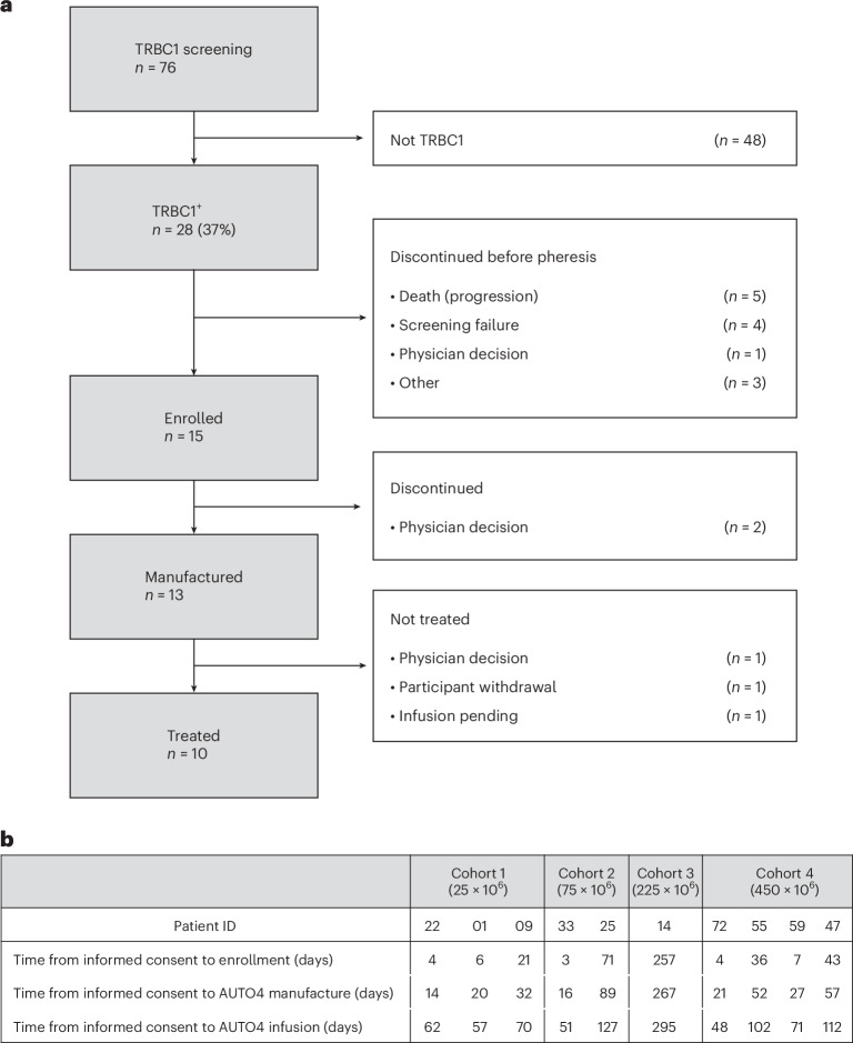
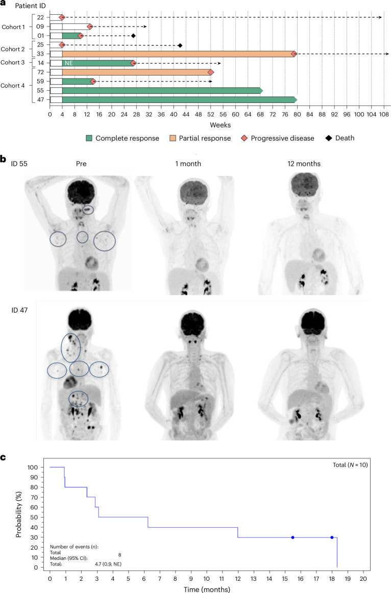
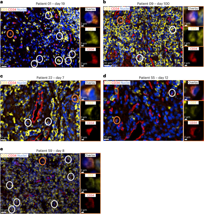
Relapsed/refractory peripheral T cell lymphomas (PTCLs) are aggressive tumors with a poor prognosis. Unlike B cell lymphomas, treatment of PTCL has not benefited from advances in immunotherapy. This is largely due to a lack of suitable target antigens that discriminate malignant from normal T cells, thus avoiding severe immunosuppression consequent to depletion of the entire T cell compartment. We recently described a targeting strategy based on the mutually exclusive expression of T cell antigen receptor beta-chain constant domain (TRBC) 1 and 2. Selective targeting of the T cell antigen receptor beta-chain expressed by the (clonal) malignancy spares normal T cells expressing the other chain. The LibraT1 study is an ongoing, multicenter, international, single-arm phase 1/2 study of TRBC1-directed autologous chimeric antigen receptor (CAR) T cells (AUTO4) in relapsed/refractory TRBC1-positive PTCL. Primary objectives were assessment of safety and tolerability of AUTO4 infusion. Key secondary endpoints included efficacy, CAR T cell expansion and persistence. Here we describe the findings from dose escalation in LibraT1 in the first ten patients, in a non-prespecified interim analysis. AUTO4 resulted in low frequency of severe immunotoxicity, with one of ten patients developing grade 3 cytokine release syndrome. Complete metabolic response was observed in four of ten evaluable patients, with remissions being durable beyond 1 year in two patients. While an absence of circulating CAR T cells was observed, CAR T cells were readily detected in lymph node biopsy samples from sites of original disease suggesting homing to tumor sites. These results support the continuing exploration of TRBC1 targeting in PTCL. ClinicalTrials.gov registration: NCT03590574 .


Conflict of interest statement

Competing interests: K.C.: consulting/advisory role: Roche, Takeda, Celgene, Atara, Gilead, KITE, Janssen, Incyte and AbbVie; speakers’ bureau: Roche, Takeda, KITE, Gilead and Incyte; conferences/travel support: Roche, Takeda, KITE, Janssen and BMS. G.I.: honoraria and travel support: Novartis, Kite/Gilead, Bristol Myers Squibb, AbbVie, Autolus, Sandoz, Miltenyi and AstraZeneca. E.T.: speaker fees: Astellas, Jazz, Kite/Gilead, Pfizer and Vertex; ad boards: Autolus, BMS/Celgene, Jazz, Janssen and Vertex. M.P.: owns stock in and has research funding and salary contribution from Autolus. D.I.: travel grants and honoraria: Kite/Gilead and Janssen. T.M.: travel grants: Amgen, Jazz, Pfizer, Bayer, Kyowa Kirin, Celgene/BMS, Kite/Gilead, Janssen and Takeda; honoraria for advisory board meetings: Kite/Gilead, Amgen, Novartis, Pfizer, Celgene/BMS, Daiichi Sankyo, Atara, Roche and Janssen; honoraria for lectures: Kite/Gilead, Takeda, Janssen, Roche, Servier, Novartis, Celgene/BMS, Pfizer and Incyte; research funding: Janssen, AstraZeneca and Novartis. T.M.: consulting/advisory role: Roche/Ventana, Haematogenix and Autolus. P.M.: Autolus: stock holder and research funding. UCL business: patent royalties. All employees of Autolus have no competing interests.

Figures

Fig. 1
Fig. 1. CONSORT diagram.
a, CONSORT diagram for AUTO4/LibraT1 study (NCT03590574). b, Timeline from informed consent to enrollment (leukapheresate), AUTO4 CAR T cell manufacturing and AUTO4 CAR T cell infusion for treated patients.
Fig. 2
Fig. 2. Clinical trial cohort.
a, Swimmer plot showing the outcome in patients who received AUTO4. Note, one patient (14) who received 225 × 106 AUTO4 CAR T cells was in CMR following bridging at time of infusion and is marked NE. Cohort 1, 25 × 106; cohort 2, 75 × 106; cohort 3, 225 × 106; cohort 4, 450 × 106. b, 18FDG PET-CT imaging before AUTO4, at month 1 and at 12 months from two participants (47 and 55) in long-term CMR following AUTO4. c, PFS based on overall response (Lugano classification). Median with 95% CI calculated from PROC LIFETEST output. Time relative to first AUTO4 treatment. 1 month = 30.4375 days. Source data
Fig. 3
Fig. 3. Lymph node biopsy.
ae, Lymph node biopsy samples after AUTO4 infusion for patient ID 01 (day 19) (a), 09 (day 100) (b), 22 (day 7) (c), 55 (day 12) (d) and 59 (day 8) (e). The main images show formalin-fixed, paraffin-embedded (FFPE) tissue sections of a T cell lymphoma stained by double IF with anti-CD34 (Q/BenD10), which stains RQR8 (red), and CD3 (yellow), which detects T cells. DAPI (blue) is used for nuclear counterstaining. CAR T cells (orange) are identified by coexpression of both Q/BenD10 and anti-CD3. The magnifications of the orange circled cells show overlay (top), CD3 staining (center) and CD34 staining (bottom). One tissue section stained per patient.
Extended Data Fig. 1
Extended Data Fig. 1. CAR T manufacturing.
Diagram of manufacturing process for AUTO4 CAR-T cells.
Extended Data Fig. 2
Extended Data Fig. 2. Drug product characterization.
a) CAR T cell product characteristics. b) Flow cytometric analysis of cryopreserved CAR-T cell drug products for patients treated using a panel to determine product exhaustion. Markers included were PD1, TM3, LAG3 and TIGIT. ‘0’ denotes cells not expressing any of the markers; ‘1’ includes cells expressing a single marker; ‘2’ includes cells expressing any two markers in combination; ‘3’ includes any three in combination or all four markers. Gating strategy in Supplementary Information. c) Flow cytometric analysis of cryopreserved CAR-T cell drug products for patients treated. Immunophenotyping panel included CD45RA and CCR7. CCR7+ CD45RA+ were considered naïve cells; CCR7+ CD45RA- T cells were considered central memory (TCM); CCR7- CD45RA- were considered Effector memory cells (TEM) and CCR7- CD45RA+ cells were considered Terminally differentiated effector memory T cell (TEMRA). Gating strategy in Supplementary Information. d) Exhaustion marker expression and CAR T cell differentiation grouped by clinical response. CR = complete response, PR = partial response, NR = non-responder. CR patients 01, 14, 47, 55 and 59. PR patients 33 and 72, NR patients 09, 22 and 25. Mean ± SD. Source data
Extended Data Fig. 3
Extended Data Fig. 3. Peripheral Blood Counts.
a) Peripheral blood counts for first 3 months are shown: Lymphocytes, Neutrophils and Platelets. b) TRBC1 and TRBC2 % in peripheral blood determined by flow cytometry. c) CD4:CD8 ratio in peripheral blood determined by flow cytometry. Source data
Extended Data Fig. 4
Extended Data Fig. 4. In vitro cytotoxicity.
In vitro cytotoxicity assay on Jurkat TRBC1 and TCR KO cell lines, and TRBC1+ and TRBC2+ healthy human T cells, with HuJovi1-hinge-TyrpTM-41BBz CAR (AUTO4) and control anti-CD19 CAR (CD8stk-CD8TM-41BBz). E:T ratios 1:1, 1:2, 1:4 and 1:8, 72h, n=6 (individual healthy PBMC donors). Mean ± SD. Two-way ANOVA with Tukey’s post-test for T cell (TRBC1) vs Jurkat (TRBC1). Exact P value in graph. Source data
Extended Data Fig. 5
Extended Data Fig. 5. Serum cytokines.
a) Cytokine levels measured in peripheral blood for TNF, GM-CSF, IFNγ, IL-2, IL-5, IL-6, IL-7, IL-8, IL-10 and IL-15. b) Peak serum cytokine concentrations grouped by clinical response. Mean ± SD. Two-way ANOVA with Tukey’s post-test. Exact P value in graph. CR = complete response, PR = partial response, NR = non-responder. CR patients 01, 14, 47, 55 and 59. PR patients 33 and 72, NR patients 09, 22 and 25. Source data
Extended Data Fig. 6
Extended Data Fig. 6. Screening biopsies.
a) Tumour PD-L1 expression score on patients screening biopsies obtained via IHC. One tissue section stained per patient. b) Representative multiplex immunofluorescent staining of screening biopsy targeting CD4 (green), CD8 (red) FOXP3 (yellow) PD-1 (magenta) and nuclei (blue). Top right, quantification of main T cell subsets (CD4+, CD8+ and dual CD4+/CD8+) in patients, grouped by clinical response. Bottom left, quantification of CD8 subsets (CD8+, CD8+/PD1+, CD8+/FOXP3+) in patients, grouped by clinical response. Bottom right, quantification of CD4 subsets (CD4+, CD4+/PD1+, CD4+/FOXP3+) in patients, grouped by clinical response. CR = complete response, PR = partial response, NR = non-responder. One tissue section stained per patient. Source data
Extended Data Fig. 7
Extended Data Fig. 7. In vitro reverse killing.
Reverse killing assay for healthy human T cell (TRBC1+ or TRBC2+) with HuJovi1-hinge-TyrpTM-41BBz CAR (AUTO4) and control anti-CD19 CAR (CD8stk-CD8TM-41BBz). E:T ratios 4:1, 1:1 and 1:4, 72h, n=6 (individual healthy PBMC donors). Mean ± SD. Two tailed multiple paired t test (Holm-Sidak method). Median (95% CI). Exact P value in graph. Source data

References

    1. Bellei, M. et al. T-cell lymphomas in South America and Europe. Rev. Bras. Hematol. Hemoter.34, 42–47 (2012). - PMC - PubMed
    1. Bellei, M. et al. The outcome of peripheral T-cell lymphoma patients failing first-line therapy: a report from the prospective, International T-Cell Project. Haematologica103, 1191–1197 (2018). - PMC - PubMed
    1. Horwitz, S. et al. Brentuximab vedotin with chemotherapy for CD30-positive peripheral T-cell lymphoma (ECHELON-2): a global, double-blind, randomised, phase 3 trial. Lancet393, 229–240 (2019). - PMC - PubMed
    1. Westin, J. R. et al. Survival with axicabtagene ciloleucel in large B-cell lymphoma. N. Engl. J. Med.389, 148–157 (2023). - PubMed
    1. Went, P. et al. Marker expression in peripheral T-cell lymphoma: a proposed clinical-pathologic prognostic score. J. Clin. Oncol.24, 2472–2479 (2006). - PubMed

Publication types

MeSH terms

Substances

Associated data

LinkOut - more resources