

Towards the clinical translation of a silver sulfide nanoparticle contrast agent: large scale production with a highly parallelized microfluidic chip
- PMID: 39528844
- PMCID: PMC11753937
- DOI: 10.1007/s00259-024-06967-5
Towards the clinical translation of a silver sulfide nanoparticle contrast agent: large scale production with a highly parallelized microfluidic chip
Abstract
Purpose: Ultrasmall silver sulfide nanoparticles (Ag2S-NP) have been identified as promising contrast agents for a number of modalities and in particular for dual-energy mammography. These Ag2S-NP have demonstrated marked advantages over clinically available agents with the ability to generate higher contrast with high biocompatibility. However, current synthesis methods for inorganic nanoparticles are low-throughput and highly time-intensive, limiting the possibility of large animal studies or eventual clinical use of this potential imaging agent.
Methods: We herein report the use of a scalable silicon microfluidic system (SSMS) for the large-scale synthesis of Ag2S-NP. Ag2S-NP produced using this system were compared to bulk synthesis and a commercially available microfluidic device through characterization, contrast generation, in vivo imaging, and clearance profiles.
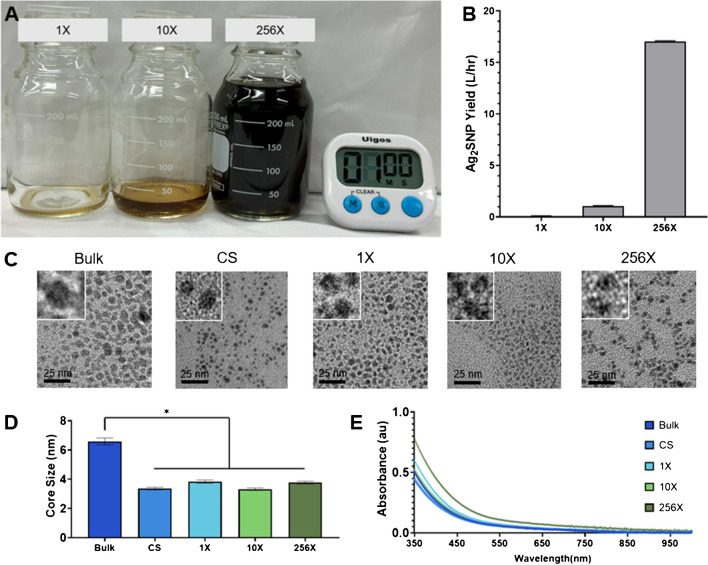
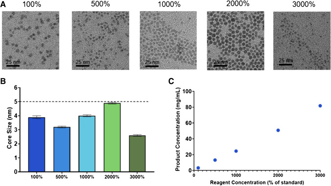
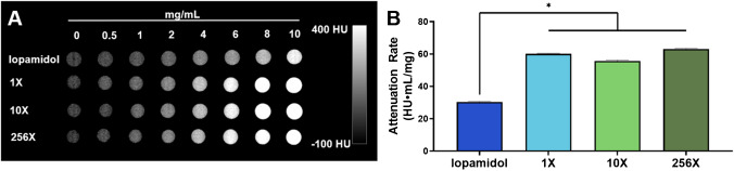
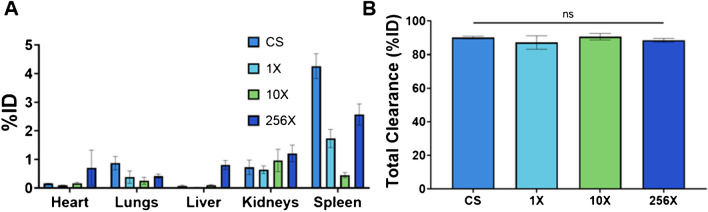
Results: Using SSMS chips with 1 channel, 10 parallelized channels, and 256 parallelized channels, we determined that the Ag2S-NP produced were of similar quality as measured by core size, concentration, UV-visible spectrometry, and in vitro contrast generation. Moreover, by combining parallelized chips with increasing reagent concentration, we were able to increase output by an overall factor of 5,100. We also found that in vivo imaging contrast generation was consistent across synthesis methods and confirmed renal clearance of the ultrasmall nanoparticles. Finally, we found best-in-class clearance of the Ag2S-NP occurred within 24 h.
Conclusions: These studies have identified a promising method for the large-scale production of Ag2S-NP, paving the way for eventual clinical translation.
Keywords: Contrast agents; High throughput; Large scale synthesis; Nanoparticles; Silver sulfide.
© 2024. The Author(s).
Conflict of interest statement
Declarations. Competing interests: The authors have no relevant financial interests to disclose. Non-financial interest: an author of this manuscript, JCH, is an editor of this journal.
Figures


Update of
-
Towards the Clinical Translation of a Silver Sulfide Nanoparticle Contrast Agent: Large Scale Production with a Highly Parallelized Microfluidic Chip.bioRxiv [Preprint]. 2023 Dec 4:2023.12.02.569706. doi: 10.1101/2023.12.02.569706. bioRxiv. 2023. Update in: Eur J Nucl Med Mol Imaging. 2025 Feb;52(3):1177-1188. doi: 10.1007/s00259-024-06967-5. PMID: 38106126 Free PMC article. Updated. Preprint.
References
-
- Liu J, Zheng X, Yan L, et al. Bismuth Sulfide Nanorods As a Precision Nanomedicine for In Vivo Multimodal Imaging-Guided Photothermal Therapy of Tumor. ACS Nano. 2015;9(1):696–707. - PubMed
-
- Liu J, Yu M, Zhou C, Zheng J. Renal Clearable Inorganic Nanoparticles: a New Frontier of Bionanotechnology. Mater Today. 2013;16(12):477–86.
-
- Yu SB, Watson AD. Metal-Based X-Ray Contrast Media. Chem Rev. 1999;99(9):2353–78. - PubMed
-
- De La Vega JC, Hafeli UO. Utilization of Nanoparticles As X-Ray Contrast Agents for Diagnostic Imaging Applications. Contrast Media Mol Imaging. 2015;10(2):81–95. - PubMed
-
- Wang B, Li R, Guo G, Xia Y. Janus and Core@Shell Gold Nanorod@Cu2-xS Supraparticles: Reactive Site Regulation Fabrication, Optical/Catalytic Synergetic Effects and Enhanced Photothermal Efficiency/Photostability. Chem Commun. 2020;56(63):8996–9. - PubMed
MeSH terms
Substances
Grants and funding
LinkOut - more resources
Full Text Sources
Miscellaneous
