

Cytokine and growth factor correlation networks associated with morbidities in extremely preterm infants
- PMID: 39529072
- PMCID: PMC11555815
- DOI: 10.1186/s12887-024-05203-1
Cytokine and growth factor correlation networks associated with morbidities in extremely preterm infants
Abstract
Background: Cytokines and growth factors (GF) have been implicated in the development of retinopathy of prematurity (ROP) and bronchopulmonary dysplasia (BPD). We hypothesize that even small coordinated changes in inflammatory proteins or GFs may reveal changes in underlying regulating mechanisms that do not induce obvious changes in concentration of individual proteins. We therefore applied correlation network analysis of serum factors to determine early characteristics of these conditions.
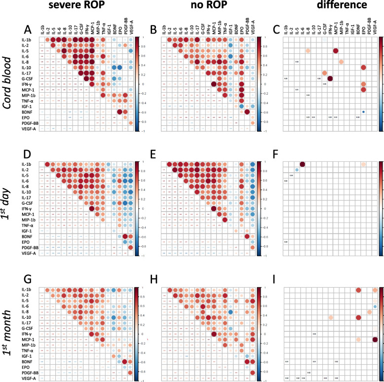
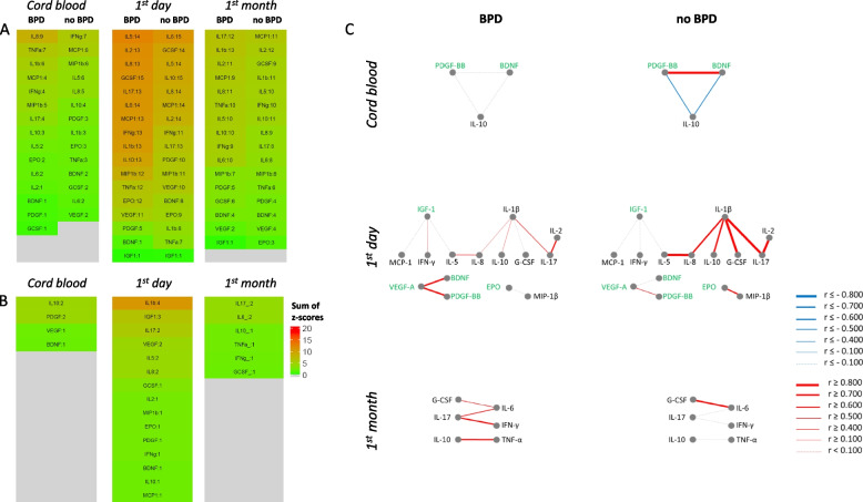
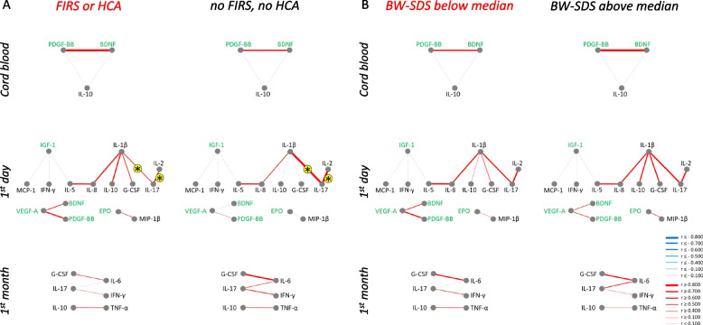
Methods: Concentrations of 17 cytokines and five GFs were measured and analysed in blood samples from cord blood, on day one and during the following month in 72 extremely preterm infants. Spearman's correlation networks distinguishing BPD and severe ROP patients from non-affected were created.
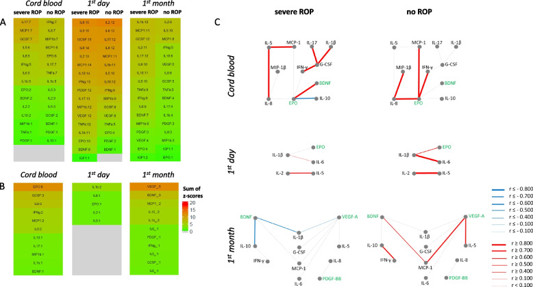
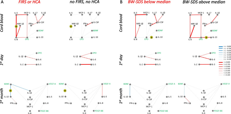
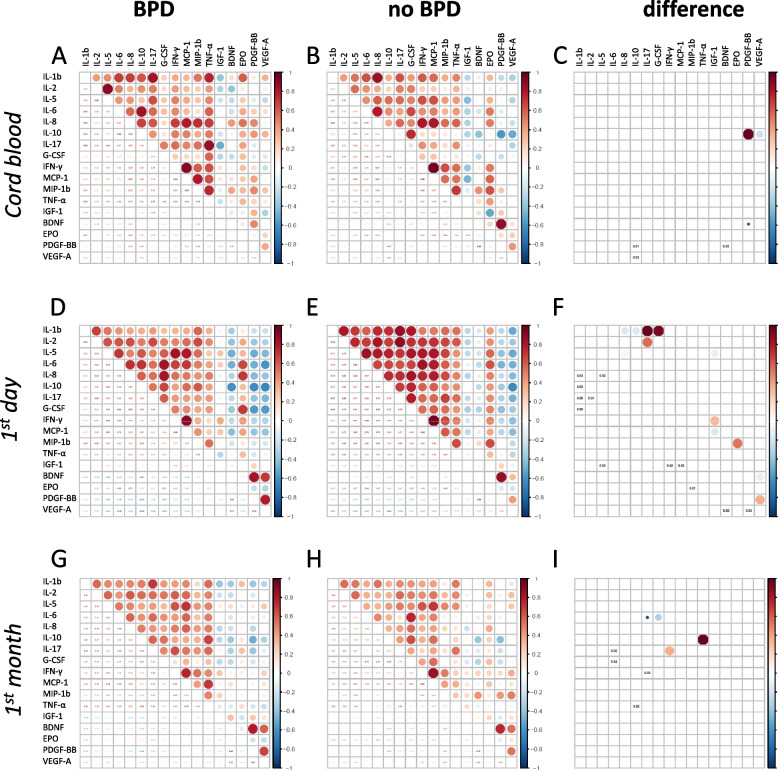
Results: Most cytokine concentrations correlated positively with each other and negatively with GFs. Very few individual cytokines differed between patients with and without ROP or BPD. However, networks of differently correlated serum factors were characteristic of the diseases and changed with time. In ROP networks, EPO, G-CSF and IL-8 (cord blood), BDNF and VEGF-A (first month) were prominent. In BPD networks, IL-1β, IGF-1 and IL-17 (day one) were noted.
Conclusions: Network analysis identifies protein signatures related to ROP or BPD in extremely preterm infants. The identified interactions between serum factors are not evident from the analysis of their individual levels, but may reveal underlying pathophysiological mechanisms in the development of these diseases.
Keywords: Bronchopulmonary dysplasia; Correlation network analysis; Cytokine interactions; Extremely preterm infants; Retinopathy of prematurity.
© 2024. The Author(s).
Conflict of interest statement
Figures
Similar articles
-
The serum thioredoxin-1 levels are not associated with bronchopulmonary dysplasia and retinopathy of prematurity.Pediatr Res. 2024 Oct;96(5):1275-1282. doi: 10.1038/s41390-024-03078-7. Epub 2024 Feb 16. Pediatr Res. 2024. PMID: 38365875 Free PMC article.
-
Postnatal serum insulin-like growth factor I deficiency is associated with retinopathy of prematurity and other complications of premature birth.Pediatrics. 2003 Nov;112(5):1016-20. doi: 10.1542/peds.112.5.1016. Pediatrics. 2003. PMID: 14595040
-
The relationship between cord blood cytokine levels and perinatal factors and retinopathy of prematurity: a gestational age-matched case-control study.Invest Ophthalmol Vis Sci. 2013 May 15;54(5):3434-9. doi: 10.1167/iovs.13-11837. Invest Ophthalmol Vis Sci. 2013. PMID: 23633656
-
Inositol in preterm infants at risk for or having respiratory distress syndrome.Cochrane Database Syst Rev. 2019 Jul 8;7(7):CD000366. doi: 10.1002/14651858.CD000366.pub4. Cochrane Database Syst Rev. 2019. PMID: 31283839 Free PMC article.
-
Retinopathy of Prematurity: Therapeutic Strategies Based on Pathophysiology.Neonatology. 2016;109(4):369-76. doi: 10.1159/000444901. Epub 2016 Jun 3. Neonatology. 2016. PMID: 27251645 Review.
Cited by
-
Safety, Efficacy and Bio-Distribution Analysis of Exosomes Derived From Human Umbilical Cord Mesenchymal Stem Cells for Effective Treatment of Bronchopulmonary Dysplasia by Intranasal Administration in Mice Model.Int J Nanomedicine. 2025 Feb 27;20:2521-2553. doi: 10.2147/IJN.S501843. eCollection 2025. Int J Nanomedicine. 2025. PMID: 40034220 Free PMC article.
-
Structural Equation Modelling of Retinopathy of Prematurity Treatment Integrating Both Physical and Clinical Effects.J Clin Med. 2025 Jan 7;14(2):297. doi: 10.3390/jcm14020297. J Clin Med. 2025. PMID: 39860303 Free PMC article.
References
-
- Serenius F, Ewald U, Farooqi A, Fellman V, Hafström M, Hellgren K, et al. Neurodevelopmental outcomes among extremely preterm infants 6.5 years after active perinatal care in Sweden. JAMA Pediatr. 2016;170(10):954–63. - PubMed
-
- Hellström A, Hård AL. Screening and novel therapies for retinopathy of prematurity - A review. Early Hum Dev. 2019;138:104846. - PubMed
-
- Bancalari E, Jain D. Bronchopulmonary Dysplasia: 50 Years after the Original Description. Neonatology. 2019;115(4):384–91. - PubMed
-
- Dammann O, Rivera JC, Chemtob S. The prenatal phase of retinopathy of prematurity. Acta Paediatr. 2021;110(9):2521–8. - PubMed
MeSH terms
Substances
LinkOut - more resources
Full Text Sources
Research Materials
Miscellaneous
