. 2024 Nov 11;22(1):697.
doi: 10.1186/s12951-024-02979-3.

Probiotic-derived extracellular vesicles alleviate AFB1-induced intestinal injury by modulating the gut microbiota and AHR activation

Affiliations

Probiotic-derived extracellular vesicles alleviate AFB1-induced intestinal injury by modulating the gut microbiota and AHR activation

Jinyan Li et al. J Nanobiotechnology. .

Abstract

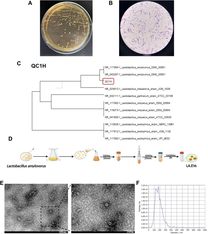
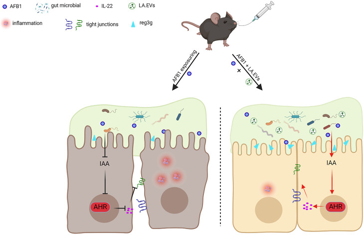
Background: Aflatoxin B1 (AFB1) is a mycotoxin that widely found in the environment and mouldy foods. AFB1 initially targets the intestine, and AFB1-induced intestinal injury cannot be ignored. Lactobacillus amylovorus (LA), a predominant species of Lactobacillus, plays a role in carbohydrate metabolism. Extracellular vesicles (EVs), small lipid membrane vesicles, are widely involved in diverse cellular processes. However, the mechanism by which Lactobacillus amylovorus-QC1H-derived EVs (LA.EVs) protect against AFB1-induced intestinal injury remains unclear.

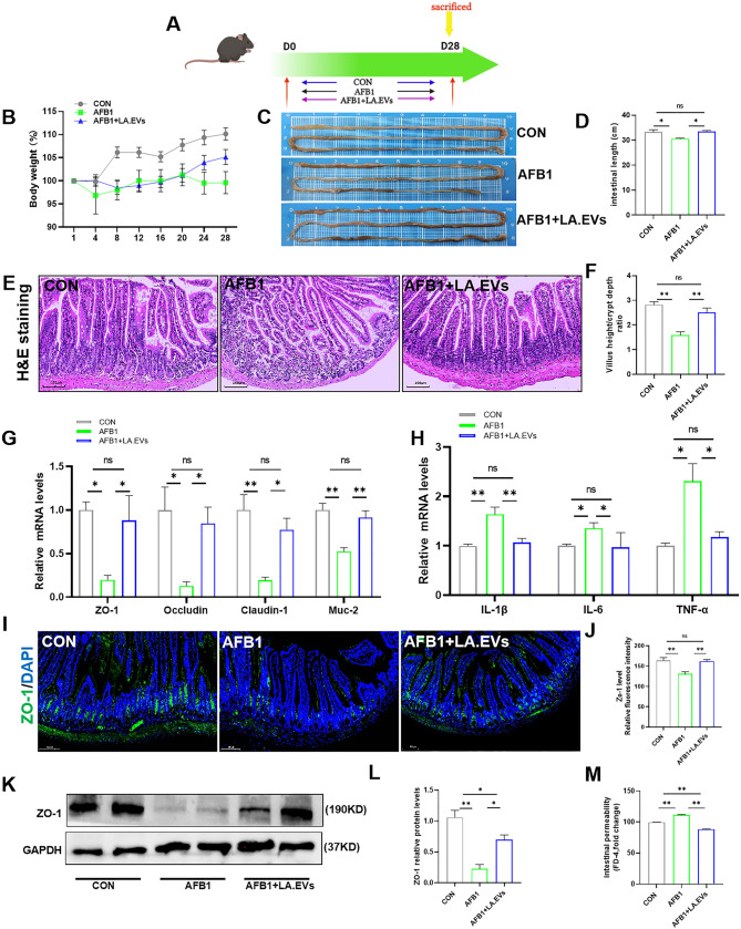
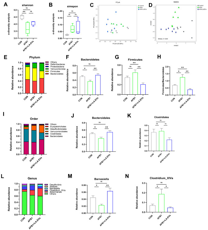
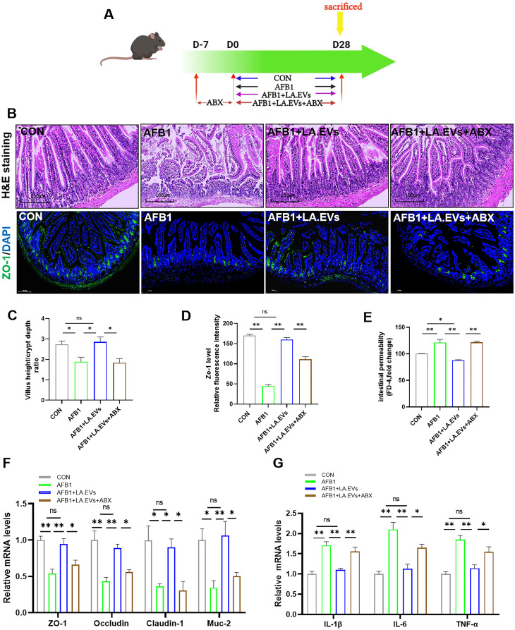
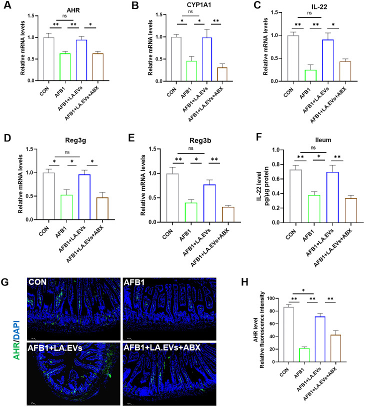
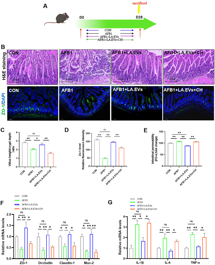
Results: In our study, a new strain named Lactobacillus amylovorus-QC1H (LA-QC1H) was isolated from pig faeces. Then, EVs derived from LA-QC1H were extracted via ultracentrifugation. Our results showed that LA.EVs significantly alleviated AFB1-induced intestinal injury by inhibiting the production of proinflammatory cytokines, decreasing intestinal permeability and increasing the expression of tight junction proteins. Moreover, 16 S rRNA analysis revealed that LA.EVs modulated AFB1-induced gut dysbiosis in mice. However, LA.EVs did not exert beneficial effects in antibiotic-treated mice. LA.EVs treatment increased intestinal levels of indole-3-acetic acid (IAA) and activated intestinal aryl hydrocarbon receptor (AHR)/interleukin-22 (IL-22) signalling in AFB1-exposed mice. Inhibition of intestinal AHR signalling markedly weakened the protective effect of LA.EVs in AFB1-exposed mice.

Conclusions: LA.EVs alleviated AFB1-induced intestinal injury by modulating the gut microbiota, activating the intestinal AHR/IL-22 signalling, reducing the inflammatory response and promoting intestinal barrier repair in mice.

Keywords: Lactobacillus amylovorus; Aflatoxin B1; Aryl hydrocarbon receptor; Extracellular vesicles; Gut microbiota.


Conflict of interest statement

Declarations Ethics approval and consent to participate Protocols of animal experiments included in this study were approved by Nanjing Agricultural University under the Laboratory Animal Care Ethics Committee for animal experiments (NJAU. No 20210624093). Informed consent was obtained from all individual participants. Consent for publication All authors consent to the publication of the article. Competing interests The authors declare no competing interests.

Figures

Fig. 1
Fig. 1
Characterization of LA and LA.EVs. (A) Colony morphology of Lactobacillus amylovorus-QC1H (LA-QC1H) on MRS agar medium. (B) Gram staining results of LA-QC1H. (C) Phylogenetic tree of LA-QC1H inferred from the 16 S rDNA gene sequences. (D) A schematic illustration of the isolation method for LA.EVs. (E) Transmission electron microscopy images of isolated EVs. The scale bar is 200 nm. (F) The particle size of LA.EVs was measured by nanoparticle tracking analysis (NTA)
Fig. 2
Fig. 2
Effects of LA.EVs treatment on inflammatory intestinal injury in AFB1-exposed mice. (A) Experimental design. (B) Body weight change. (C) Typical photos of intestines in the three groups. (D) Corresponding intestinal length was measured. (E, F) Representative image of H&E staining of ileum sections (scale bar = 100 μm) and statistical analysis of the villus height/crypt depth ratio. (G) Relative mRNA levels of tight junction proteins and muc-2 were measured in the ileum by qRT-PCR. (H) IL-1β, IL-6, and TNF-α mRNA abundances in the ileum were measured by real-time PCR analysis. (I, J) Representative immunofluorescence staining of ZO-1 antibody in the ileum (scale bar = 100 μm). (K, L) Relative protein abundances of ZO-1 in the intestine. (M) Intestinal permeability was measured by serum FD-4 concentration (n = 5). Values are expressed as the mean ± SEM. *p < 0.05, **p < 0.01, ns, the difference is not significant
Fig. 3
Fig. 3
Effects of LA.EVs treatment of the intestinal microbiota in AFB1-exposed mice. (A, B) Shannon and Simpson indices in ɑ-diversity analysis. (C, D) Beta diversity was assessed by PCoA and NMDS analysis for each group. (E-H) Microbiota compositions at the phylum level. (I-K) Microbiota compositions at the order level. (L-N) Microbiota compositions at the genus level (n = 5). Values are expressed as the mean ± SEM. *p < 0.05, **p < 0.01, ns, the difference is not significant
Fig. 4
Fig. 4
Effects of LA.EVs treatment of the ileum AHR/IL-22 axis in AFB1-exposed mice. (A) The ileum IAA levels were measured by ELISA. (B-F) Relative mRNA levels of AHR, CYP1A1, IL-22, Reg3g and Reg3b were measured in the ileum by qRT-PCR. (G) Ileum IL-22 levels were measured by ELISA. (H, I) Image of AHR IF staining in the ileum (scale bar = 100 μm). (J, K) Relative protein abundances of AHR in the intestine (n = 5). Values are expressed as the mean ± SEM. *p < 0.05, **p < 0.01, ns, the difference is not significant
Fig. 5
Fig. 5
Effects of LA.EVs treatment on AFB1-induced intestinal injury in ABX mice. (A) Experimental design. (B) Representative image of H&E and ZO-1 antibody immunofluorescence staining of the ileum (scale bar = 100 μm). (C) Statistical analysis of the villus height/crypt depth ratio. (D) Statistical analysis of relative fluorescence intensity. (E) Intestinal permeability was assessed by serum FD-4 concentration. (F) Tight junction proteins and muc-2 mRNA abundances in the ileum were measured by qRT-PCR analysis, and relative gene expression was normalized to GAPDH. (G) Relative mRNA levels of proinflammatory factors were measured in the ileum by qRT‒PCR (n = 5). Values are expressed as the mean ± SEM. *p < 0.05, **p < 0.01, ns, the difference is not significant
Fig. 6
Fig. 6
Effects of LA.EVs treatment of the ileum AHR/IL-22 signalling in antibiotic-treated mice. (A-E) AHR, CYP1A1, IL-22, Reg3g and Reg3b mRNA abundances in the intestine were measured by real-time PCR analysis. (F) Ileum IL-22 levels were measured by ELISA. (G, H) Representative image of AHR IF staining in the ileum (scale bar = 100 μm) (n = 5). Values are expressed as the mean ± SEM. *p < 0.05, **p < 0.01, ns, the difference is not significant
Fig. 7
Fig. 7
Inhibition of intestinal AHR activation abolished the protective effects of LA.EVs. (A) Experimental design. (B) Representative image of H&E and ZO-1 antibody IF staining of ileum sections (scale bar = 100 μm). (C) Statistical analysis of the villus height/crypt depth ratio. (D) Statistical analysis of relative fluorescence intensity. (E) Intestinal permeability was assessed by serum FD-4 concentration. (F) Tight junction proteins and muc-2 mRNA abundances in the ileum were measured by real-time PCR analysis. (G) Relative mRNA expression levels of IL-1β, IL-6 and TNF-α were measured in the ileum by qRT-PCR (n = 5). Values are expressed as the mean ± SEM. *p < 0.05, **p < 0.01, ns, the difference is not significant
Fig. 8
Fig. 8
A proposed model of LA.EVs protect against aflatoxin B1-induced inflammatory intestinal injury by remodelling the gut microbiota and activating intestinal AHR/IL-22 signalling in mice

References

    1. Zhang N-Y, Qi M, Zhao L, Zhu M-K, Guo J, Liu J, Gu C-Q, Rajput SA, Krumm CS, Qi D-S, Sun L-H. Curcumin prevents aflatoxin B₁ hepatoxicity by inhibition of cytochrome P450 isozymes in Chick Liver. Toxins 2016;8. - PMC - PubMed
    1. Cheng L, Qin Y, Hu X, Ren L, Zhang C, Wang X, Wang W, Zhang Z, Hao J, Guo M, et al. Melatonin protects in vitro matured porcine oocytes from toxicity of aflatoxin B1. J Pineal Res. 2019;66:e12543. - PubMed
    1. Pauletto M, Giantin M, Tolosi R, Bassan I, Barbarossa A, Zaghini A, Dacasto M. Discovering the Protective effects of Resveratrol on aflatoxin B1-Induced toxicity: a whole transcriptomic study in a bovine hepatocyte cell line. Antioxidants; 2021. p. 10. (Basel, Switzerland). - PMC - PubMed
    1. Pang VF, Chiang C-F, Chang C-C. The in vitro effects of aflatoxin B1 on physiological functions of swine alveolar macrophages. Veterinary Med Sci. 2020;6:919–25. - PMC - PubMed
    1. Yang C, Song G, Lim W. Effects of mycotoxin-contaminated feed on farm animals. J Hazard Mater. 2020;389:122087. - PubMed

LinkOut - more resources