. 2024 Nov 11;22(1):1017.
doi: 10.1186/s12967-024-05829-w.

Compromised C3b-VSIG4 axis between decidual NK cells and macrophages contributes to recurrent spontaneous abortion

Affiliations

Compromised C3b-VSIG4 axis between decidual NK cells and macrophages contributes to recurrent spontaneous abortion

Siao Chen et al. J Transl Med. .

Abstract

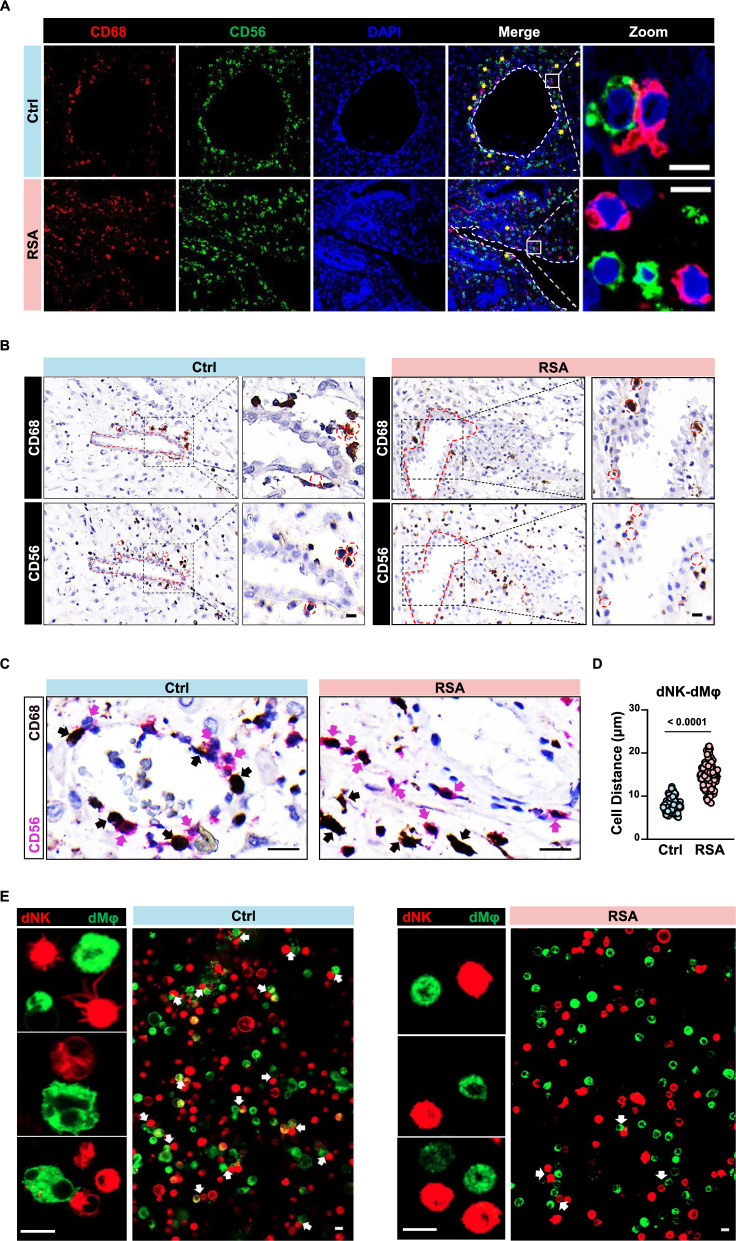
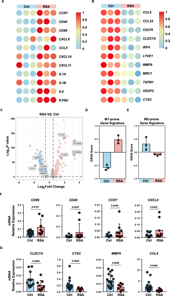
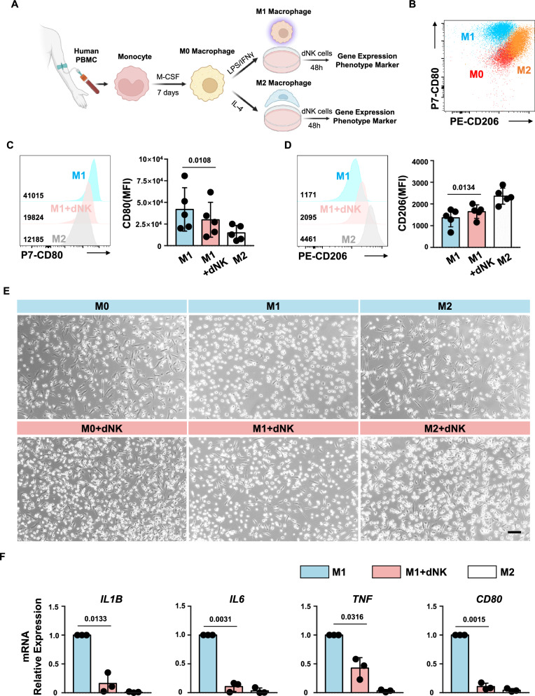
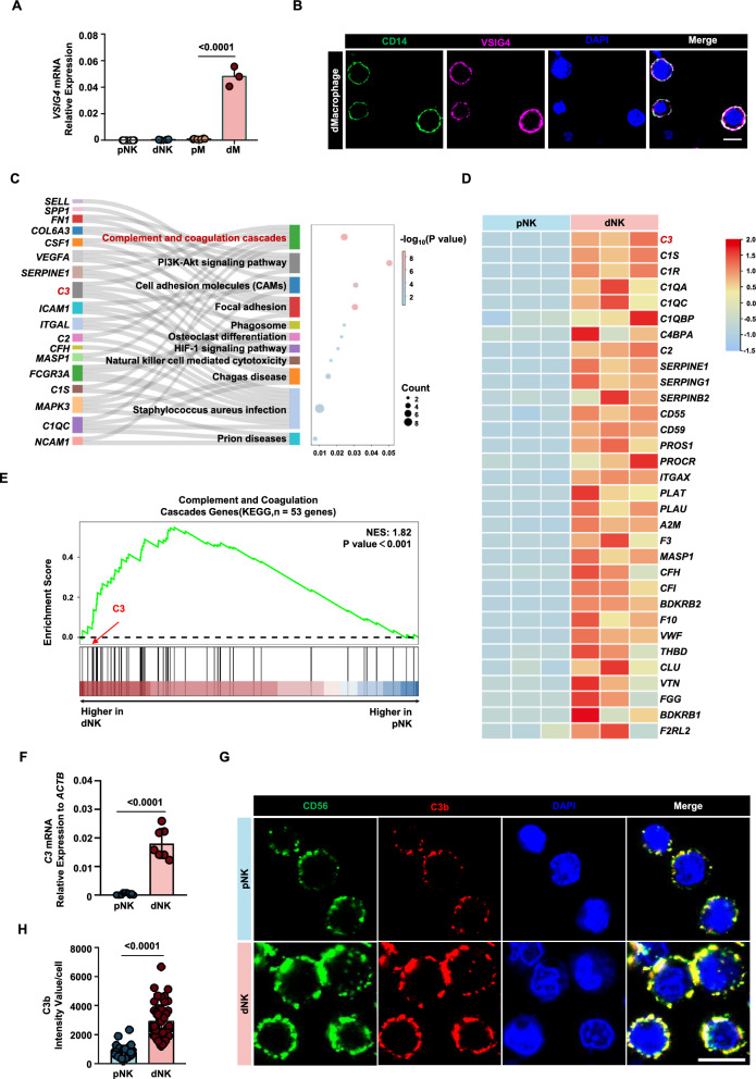
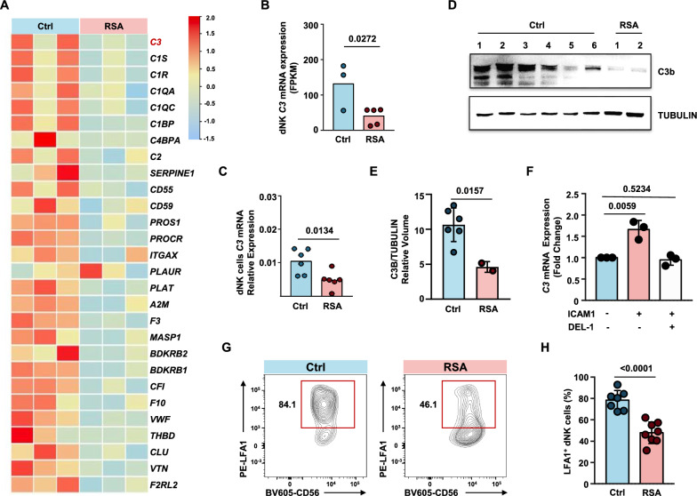
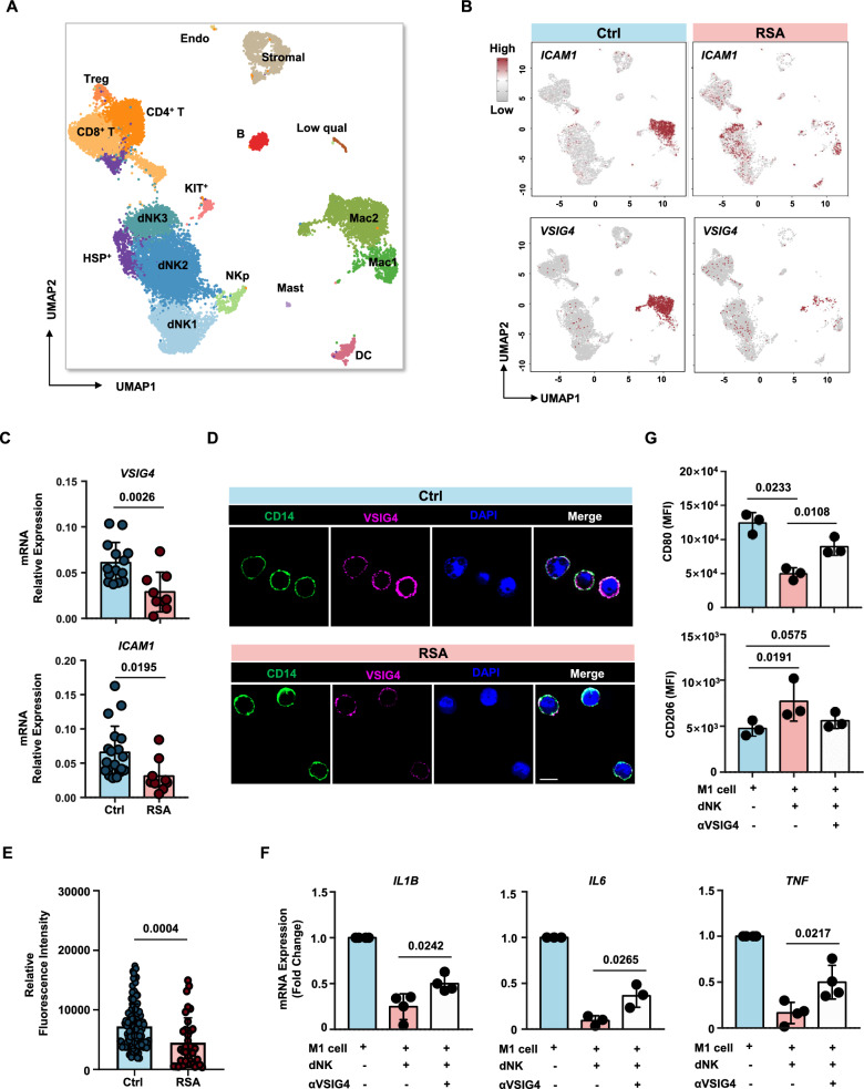
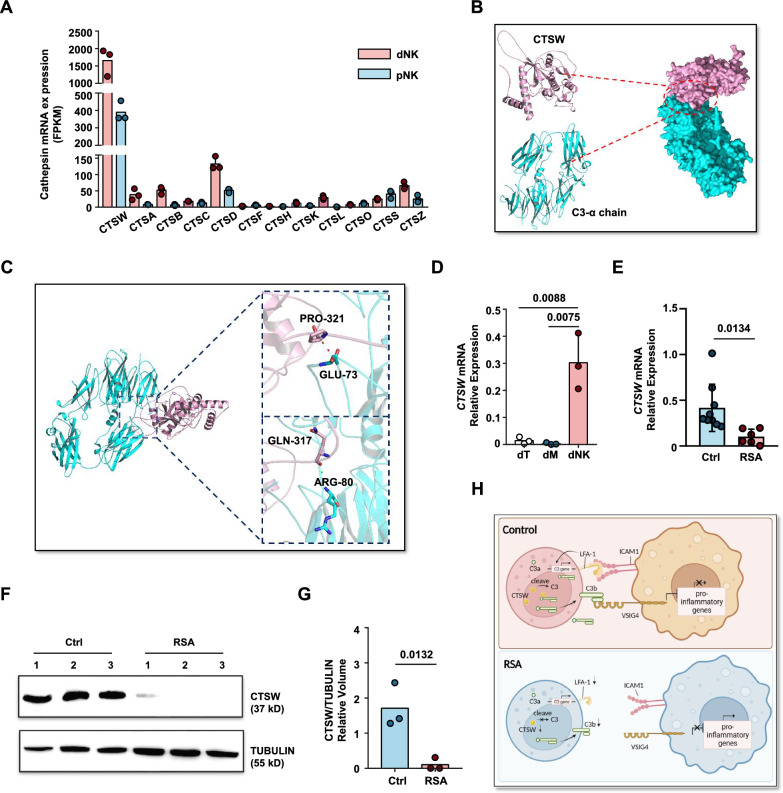
NK cells and macrophages constitute the predominant immune cell subsets in the decidua during the first trimester of pregnancy, with macrophages typically adopting an anti-inflammatory phenotype. Conversely, in the third trimester, macrophages undergo a shift towards a pro-inflammatory phenotype concurrent with a reduction in NK cell numbers. The direct regulatory impact of NK cells on macrophage phenotype remains poorly explored. In our investigation, we observed that ICAM1+ macrophages stimulate the expression of intracellular C3 in LFA1+ decidual NK cells. Notably, Cathepsin W within NK cells exhibit the potential to generate active C3b fragments, effectively inhibit the proinflammatory phenotype of macrophages by binding to VSIG4. Our study unveils a direct regulatory mechanism orchestrated by decidual NK cells over macrophages, providing a potential pathogenic explanation for recurrent spontaneous abortion.

Keywords: Complement; Decidual natural killer cell; Macrophage; Recurrent spontaneous abortion; VSIG4.


Conflict of interest statement

Declarations Ethics approval and consent to participate All human samples used in the present study were obtained under the approval of the Ethics Committee of the University of Science and Technology of China (2022KY-015; Hefei, China). Competing interests The authors declare no potential conflicts of interest.

Figures

Fig. 1
Fig. 1
Reduced cell-to-cell interactions between decidual NK cells and macrophages in RSA patients. A Immunofluorescence staining demonstrating the co-localization of decidual NK cells and macrophages in situ in decidua tissues from normal pregnancies and RSA patients. Scale bar = 5 μm. B Representative immunohistochemical staining of CD68 and CD56 in the serial sections from the same normal pregnant woman or RSA patient. Scale bar = 5 μm. C Dual-color immunohistochemistry depicting cells expressing CD68 (pink) or CD56 (brown) within the same decidual tissue section. Scale bar = 10 μm. D Statistical analysis of cell-to-cell distances between NK cells and macrophages in decidual tissues from normal pregnancies (n = 3) and RSA patients (n = 3). Thirty cell pairs were randomly selected per tissue section. P values have been determined by two-tailed unpaired t-test. E Live cell fluorescence staining showing in vitro binding interactions between FACS-sorted decidual NK cells and macrophages from normal pregnancies or RSA patient. Scale bar = 5 μm
Fig. 2
Fig. 2
M1-prone gene expression signature in RSA patients. A, B Heatmap showing expression levels of DEGs selected for known M1(A) or M2(B) gene markers in decidual macrophages sorted from normal pregnancies (n = 3) and RSA patients (n = 3). C Volcano plot showing DEGs in macrophages single cell sequencing from NP and RSA. D, E GSVA scores were calculated from the macrophages RNA-seq dataset using the gene lists indicated M1(D) and M2(E) phenotype. F, G Validation of M1 or M2 gene expression in normal macrophages and RSA patient macrophages through qPCR analysis. P values have been determined by two-tailed unpaired t-test
Fig. 3
Fig. 3
dNK cells from normal pregnancy inhibit pro-inflammatory macrophages. A Schematic representation of macrophage induction and co-culture system. B Flow cytometry plots depicting distinct subsets of induced macrophages. C, D Representative histogram (left) and expression level (right) of CD80 (C) or CD206 (D) in macrophages after co-culture with dNK cells from normal pregnancies. Experiments were repeated for 5 independent times. P values have been determined by two-tailed paired t-test. E Microscopic images illustrating changes in the cellular morphology of M1 macrophages following co-culture with dNK cells. Scale bar = 100 μm. F Gene expression levels of IL1B, IL6, TNF, and CD80 in macrophages co-cultured with normal pregnancy dNK cells. Experiments were repeated for 3 independent times. P values have been determined by two-tailed unpaired t-test
Fig. 4
Fig. 4
dNK cells from normal pregnancy express the natural ligand of VSIG4. A Gene expression levels of VSIG4 in peripheral blood NK cells (pNK), peripheral blood monocytes (pM), decidual macrophages (dM), and decidual NK cells (dNK). B Fluorescence image depicting the expression of VSIG4 protein on decidual macrophages. C Biological processes that are significantly enriched in Sankey dot pathway enrichment analysis of the upregulated genes of dNK relative to pNK. D Heat map of differential genes in complement and coagulation cascade pathways, n = 3 per group. E Gene set enrichment analysis (GSEA) revealed an increase in complement and coagulation cascade pathways (enrichment plot: COMPLEMENT AND COAGULATION CASCADES PATHWAYS, HSA04610) in dNK cells compared with pNK cells, n = 3 per group. F qPCR validation of the differential expression of the C3 gene in dNK cells and pNK cells. G CLSM images showing the secretion of C3b in pNKs and dNKs. The images were captured using the same parameters. Scale bars = 5 μm. H Statistical histogram of C3b mean fluorescence intensity of each decidual or peripheral NK cell. P values have been determined by two-tailed unpaired t-test
Fig. 5
Fig. 5
Weakened LFA1-C3 signaling in dNK cells in RSA patients. A Heat map of differential genes in complement and coagulation cascade pathways, n = 3 per group. B Comparison of FPKM values of C3 (complement C3) in dNK cells from RNA-seq data. Unpaired t-test. C qPCR assessment of C3 gene expression levels in decidual NK cells from normal pregnancies and RSA patients (n = 6 per group). D Western blot analysis of C3 protein expression levels in decidual NK cells from normal pregnancies and RSA patients. E Relative volume quantification of C3B protein and the internal control protein TUBULIN using ImageLab software. F qPCR analysis of C3 gene expression in dNK cells stimulated with ICAM1 protein, or co-stimulated with ICAM1 protein and the LFA1-ICAM1 binding blocking protein DEL-1 for 24 h. G Representative contour plots of LFA1+ dNK cells gated on CD45+CD3CD56+ cells. H Statistical chart presenting the proportions of LFA1+ dNK cells across normal pregnancies group and RSA group. P values have been determined by two-tailed unpaired t-test
Fig. 6
Fig. 6
VSIG4 blockade impairs C3b induced macrophages polarization. A Uniform manifold approximation and projection (UMAP) of single-cell RNA-seq from human decidual immune cells isolated from patients with RSA and healthy controls. The single-cell RNA sequencing data used in this study were previously published by Guo [9]. B Feature plot showed ICAM1 and VSIG4 expression in the different clusters between Ctrl vs. RSA as defined in (A). C Gene expression levels of VSIG4 and ICAM1 in decidual macrophages from normal pregnancies and RSA patients. D CLSM images showing the expression of VSIG4 protein in decidual macrophages from normal pregnancies and RSA patients. Scale bars = 5 μm. E Statistical histogram of VSIG4 mean fluorescence intensity of each decidual macrophages from normal pregnancies (n = 3) and RSA patients (n = 3). F Gene expression levels of IL1B, IL6 and TNF in macrophages with or without the addition of VSIG4 blocking antibody during co-culture with dNK. G Representative mean fluorescence intensity of CD80 (upper panel) or CD206 (lower panel) in macrophages with or without the addition of VSIG4 blocking antibody during co-culture with dNK. Experiments were repeated for 3 independent times. P values have been determined by two-tailed paired t-test
Fig. 7
Fig. 7
C3 cleavage potential of cathepsin W. A FPKM values of cathepsin family genes in pNK cells and dNK cell RNA-seq data. B Schematic representation of the interaction between CTSW protein and C3 α-chain protein. C Schematic enlargement of the binding site between CTSW protein and C3 α-chain protein as simulated by ZDOCK. D Gene expression levels of CTSW in the three major immune cell subtypes of the decidua: NK cells, macrophages, and T cells. E Gene expression levels of CTSW in decidua NK cells from normal pregnancy and RSA patients. F Western blot analysis of CTSW protein expression in decidua NK cells from normal pregnancy and RSA patients, TUBULIN expression was measured as loading control. Experiments were repeated for 2 independent times. G Relative volume quantification of CTSW protein and the loading control protein TUBULIN using ImageLab software. H Graphic abstract of this article. Created with BioRender.com

References

    1. Trundley A, Moffett A. Human uterine leukocytes and pregnancy. Tissue Antigens. 2004. 10.1111/j.1399-0039.2004.00170.x. - PubMed
    1. Bulmer JN, Williams PJ, Lash GE. Immune cells in the placental bed. Int J Dev Biol. 2010;54:281–94. 10.1387/ijdb.082763jb. - PubMed
    1. Koopman LA, et al. Human decidual natural killer cells are a unique NK cell subset with immunomodulatory potential. J Exp Med. 2003;198:1201–12. 10.1084/jem.20030305. - PMC - PubMed
    1. Manaster I, Mandelboim O. The unique properties of uterine NK cells. Am J Reprod Immunol. 2010;63:434–44. 10.1111/j.1600-0897.2009.00794.x. - PubMed
    1. Hanna J, et al. Decidual NK cells regulate key developmental processes at the human fetal-maternal interface. Nat Med. 2006;12:1065–74. 10.1038/nm1452. - PubMed

LinkOut - more resources