. 2024 Nov 11;17(1):461.
doi: 10.1186/s13071-024-06489-x.

Distinct neutrophil effector functions in response to different isolates of Leishmania aethiopica

Affiliations

Distinct neutrophil effector functions in response to different isolates of Leishmania aethiopica

E Adem et al. Parasit Vectors. .

Abstract

Background: In Ethiopia, cutaneous leishmaniasis is mainly caused by Leishmania (L.) aethiopica parasites and presents in three main clinical forms. It is still not clear if the host immune response plays a role in the development of these different presentations. Since neutrophils are likely to be one of the first immune cells present at the site of the sand fly bite, we set up an in vitro model of infection of neutrophils with L. aethiopica and assessed some of the main neutrophil effector functions: association with and internalisation of parasites, apoptosis and ROS production. We used three freshly isolated clinical isolates and one isolate that has been kept in culture for decades.

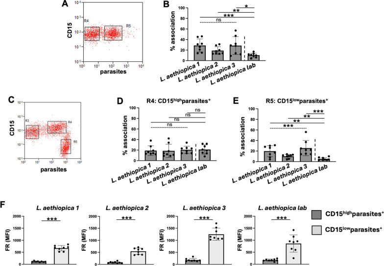
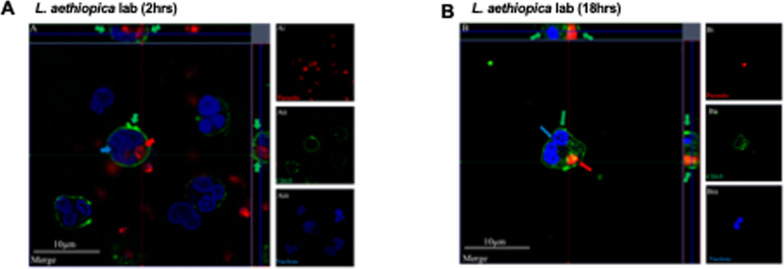
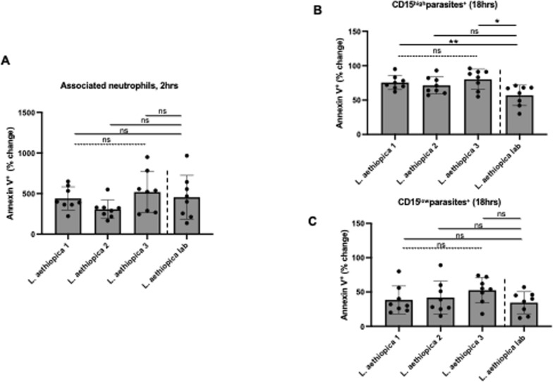
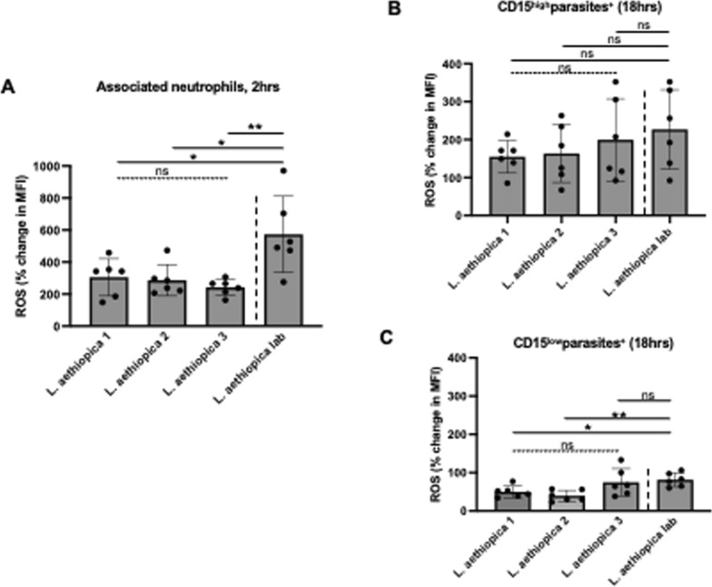
Results: Our results showed by flow cytometry that all four L. aethiopica isolates had the ability to associate with neutrophils. The three clinical isolates of L. aethiopica associated more efficiently with neutrophils than the long-term cultured L. aethiopica. At 18 h, two distinct populations of neutrophils were identified that associated with L. aethiopica, CD15high and CD15low neutrophils. Confocal microscopy demonstrated that all isolates can be internalised. Our results also showed that all parasites induced apoptosis in L. aethiopica-associated neutrophils. Moreover, our results showed that after 2 h, L. aethiopica-associated neutrophils upregulated their production of ROS, but to a greater extent with the long-term cultured L. aethiopica. After 18 h of incubation, CD15lowparasite+ showed an impaired ability to produce ROS compared to CD15highparasite+.

Conclusions: Using this in vitro model, our results show that different L. aethiopica parasite isolates, most notably long-term cultured parasites, had differential effects on neutrophil effector functions.

Keywords: Leishmania aethiopica; Apoptosis; Neutrophils; Phagocytosis; ROS.


Conflict of interest statement

Declarations Ethics approval and consent to participate Ethical approval was obtained from the Faculty Ethic Committee University of Greenwich (FES-FREC-20-01.04.08.CA). Informed written consent was obtained from each participant. Consent for publication Not applicable. Competing interests The authors declare no competing interests.

Figures

Fig. 1
Fig. 1
Association of neutrophils with different isolates of Leishmania aethiopica; 1 × 105 cells/ml neutrophils were co-cultured with 1 × 106 cells/ml FR labelled L. aethiopica isolates for 2 h (A, B) and 18 h (CF). The percentages of neutrophils associated with L. aethiopica were determined by flow cytometry. A Dot plot showing neutrophils unassociated (gate R4) and associated (gate R5) with L. aethiopica. B % of neutrophils associated with the four different isolates of L. aethiopica. C Dot plot showing the three different population of neutrophils: CD15intermediate (int)parasite (gate R3), CD15highparasite+ (gate R4) and CD15lowparasite+ (gate R5). D % of CD15highparasite+ associated with the four different isolates of L. aethiopica. E % of CD15lowparasite+ associated with the four different isolates of L. aethiopica. F Comparison in FR MFI between CD15highparasite+ and CD15lowparasite+ for each L. aethiopica. Data are presented as scatter plot with bar (mean with standard deviation), with each dot representing the value for one experiment. Statistical differences were determined using Kruskal-Wallis (dotted line) and Mann-Whitney (solid line) tests
Fig. 2
Fig. 2
Internalisation of Leishmania aethiopica by neutrophils; 1 × 105 cells/ml neutrophils were co-cultured with 1 × 106 cells/ml FR labelled L. aethiopica lab for 2 h (A) and 18 h (B) and cells were labelled as described in “Methods”. The red arrows point to the parasite (FR), the green arrows to the CD15 (Alexa Fluor 555) and the blue arrow to the nucleus (DAPI). These are representative images of at least three independent experiments
Fig. 3
Fig. 3
% change in apoptotic neutrophils between the different parasite isolates; 1 × 105 cells/ml neutrophils were co-cultured with 1 × 106 cells/ml FR labelled L. aethiopica isolates for 2 h (A) and 18 h (B, C). The % change was measured by deducting the % apoptotic (as defined by Annexin V+7-AAD) CD15high neutrophils co-cultured with the parasites from the % of apoptotic neutrophils cultured in the absence of parasites. Data are presented as scatter plot with bar (mean with standard deviation), with each dot representing the value for one experiment. Statistical differences were determined using Kruskal-Wallis (dotted line) and Mann-Whitney (solid line) tests
Fig. 4
Fig. 4
% change in ROS MFI in neutrophils between the different parasite isolates; 1 × 105 cells/ml neutrophils were co-cultured with 1 × 106 cells/ml FR labelled L. aethiopica isolates for 2 h (A) and 18 h (B, C). The % change was measured by deducting the ROS MFI in the neutrophils co-cultured with the parasites from the ROS MFI in the neutrophils cultured in the absence of parasites. Data are presented as scatter plot with bar (mean with standard deviation), with each dot representing the value for one experiment. Statistical differences were determined using Kruskal-Wallis (dotted line) and Mann-Whitney (solid line) tests

References

    1. Ruiz-Postigo JA, Jain S, Madjou S, Virrey Agua JF, Maia-Elkhoury AN, Valadas S, et al. Global leishmaniasis surveillance, 2022: assessing trends over the past 10 years. Wkly Epidemiol Rec. 2023;40:471–87.
    1. Yizengaw E, Gashaw B, Yimer M, Takele Y, Nibret E, Yismaw G, et al. Demographic characteristics and clinical features of patients presenting with different forms of cutaneous leishmaniasis, in Lay Gayint, northern Ethiopia. PLoS Negl Trop Dis. 2024;18:e0012409. - PMC - PubMed
    1. van Henten S, Adriaensen W, Fikre H, Akuffo H, Diro E, Hailu A, et al. Cutaneous leishmaniasis due to Leishmania aethiopica. EClinicalMedicine. 2018;6:69–81. - PMC - PubMed
    1. Yizengaw E, Takele Y, Franssen SU, Gashaw B, Yimer M, Abera A, et al. Parasite genetic variation and systemic immune responses are not associated with different clinical presentations of cutaneous leishmaniasis caused by Leishmania aethiopica. BioRxiv. 2024. 10.1101/2024.04.19.590259. - PMC - PubMed
    1. de Carvalho RVH, Lima-Junior DS, da Silva MVG, Dilucca M, Rodrigues TS, Horta CV, et al. Leishmania RNA virus exacerbates leishmaniasis by subverting innate immunity via TLR3-mediated NLRP3 inflammasome inhibition. Nat Commun. 2019;10:5273. - PMC - PubMed

Substances

LinkOut - more resources