

Histopathological and radiological evaluation of the efficacy of hydroxyapatite-boric acid and hydroxyapatite-magnesium coated Kirschner wires on fracture healing in femoral diaphyseal fractures: an experimental study
- PMID: 39529159
- PMCID: PMC11552370
- DOI: 10.1186/s13018-024-05228-3
Histopathological and radiological evaluation of the efficacy of hydroxyapatite-boric acid and hydroxyapatite-magnesium coated Kirschner wires on fracture healing in femoral diaphyseal fractures: an experimental study
Abstract
Background: Biomaterials used in fracture healing hold a significant place in orthopedics. This study aimed to develop biomaterials coated with hydroxyapatite (HA), boric acid (BA), and magnesium (Mg) and investigate their effects on fracture healing.
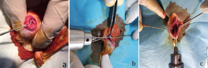
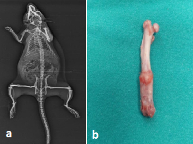
Methods: Sixty female Wistar Albino rats were included in the study. The subjects were randomized into five groups. Cytotoxicity tests were performed on HA, BA, and Mg, and cell viability rates were calculated. Coatings were applied to Kirschner (K) wires at determined ratios. Group I was the control group with a steel K wire, Group II used HA-coated K wires, Group III used HA + BA-coated K wires, Group IV used HA + BA + Mg-coated K wires, and Group V used HA + Mg-coated K wires. A fracture was induced in the right femur of the subjects, followed by fixation with intramedullary K wires. The subjects were randomly divided into equal numbers and sacrificed at 6 and 12 weeks. Radiological and histopathological evaluations were performed.
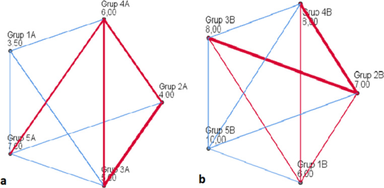
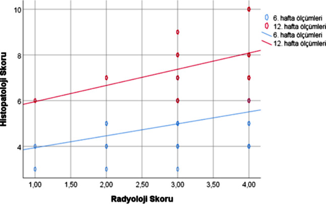
Results: In direct cytotoxicity tests, the highest viability rate was observed in Group IV, while in indirect cytotoxicity tests, it was highest in Group II. In radiological evaluation at the 6th week, the highest scores were in Groups IV and V, while the lowest was in Group III. At the 12th week, the highest scores were in Groups II and V, while the lowest was in Group I. No significant differences were found between the groups (p = 0.837, p = 0.0479). In histopathological evaluation, a significant difference was observed between the groups (p < 0.001), with the highest scores in Group V. A correlation was found between the radiological and histopathological scores (p < 0.001, r = 0.438).
Conclusion: It was found that HA + Mg significantly improved histological outcomes in fracture healing. Good histological results can be achieved with the use of Mg-containing implants in both early and late-stage fracture healing. Coating the biomaterials used in fracture fixation with Mg may lead to positive outcomes in fracture healing.
Keywords: Biomaterial; Boric acid; Fracture healing; Hydroxyapatite; Magnesium; Nonunion.
© 2024. The Author(s).
Conflict of interest statement
Figures
References
-
- Schindeler BPDG, McDonald A. Bone remodeling during fracture repair: the cellular picture. Semin Cell Dev Biol. 2008;19(5):459–66. 10.1016/j.semcdb.2008.07.004. - PubMed
-
- Calori TL, Albisetti G, Agus W, Iori A, S. Risk factors contributing to fracture non-unions. Injury. 2007;38(8)–11. - PubMed
-
- Cirakli A. Effects of cilostazol on fracture healing: an experimental study. Acta Orthop Traumatol Turc. 2016. 10.3944/AOTT.2016.15.0211. - PubMed
-
- Murugan S, Ramakrishna R. Development of nanocomposites for bone grafting. Compos Sci Technol, 65, pp. 2385–406.
MeSH terms
Substances
LinkOut - more resources
Full Text Sources
Medical
