. 2024 Nov 11;14(1):27.
doi: 10.1186/s13395-024-00359-x.

3D-environment and muscle contraction regulate the heterogeneity of myonuclei

Affiliations

3D-environment and muscle contraction regulate the heterogeneity of myonuclei

Rosa Nicolas et al. Skelet Muscle. .

Abstract

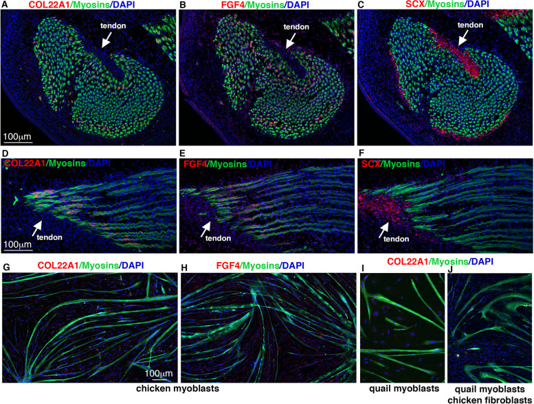
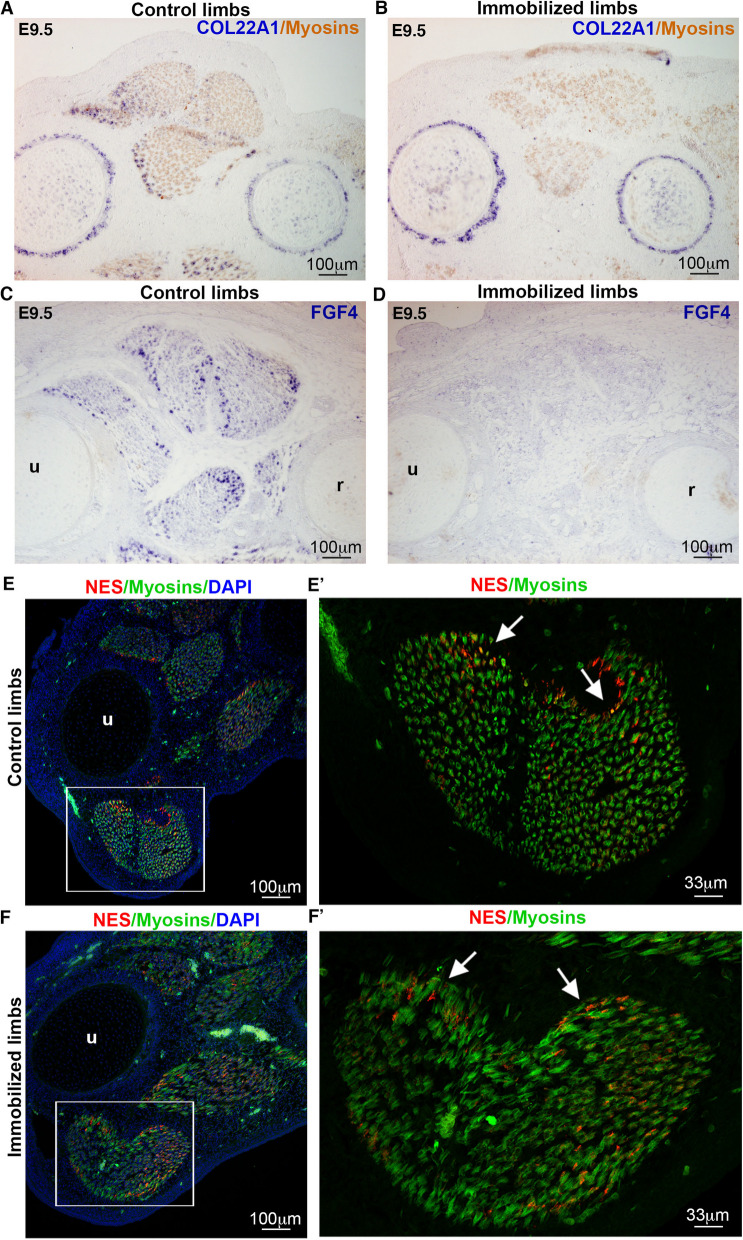
Skeletal muscle formation involves tight interactions between muscle cells and associated connective tissue fibroblasts. Every muscle displays the same type of organisation, they are innervated in the middle and attached at both extremities to tendons. Myonuclei are heterogeneous along myotubes and regionalised according to these middle and tip domains. During development, as soon as myotubes are formed, myonuclei at muscle tips facing developing tendons display their own molecular program. In addition to molecular heterogeneity, a subset of tip myonuclei has a fibroblastic origin different to the classical somitic origin, highlighting a cellular heterogeneity of myonuclei in foetal myotubes. To gain insights on the functional relevance of myonucleus heterogeneity during limb development, we used 2D culture and co-culture systems to dissociate autonomous processes (occurring in 2D-cultures) from 3D-environment of tissue development. We also assessed the role of muscle contraction in myonucleus heterogeneity in paralysed limb muscles. The regionalisation of cellular heterogeneity was not observed in 2D cell culture systems and paralyzed muscles. The molecular signature of MTJ myonuclei was lost in a dish and paralysed muscles indicating a requirement of 3D-enviroment and muscle contraction for MTJ formation. Tip genes that maintain a regionalized expression at myotube tips in cultures are linked to sarcomeres. The behaviour of regionalized markers in cultured myotubes and paralyzed muscles allows us to speculate whether the genes intervene in myogenesis, myotube attachment or MTJ formation.

Keywords: Cell cultures; Chicken; Embryos; Fibroblast; Heterogeneity; Immobilization; Limbs; Myoblast; Myonuclei; Myotendinous junction; Quail; Regionalisation.


Conflict of interest statement

Declarations Ethics approval and consent to participate All experiments with chicken embryos were performed before E10 (embryonic day 10 of developpement) and consequently are not submitted to a licensing committee, in accordance with European guidelines and regulations. Competing of interests The authors declare no competing interests.

Figures

Fig. 1
Fig. 1
Loss of regionalisation of fusion-associated genes in cultured myotubes. A-D Fluorescent in situ hybridization to adjacent transverse muscle sections from E10 chicken embryos with TMEM8C (A), MYOG (B), MYOD (C) and SCX (D) probes (red), followed by an immunolabelling of myosins (green) (D), combined with DAPI staining (nuclei, blue). White dashed lines correspond to the muscle/tendon interface. E-J Primary cultures of limb myoblasts from E10 quail embryos. E,F Myoblasts and myotubes labelled with PAX7 (muscle progenitors, red) and MF20 (myosins, green) antibodies (E), or with MYOG (red) and MF20 (myosins, green) antibodies (F), combined with DAPI staining (nuclei, blue). G Percentage of PAX7-positive nuclei versus total nuclei. Percentage of MYOG-positive nuclei versus total nuclei. Fusion index. Graphs show mean ± s.d. H-J Fluorescent in situ hybridization to quail myoblast cultures with TMEM8C (H), MYOG (I) and MYOD (J) probes (red) followed by an immunolabelling of myosins (green) combined with DAPI staining (nuclei, blue). K-M Fluorescent in situ hybridization to chicken myoblast cultures with TMEM8C (K), MYOG (L) and MYOD (M) probes (red) followed by an immunolabelling of myosins (green) combined with DAPI staining (nuclei, blue). (K’-M’) are high magnification of squared regions in (K-M)
Fig. 2
Fig. 2
Fibroblasts promote myoblast differentiation in fibroblast/myoblast co-cultures. A Percentage of quail myoblasts and chicken fibroblasts in co-cultures after five days of cultures. Representative field of quail nuclei (QCPN antibody, red) and chicken nuclei (DAPI, blue). B Quail myoblast / chicken fibroblast co-cultures immunolabelled to PAX7 (PAX7 antibody, red) and myosins (QCPN antibody, green) combined with DAPI staining (blue). C Quail myoblast / chicken fibroblast co-cultures immunolabelled to MYOG (red) and myosins (QCPN antibody, green) combined with DAPI staining (blue). D,E Percentage of PAX7-positive nuclei (D) and of MYOG-positive nuclei (E) versus total nuclei in fibroblast/myoblast co-cultures. F Fusion index of quail cells in co-cultures. Graphs show mean ± s.d. G,H Myoblast cultures (G) and myoblast/fibroblast co-cultures (H) labelled with the MF20 antibody (myosins, green), combined with DAPI staining (nuclei, blue). I Quantification of myotube area per surface unit in myoblast cultures and myoblast/fibroblast co-cultures. Graph shows mean ± s.d. J,K Fluorescent in situ hybridization to quail myoblast cultures (J) and to quail myoblast / chicken fibroblast co-cultures (K) with TMEM8C probe (red) followed by an immunolabelling of myosins (green) combined with DAPI staining (nuclei, blue)
Fig. 3
Fig. 3
Recruitment of fibroblast nuclei into myotubes in fibroblast/myoblast co-cultures. (A,A’,A”) Quail myoblast and chicken fibroblast and co-cultures immunolabelled to quail nuclei (QCPN antibody, red) and myosins (MF20 antibody, green), combined with DAPI staining (nuclei, blue). A Left panel shows quail nuclei/Myosins/DAPI, while right panels (A’,A”) are high magnification squared in A, showing quail nuclei/Myosins/DAPI staining (A’) and quail nuclei/Myosins (A”). Arrows point to chicken fibroblast myonuclei. B Percentage of chicken fibroblast myonuclei versus quail and chicken myonuclei and versus total chicken nuclei. Graph shows mean ± s.d. (C,C’,C”) Chicken myoblast and quail fibroblast and co-cultures immunolabelled to quail nuclei (QCPN antibody, red) and myosins (MF20 antibody, green), combined with DAPI staining (nuclei, blue). C Left panel shows quail nuclei/Myosins/DAPI, while right panels (C’,C”) are high magnification squared in C, showing quail nuclei/Myosins/DAPI staining (C’) and Myosins/DAPI (C”). Arrows point to quail fibroblast myonuclei. D Percentage of quail fibroblast myonuclei versus chicken and quail myonuclei and versus total quail nuclei. Graph shows mean ± s.d. (E,E’,E”,F,F’,F”) Fluorescent in situ hybridization to quail myoblast / chicken fibroblast co-cultures with MYOG (E,E’,E”) or TMEM8C (F,F’,F”) probes (red) followed by an immunolabelling of myosins (green) and of quail cells (grey) combined with DAPI staining (nuclei, blue). Arrows point to chicken fibroblast myonuclei expressing MYOG (E,E’,E”) and TMEM8C (F,F’,F”)
Fig. 4
Fig. 4
BMP signalling regulates the incorporation of fibroblast nuclei into myotubes. A Fluorescent in situ hybridization to longitudinal sections of limb muscles with BMP4 probe (red) followed by an immunohistochimistry with the MF20 antibody (myosins, green) combined with DAPI staining (nuclei, blue). B,B’ Immunohistochemistry to adjacent longitudinal sections of limb muscle sections with pSMAD1/5/9 (red) and MF20 (myosins, green) antibodies combined with DAPI staining (nuclei, blue). C Immunohistochemistry to transverse sections of limb muscles with pSMAD1/5/9 (red) and MF20 (myosins, green) antibodies). D,D’,D” Immunohistochemistry to quail myoblast cultures with pSMAD1/5/9 (red), PAX7 (grey) and MF20 (myosins, green) antibodies combined with DAPI staining (nuclei, blue). E-J BMPR1Aca-transfected chicken fibroblasts (E), BMP4-transfected chicken fibroblasts (F), BMPR1Adn-transfected chicken fibroblasts (H) or NOGGIN-transfected chicken fibroblasts (I) were co-cultured with quail myoblasts; and labelled with the QCPN (quail nuclei, red), MF20 antibody (myosins, green) combined with DAPI staining (nuclei, blue). G Percentage of chicken fibroblast myonuclei within myotubes in control-, BMPR1Aca-, BMP4-transfected chicken fibroblasts cultured with quail myoblasts, (BMP gain-of-function experiments). J Percentage of chicken fibroblast myonuclei within myotubes in control-, BMPR1Adn-, NOGGIN-transfected chicken fibroblasts cultured with quail myoblasts, (BMP loss-of-function experiments). (G,J) Graphs shows mean ± s.d
Fig. 5
Fig. 5
The COL22A1 and FGF4 expression in tip myonuclei of limb muscles is lost in cultured myotubes. A-F Gene expression for muscle tip genes (COL22A1, FGF4) and tendon gene (SCX) in adjacent transverse (A-C) and longitudinal (D-F) muscle sections from E10 chicken limbs with fluorescent in situ hybridization with COL22A1 (A,D), FGF4 (B,E) and SCX (C,F) probes (red), followed by an immunohistochemistry with the MF20 antibody to label myosins (green) combined with DAPI staining (blue). Arrows point to tendons and muscle/tendon interface. G-J Fluorescent in situ hybridization to chicken myoblast cultures (G,H), quail myoblast cultures (I) and quail myoblast / chicken fibroblast co-cultures (J) with COL22A1 (G,I,J) and FGF4 (H) probes (red) followed by an immunolabelling of myosins (green) combined with DAPI staining (nuclei, blue)
Fig. 6
Fig. 6
The regionalized expression of NES, ANKRD1 and MEF2C in tip myonuclei of limb muscles is maintained in cultured myotubes. A-D Gene expression for muscle tip genes (NES, ANKRD1, MEF2C) and tendon gene (SCX) in transverse muscle sections from E10 chicken limbs with fluorescent in situ hybridization for NES probe (A) and with colorimetric in situ hybridization for ANKRD1 (B), MEF2C (C) and SCX (D) probes (dark grey), followed by an immunohistochemistry with the MF20 antibody to label myosins (green). E Fluorescent in situ hybridization to quail myoblast cultures with NES probe (red) followed by an immunolabelling of myosins (green) combined with DAPI staining (nuclei, blue). F-G Fluorescent in situ hybridization to chicken myoblast cultures with ANKRD1 (F), MEF2C (G), MYOG (H) probes (red) followed by an immunolabelling of myosins (green) combined with DAPI staining (nuclei, blue). E-H’ are high magnifications of squared areas in (E–H). Arrows point to NES (E’), ANKRD1 (F’) and MEF2C (G’) expression at myotube tips in low myosins-expressing zones, while MYOG transcripts (F’) are expressed in myosins-positive zones
Fig. 7
Fig. 7
The regionalized expression of NES, ANKRD1 and MEF2C in cultured myotubes is not changed in the presence of fibroblasts. A-C Fluorescent in situ hybridization to quail myoblast / chicken fibroblast co-cultures with the NES (A), ANKRD1 (B) and MEF2C (C) probes (red), followed by an immunohistochemistry with the MF20 antibody to label myosins (green) combined with DAPI staining (blue). D,D’ High magnification of the area squared in A, showing in the top panel, (NES transcripts, red, myosins, green) and DAPI staining, blue) combined with immunolabelling of quail nuclei (grey) (D), and in the bottom panel, immunolabelling of quail nuclei (grey) combined with DAPI staining, (blue) (D’). Arrowheads point to chicken fibroblast myonuclei expressing NES, while arrows point to point to chicken fibroblast myonuclei not expressing NES. E Fluorescent in situ hybridization to quail myoblast / chicken fibroblast co-cultures with NES probes (red) followed by an immunolabelling of myosins (green) and of quail nuclei (grey), combined with DAPI staining (nuclei, blue). F,F’,F” High magnification of the area squared in E, showing NES transcripts, (red), myosins, (green) and DAPI staining (blue) combined with immunolabelling of quail nuclei (grey) (F), showing myosins, (green) and DAPI staining (blue) combined with immunolabelling of quail nuclei (grey) (F’) and showing NES transcripts, (red), DAPI staining (blue) combined with immunolabelling of quail nuclei (grey) (F”). (F,F’,F”) Arrows points to a chicken fibroblast myonuclei with low/residual levels of NES incorporated into a quail myotube
Fig. 8
Fig. 8
Inhibition of muscle contraction modified the expression of genes expressed in tip myonuclei of foetal muscles. Chicken embryos were treated with DMB at E7.5 to block muscle contraction and analysed 48 h after treatment at E9.5 (N = 3). A-D In situ hybridization to transverse limbs sections at the level of the zeugopod of control (A,C) and DMB-treated (B,D) E9.5 chicken embryos, with COL22A1 (A,B) and FGF4 (C,D) probes (blue) followed by immunostaining with the MF20 antibody to label myosins (light brown). The expression of COL22A1 and FGF4 was lost in paralyzed muscles (B,D) compared to control muscles (A,C). Note that COL22A1 expression is maintained in cartilage and perichondrium in paralyzed limbs (B) as in control limbs (A). E, E’, F,F’ Fluorescent situ hybridization to transverse limbs sections at the level of the zeugopod of control (E,E’) or DMB-treated (F,F’) E9.5 chicken embryos, with NES probe (red) followed by immunostaining with the MF20 antibody to label myosins (green) and stained with DAPI (blue). (E’,F’) are high magnification of the FCU muscles of E (control) and F (DMB-treated) limbs. u, ulna, r, radius
Fig. 9
Fig. 9
Schematic of molecular and cellular heterogeneity of myonuclei in a dish. In foetal limb muscles, COL22A1, FGF4, NES, ANKRD1 and MEF2C transcripts are located in tip myonuclei. In a dish, the expression of COL22A1 and FGF4 is lost, while the expression of NES, ANKRD1 and MEF2C is maintained in tips of cultured foetal myotubes. In limb muscles, fibroblast incorporation is observed preferentially at muscle tips, while there is no regionalisation of fibroblast incorporation in myotubes in fibroblast/myoblast co-cultures. Myotubes are in pink and tendons in green. Tip gene expression is schematized in purple. Myoblast nuclei and myonuclei are in red and fibroblast nuclei are in green

References

    1. Chevallier A, Kieny M, Mauger A. Limb-somite relationship: origin of the limb musculature. J Embryol Exp Morphol. 1977;41:245–58. - PubMed
    1. Christ B, Jacob HJ, Jacob M. Experimental analysis of the origin of the wing musculature in avian embryos. Anat Embryol (Berl). 1977;150:171–86. - PubMed
    1. Nassari S, Duprez D, Fournier-Thibault C. Non-myogenic Contribution to Muscle Development and Homeostasis: The Role of Connective Tissues. Front Cell Dev Biol. 2017;5:22. - PMC - PubMed
    1. Helmbacher F, Stricker S. Tissue cross talks governing limb muscle development and regeneration. Semin Cell Dev Biol. 2020;104:14–30. - PubMed
    1. Kardon G. Muscle and tendon morphogenesis in the avian hind limb. Development. 1998;125:4019–32. - PubMed

LinkOut - more resources