

Lymph node involvement in secondary breast angiosarcoma - a case presentation
- PMID: 39529346
- PMCID: PMC11657328
- DOI: 10.47162/RJME.65.3.15
Lymph node involvement in secondary breast angiosarcoma - a case presentation
Abstract
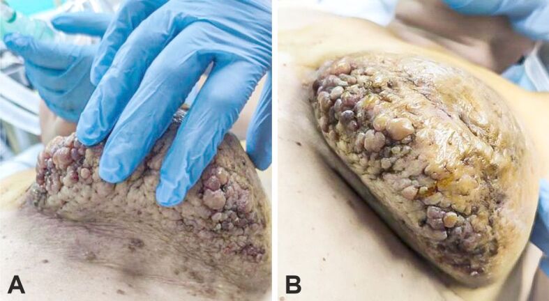
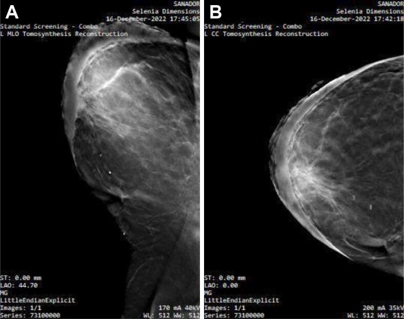
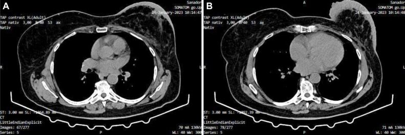
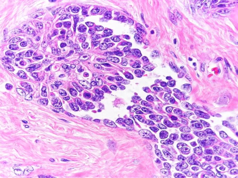
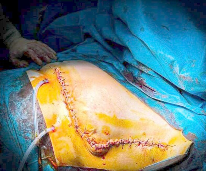
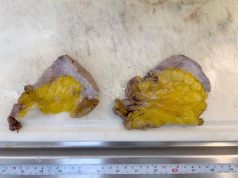
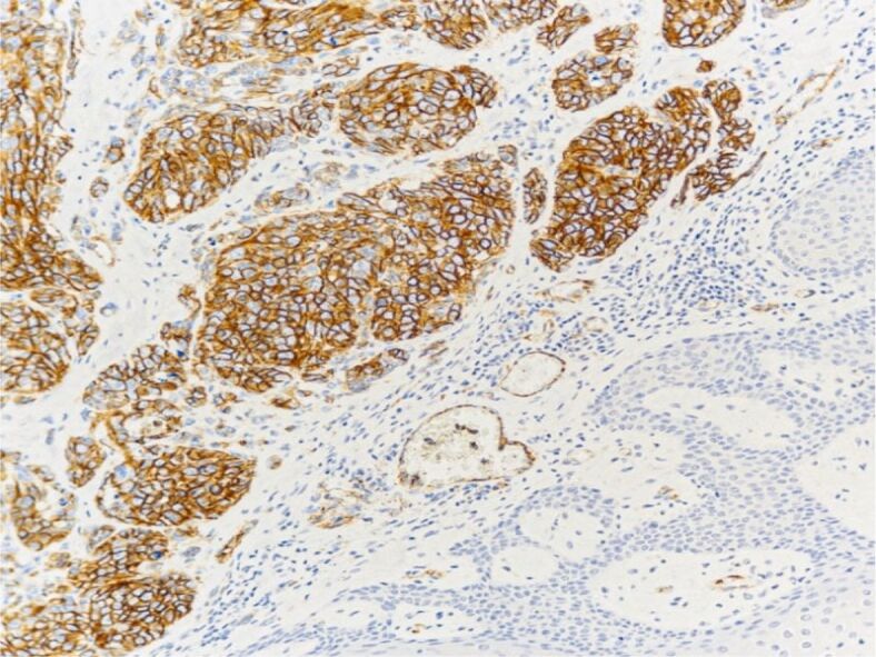
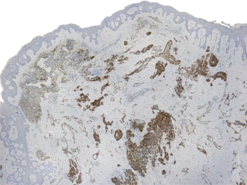
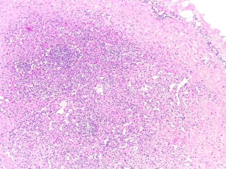
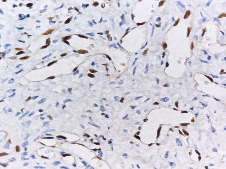
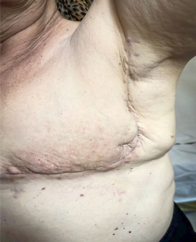
Angiosarcoma represents a group of rare tumors originating from vascular and lymphatic endothelial cells, characterized by marked aggressiveness, rapid growth and poor clinical outcome. The incidence of breast angiosarcoma accounts for approximately 0.05% of all malignant breast tumors and less than 1% of all sarcomas. In this article, we report the case of a 67-year-old female patient who presented to our Clinic due to a rapidly evolving, non-painful, vegetating mass, encompassing almost her entire left breast. Imaging studies revealed diffuse skin thickening in all quadrants and an intensely opaque axillary lymph node (LN). Interestingly, the patient had prior medical history of breast carcinoma treated conservatively in 2007 with limited breast resection and left axillary lymphadenectomy, followed by post-operative chemotherapy and radiotherapy. At the current presentation, we performed a radical mastectomy with ipsilateral lymphadenectomy. The histopathological examination revealed a secondary angiosarcoma with LN involvement mimicking an atypical vascular lesion. In this article, we report the clinicopathological particularities of this case and discuss the challenge of diagnosing LN involvement in angiosarcoma.
Keywords: breast angiosarcoma; epithelioid angiosarcoma; secondary angiosarcoma.
Conflict of interest statement
The authors declare that they have no conflict of interests.
Figures
Similar articles
-
Innovative approach to lymphadenectomy in breast sarcoma.Bull Cancer. 2022 Oct;109(10):1017-1028. doi: 10.1016/j.bulcan.2022.04.012. Epub 2022 Jun 15. Bull Cancer. 2022. PMID: 35717223
-
Primary angiosarcoma of the breast: do enlarged axillary nodes matter?Breast J. 2006 Jul-Aug;12(4):371-4. doi: 10.1111/j.1075-122X.2006.00280.x. Breast J. 2006. PMID: 16848851
-
[Primary angiosarcoma of the breast].Zentralbl Gynakol. 1996;118(5):295-8. Zentralbl Gynakol. 1996. PMID: 8701627 German.
-
Cutaneous angiosarcoma and atypical vascular lesions of the skin and breast after radiation therapy for breast carcinoma.Am J Clin Pathol. 1994 Dec;102(6):757-63. doi: 10.1093/ajcp/102.6.757. Am J Clin Pathol. 1994. PMID: 7801888 Review.
-
[Breast angiosarcoma induced by radiotherapy - surgical treatment options and review of literature].Rozhl Chir. 2017 Fall;96(8):353-358. Rozhl Chir. 2017. PMID: 29058926 Review. Czech.
References
-
- Yan M, Gilmore H, Bomeisl P, Harbhajanka A. Clinicopathologic and immunohistochemical study of breast angiosarcoma. Ann Diagn Pathol. 2021;54:151795–151795. - PubMed
-
- Elder DR , Massi D , Scolyer RA , Willemze R . WHO Classification of skin tumours . 4 . Lyon, France : International Agency for Research on Cancer (IARC) Press ; 2018 . pp. 335 – 336 .
-
- Tan PH , Ellis I , Allison K , Brogi E , Fox SB , Lakhani S , Lazar AJ , Morris EA , Sahin A , Salgado R , Sapino A , Sasano H , Schnitt S , Sotiriou C , van Diest P , White VA , Lokuhetty D , Cree IA . World Health Organization (WHO) Classification of Tumours Editorial Board . WHO Classification of Tumours . 5 . Lyon, France : International Agency for Research on Cancer (IARC) Press ; 2019 . Breast tumors ; pp. 197 – 199 .
-
- Marchal C, Weber B, de Lafontan, Resbeut M, Mignotte H, du Chatelard, Cutuli B, Reme-Saumon M, Broussier-Leroux A, Chaplain G, Lesaunier F, Dilhuydy JM, Lagrange JL. Nine breast angiosarcomas after conservative treatment for breast carcinoma: a survey from French comprehensive Cancer Centers. Int J Radiat Oncol Biol Phys. 1999;44(1):113–119. - PubMed
Publication types
MeSH terms
Supplementary concepts
LinkOut - more resources
Full Text Sources
Medical
