. 2024 Oct 28:12:1476370.
doi: 10.3389/fbioe.2024.1476370. eCollection 2024.

Accelerated innervation of biofabricated skeletal muscle implants containing a neurotrophic factor delivery system

Affiliations

Accelerated innervation of biofabricated skeletal muscle implants containing a neurotrophic factor delivery system

Vladimir Mashanov et al. Front Bioeng Biotechnol. .

Abstract

Introduction: Volumetric muscle loss (VML) is one of the most severe and debilitating conditions in orthopedic and regenerative medicine. Current treatment modalities often fail to restore the normal structure and function of the damaged skeletal muscle. Bioengineered tissue constructs using the patient's own cells have emerged as a promising alternative treatment option, showing positive outcomes in fostering new muscle tissue formation. However, achieving timely and proper innervation of the implanted muscle constructs remains a significant challenge. In this study, we present a clinically relevant strategy aimed at enhancing and sustaining the natural regenerative response of peripheral nerves to accelerate the innervation of biofabricated skeletal muscle implants.

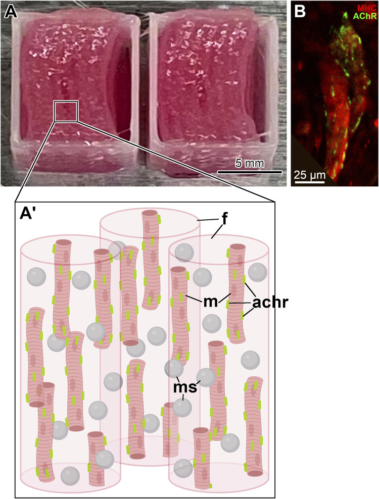
Methods: We previously developed a controlled-release neurotrophic factor delivery system using poly (lactic-co-glycolic acid) (PLGA) microspheres encapsulating ciliary neurotrophic factor (CNTF) and glial cell line-derived neurotrophic factor (GDNF). Here, we incorporate this neurotrophic factor delivery system into bioprinted muscle constructs to facilitate innervation in vivo.

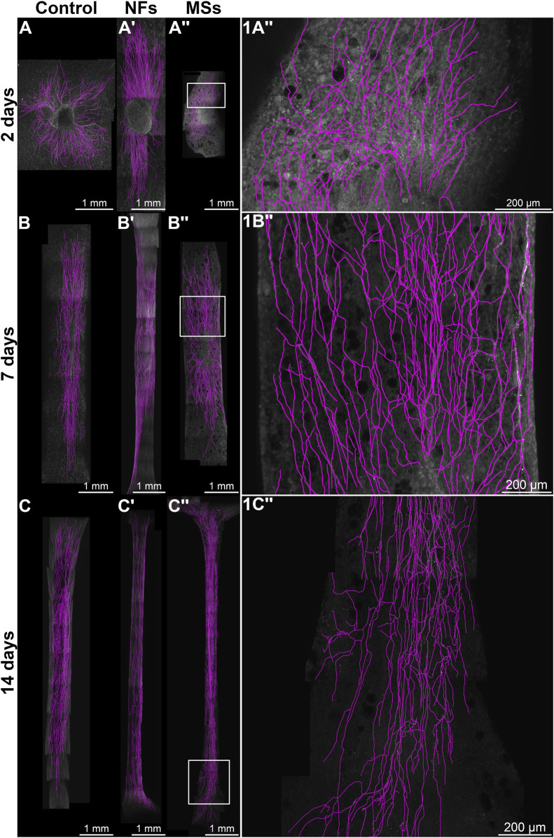
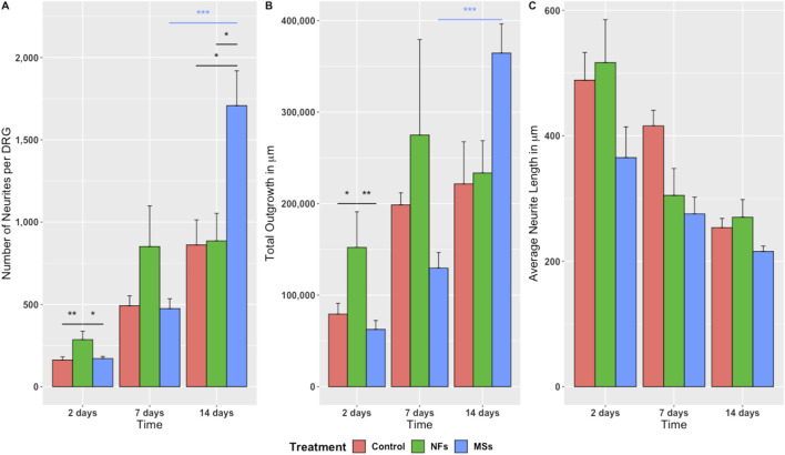
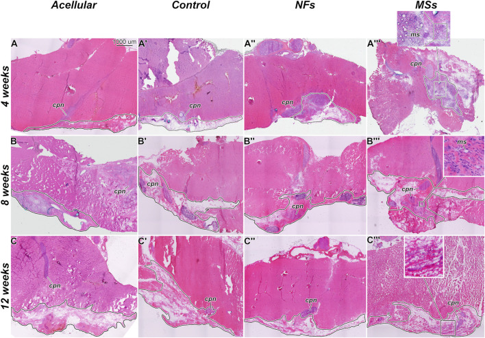
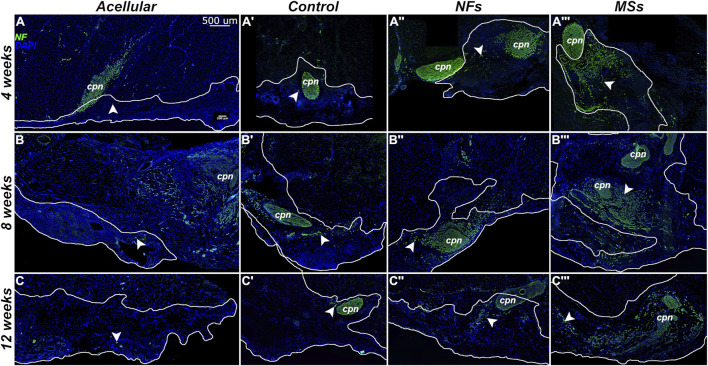
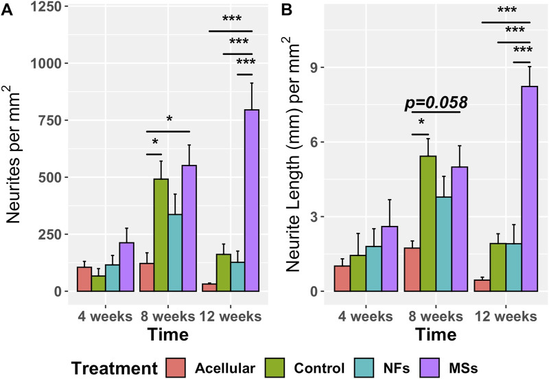
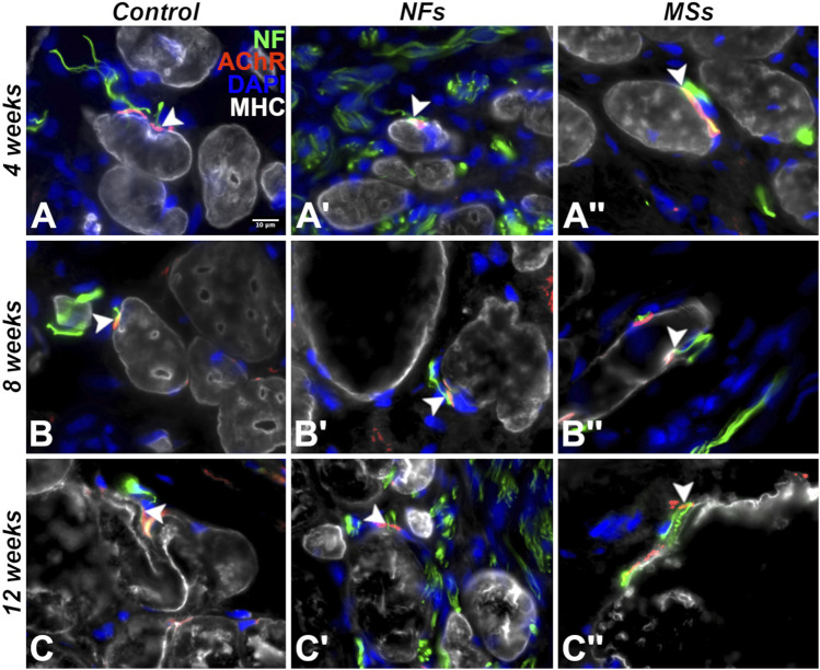
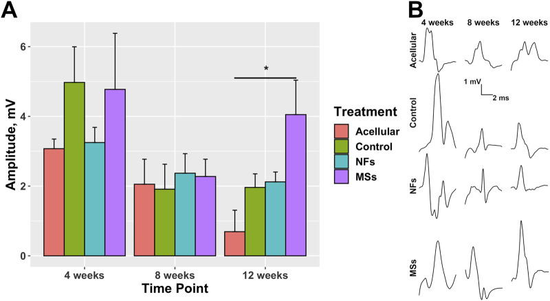
Results: Our results demonstrate that the neurotrophic factors released from the microspheres provide a chemical cue, significantly enhancing the neurite sprouting and functional innervation of the muscle cells in the biofabricated muscle construct within 12 weeks post-implantation.

Discussion: Our approach provides a clinically applicable treatment option for VML through accelerated innervation of biomanufactured muscle implants and subsequent improvements in functionality.

Keywords: CNTF; GDNF; bioengineered skeletal muscle; controlled-release delivery system; innervation; neurotrophic factors.


Conflict of interest statement

The authors declare that the research was conducted in the absence of any commercial or financial relationships that could be construed as a potential conflict of interest.

Figures

FIGURE 1
FIGURE 1
Bioprinted skeletal muscle constructs. (A) A pair of fully printed skeletal muscle constructs with a neurotrophic factor delivery system. They have dimensions of 10 mm × 10 mm × 4 mm (W × L × H) and are composed of eight layers of cell-laden fibrinogen-based bioink with sacrificial bioink spacers. The cell-laden bioink contains CNTF/GDNF-loaded microspheres. In addition, an anchoring structure composed of PCL is deposited as a square frame at the periphery of the implant. (A′) Diagram of the microscale organization of the muscle construct. Created with BioRender. Abbreviations: achr–AChR cluster; f – fibrin hydrogel; m–muscle cell; ms–microsphere. (B) Differentiated muscle cells (red) with induced pre-formed AChR clusters (green) in the muscle construct prior to implantation. The sample was immunostained with antibodies against myosin heavy chain (MHC, red) and AChR (green). The image is a maximum intensity Z-projection of a confocal stack.
FIGURE 2
FIGURE 2
Representative micrographs of chick embryo dorsal root ganglia (DRGs) cultured on 3D bioprinted skeletal muscle constructs in the in vitro neurite outgrowth assay. In the Control group (A–C), the constructs contained no neurotrophic factors. The microsphere-free constructs in the NFs group (A′–C′) contained CNTF and GDNF directly mixed into the hydrogel of the construct. The constructs in the MSs group (A′′–C′′) contained the matching load of the neurotrophic factors encapsulated in PLGA microspheres. The ganglia were cultured on the muscle constructs for 2 days (A, A′′), 7 days (B, B′′), and 14 days (C, C′′). Micrographs in the right column (1A′′–1C′′) are representative high-magnification images [of the boxed areas in (A′′–C′′), respectively]. The samples were then fixed and immunostained with anti-neurofilament antibodies. All images are maximum intensity Z-projections of confocal stacks. The neurites were traced with the NeuronJ plugin in the Fiji/ImageJ software and are shown in magenta.
FIGURE 3
FIGURE 3
Quantitative assessment of neurite sprouting from the chick embryo DRGs grown on 3D bioprinted skeletal muscle constructs. Three metrics were used: (A) Number of neurites sprouting from individual ganglia; (B) Total outgrowth (the sum total of neurite length); (C) Average neurite length. The significance level annotations are shown in black for the treatment cohorts at each time point and in the respective color for each treatment cohort across time points. Three to eight ganglia were quantified per treatment cohort at each time point. The freely dissolved neurotrophic factors (NFs) induced higher neurite sprouting on day 2. This effect was, however, unsustainable. In contrast, the neurotrophic factors delivered in microspheres (MSs) induced the sprouting of more neurites per ganglion and higher total neurite outgrowth by day 14. * p<0.05, ** p<0.01, *** p<0.001 .
FIGURE 4
FIGURE 4
Representative micrographs of the histological organization of the tissue samples from the in vivo transposed nerve study at 4 weeks (A–A‴), 8 weeks (B–B‴), and 12 weeks (C–C‴) post-implantation. Hematoxylin and eosin staining. The Acellular cohort involves fibrin hydrogel implants that contain no cells and no neurotrophic factors. The Control group involves biofabricated muscle implants with 30 million hMPCs per mL of hydrogel, but no extrinsic neurotrophic factors. The third group (NFs) contains CNTF and GDNF freely dissolved in the extracellular matrix of the implant. The fourth group (MSs) contains the matching load of the neurotrophic factors encapsulated in PLGA microspheres suspended throughout the hydrogel of the construct. The implanted constructs are outlined. Note that in the MSs cohort, the microspheres are abundant in the extracellular matrix of the implant at 4 weeks post-implantation. After 8 weeks, they become much smaller in size and are no longer detectable after 12 weeks. This indicates that the microspheres slowly dissolve in time and release the encapsulated CNTF and GDNF. cpn–transposed common peroneal nerve; ms–CNTF/GDNF-loaded PLGA microspheres.
FIGURE 5
FIGURE 5
Representative micrographs of the immunostained samples from the transposed nerve study at 4 weeks (A–A‴), 8 weeks (B–B‴), and 12 weeks (C–C‴) post-implantation. Sections were immunostained with an anti-neurofilament (NF) antibody (green) to visualize the sprouting neurites (arrowheads). The implanted constructs are outlined. The nuclei are stained with DAPI (blue). cpn–transposed common peroneal nerve.
FIGURE 6
FIGURE 6
Quantitative assessment of neurite sprouting from the transposed host common peroneal nerve in the implanted skeletal muscle constructs at 4, 8, and 12 weeks post-implantation. The Acellular cohort involves fibrin hydrogel implants with no cells and no extrinsic neurotrophic factors. The Control group contains cellularized biofabricated muscle implants without any extrinsic neurotrophic factors. The third group (NFs) contains CNTF and GDNF freely dissolved in the extracellular matrix of the implant. The fourth group (MSs) contains the matching load of the neurotrophic factors encapsulated in PLGA microspheres suspended throughout the hydrogel of the construct. There is no statistically significant difference among the treatment cohorts at 4 weeks post-implantation. At 8 weeks, the sprouting in the Control and MSs cohorts was higher than in the Acellular cohort. At 12 weeks, the constructs in the MSs cohort showed more extensive sprouting than any of the other three cohorts, indicating that only the CNTF/GDNF delivered in PLGA microspheres were capable of sustaining long-term neurite outgrowth. Three to four animals were analyzed in each treatment group at all three time points. * p<0.05, *** p<0.001 .
FIGURE 7
FIGURE 7
Representative micrographs of differentiation and innervation of skeletal muscle cells in the implants in the in vivo transposed nerve study at 4 weeks (A–A′′), 8 weeks (B–B′′), and 12 weeks (C–C′′) post-implantation. Triple immunostaining with an AChR antibody (red), an anti-neurofilament (NF) antibody (green), and anti-myosin heavy chain (MHC) antibody (white). The nuclei were stained with DAPI (blue). White arrowheads indicate neuromuscular junctions. As early as 4 weeks post-implantation, the cells in the implant display the proper skeletal muscle cell phenotype and form neuromuscular junctions with the sprouting neurites of the host. The muscle cells in the implant maintained their phenotype and stayed innervated throughout the duration of the experiment (12 weeks post-implantation).
FIGURE 8
FIGURE 8
Compound muscle action potential (CMAP) in the transposed nerve model study at 4, 8, and 12 weeks post-implantation. The Acellular cohort received bioprinted implants containing only fibrin hydrogel with no cells and no intrinsic neurotrophic factors. The Control group contains cellularized biofabricated muscle implants without any extrinsic neurotrophic factors. The third group (NFs) contains CNTF and GDNF freely dissolved in the extracellular matrix of the cellularized implant. The fourth group (MSs) contains the matching load of the neurotrophic factors encapsulated in PLGA microspheres suspended throughout the extracellular matrix of the construct. (A) CMAP amplitude. At 12 weeks post-implantation, the CMAP recorded from the constructs of the MSs cohort shows a trend to be higher than in the other two cohorts with cellularized constructs (the Control and NFs cohorts), and was the only one showing a statistically significant difference from the Acellular cohort. These data indicate that the CNTF and GDNF released from the PLGA microspheres facilitate innervation of the skeletal muscle implants after 12 weeks. This effect requires the microsphere-based neurotrophic factor delivery system and cannot be achieved by implementing the neurotrophic factors in the freely dissolved form. Three animals were analyzed in each treatment cohort (n = 3) * p<0.05 (B) Representative waveforms.

References

    1. Ahuja N., Awad K., Peper S., Brotto M., Varanasi V. (2021). Mini review: biomaterials in repair and regeneration of nerve in a volumetric muscle loss. Neurosci. Lett. 762, 136145. 10.1016/j.neulet.2021.136145 - DOI - PMC - PubMed
    1. Boyd J., Gordon T. (2003). Glial cell line-derived neurotrophic factor and brain-derived neurotrophic factor sustain the axonal regeneration of chronically axotomized motoneurons in vivo . Exp. Neurol. 183, 610–619. 10.1016/s0014-4886(03)00183-3 - DOI - PubMed
    1. Bruneau E. G., Macpherson P. C., Goldman D., Hume R. I., Akaaboune M. (2005). The effect of agrin and laminin on acetylcholine receptor dynamics in vitro . Dev. Biol. 288, 248–258. 10.1016/j.ydbio.2005.09.041 - DOI - PubMed
    1. Chan K. M., Gordon T., Zochodne D. W., Power H. A. (2014). Improving peripheral nerve regeneration: from molecular mechanisms to potential therapeutic targets. Exp. Neurol. 261, 826–835. 10.1016/j.expneurol.2014.09.006 - DOI - PubMed
    1. Das S., Browne K. D., Laimo F. A., Maggiore J. C., Hilman M. C., Kaisaier H., et al. (2020). Pre-innervated tissue-engineered muscle promotes a pro-regenerative microenvironment following volumetric muscle loss. Commun. Biol. 3, 1–14. 10.1038/s42003-020-1056-4 - DOI - PMC - PubMed

LinkOut - more resources