. 2025 Mar 14;117(3):qiae239.
doi: 10.1093/jleuko/qiae239.

Neutrophil swarming is crucial for limiting oral mucosal infection by Candida albicans

Affiliations

Neutrophil swarming is crucial for limiting oral mucosal infection by Candida albicans

Darpan Saraswat et al. J Leukoc Biol. .

Abstract

Oral mucosal colonization by Candida albicans is benign in healthy people but progresses to deeper infection, known as oropharyngeal candidiasis, that may become disseminated when combined with immunosuppression. Cortisone use and neutropenia are risk factors for invasive mucosal fungal infections; however, the mechanisms are poorly understood. Here, we identify in vivo neutrophil functional complexes known as swarms that are crucial for preventing C. albicans epithelial invasion. Anti-Ly6G antibody treatment impaired swarm formation and increased fungal infection depth, confirming the role of neutrophil swarms in limiting C. albicans invasion. Neutrophil swarm function could be disrupted by administration of resolvins, and required BLT1 (leukotriene B4 receptor 1) expression so that administration of a leukotriene synthesis inhibitor reduced neutrophil swarm size permitting C. albicans invasion beyond the basement membrane. Cortisone treatment similarly reduced neutrophil swarming behavior and BLT1 expression and delayed expression of epithelial cytokines and chemokines. Thus, swarm structures have an important function in preventing deep invasion by C. albicans within the oral mucosa and represent a mechanism for increased disease severity under immune deficient clinical settings.

Keywords: corticosteroids; epithelial invasion; neutrophil; oral candidiasis; swarming.


Conflict of interest statement

Conflict of interest statement. None declared.

Figures

Fig. 1.
Fig. 1.
IS impairs inflammatory cell recruitment and alters epithelial morphology. Cortisone-treated mice (IS) and IC mice were infected sublingually with 107 CFU of Candida albicans (Ca), and tongue, stomach, cecum, liver, and stool were collected from 1 to 21 dpi (5 to 7 mice/group, per time point) or sham infected mice (S). (A–C) Representative colocalized tongue sections from IC and IS mice after Ca infection (black arrows) in IC and IS mice stained for periodic acid Schiff (top) and hematoxylin and eosin (bottom) (n = 3 mice/group per time point). (A) IC mice show early and robust intraepithelial recruitment of inflammatory cells to the areas of Ca invasion (black arrows) as compared with IS mice (white dotted area) (1 dpi). (B) At 3 dpi, IS mice had increased Ca infection (black arrows) and inflammatory cell recruitment that was dispersed throughout the cellular epithelium (CT) (yellow arrows). (C) At 5 dpi, Ca infection is resolved in IC mice, while IS mice have increased Ca (black arrows) and inflammatory cell infiltrate (yellow arrows) that was found around areas of detachment of CS from underlying cellular epithelium. (D) Percentage of weight loss after Ca infection was compared with preinfection weight in each group. (E) Number of CFU/g tongue tissue in IC and IS mice showing rapid clearance of Ca in IC mice and a persistence of infection in IS mice. (F) Dissemination of Ca in various organs of IC and IS mice. **P < 0.01; ***P < 0.001; ****P < 0.0001 (Mann-Whitney or 2-tailed t-test). Scale bar = 100 μm. cEp = cellular epithelium.
Fig. 2.
Fig. 2.
Oral Candida albicans infection results in recruitment of Ly6G+ neutrophils and expression of G-CSF, IL-1β, KC(CXCL1), and MIP-2 (CXCL2) at 1 dpi that is delayed by cortisone. Neutrophil depletion with anti-Ly6G leads to significantly increased oral infection levels and infection depth in both IC and IS animals. (A) Representative slides showing recruitment of Ly6G+ into the tongue epithelium (Ep) of IC and IS mice. (B) Changes in recruitment of Ly6G+ granulocytes/mm2 (×103) from IC and IS mice as compared with sham (S) at 1 to 5 dpi (n = 3/group). (C) Freshly extracted tongue tissues were dissected to separate Ep and CT and protein were extracted. Kinetics of G-CSF, IL-1β, KC(CXCL1), and MIP-2 (CXCL2) (measured by Bio-Plex assay) in IC and IS mice from 1 to 5 dpi as compared with S mice (n = 5 mice/group per time point). (D) Neutrophil depletion by anti Ly6G resulted in significantly increased fungal infection load (CFU) (E) and increased invasion depth into the CT (F) in both IC and IS animals. *P < 0.05; **P < 0.01; ***P < 0.001; ****P < 0.0001 (Mann-Whitney or 2-tailed t-test and analysis of variance and Tukey's multiple comparisons). Scale bar = 50 μm. n.s. = not significant.
Fig. 3.
Fig. 3.
Dynamics of neutrophil swarm formation and resolution induced by oral fungal infection. Time course experiment showing neutrophil recruitment and resolution following oral Candida albicans (Ca) infection. (A–E) At 16 h post–Ca infection, Ly6G+ neutrophils were observed migrating from the CT (green dotted line) into the epithelium (B), while in sham-infected mice neutrophils are completely absent (A). (C) Multiple neutrophil swarms are formed on tongue epithelium, as demarcated by the white dotted line. Image inserts show a higher magnification image showing the morphology of neutrophil swarms containing fungal cells at 1 dpi. (D) neutrophil swarm resolution was initiated by 2 dpi, and neutrophils are found migrating away from the infection site. (E) At 3 dpi, only few neutrophils and fungal cells remain on the superior surface of tongue epithelium. (F) Coordinated time-dependent increase and decrease in neutrophil mean swarm area and CFU log10/g of tongue tissue (n = 3 to 4 mice/group). Mean ± SEM is shown. *P < 0.05; **P < 0.01; ****P < 0.0001 (1-way analysis of variance Tukey post-test). Scale bar = 25 µm.
Fig. 4.
Fig. 4.
Swarm resolution lipid mediator resolvin treatment attenuates neutrophil swarm formation in epithelium infected with Candida albicans (Ca). Neutrophil swarm formation was reduced following administration of proresolving mediator (RevE1, RevD1, and RevD2). (A) Resolvins reduced the size of swarm formation at 1 and 2 dpi, whereas control mice showed intact neutrophil swarms. (B) Resolvin treatment resulted in significant reduction in mean swarm area on 1 and 2 dpi compared with the control group (n = 3 to 4 mice/group). Mean ± SEM is shown. *P < 0.05; **P < 0.01; ***P < 0.001 (2-way analysis of variance Holm-Sidak post-test). Scale bar = 25µm. MFI = mean fluorescence intensity; ns = not significant.
Fig. 5.
Fig. 5.
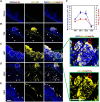
Neutrophil swarming and neutrophil BLT1 expression are reduced by immunosuppression. Epithelial infiltration of Ly6G+ cells (yellow) in IC and IS mice at 1 to 5 dpi and in sham-infected mice. First image in each panel showing the association between neutrophil swarms (demarcated by white dotted line) and Candida albicans (Ca) (red). Second image in each panel showing BLT1 expression in neutrophil and epithelial cells. Multiple intraepithelial Ly6G+ BLT1+ neutrophil swarms in IC mice, whereas IS mice show minimal swarm formation and less BLT1 expression in neutrophils. Image inserts show higher magnification image to show morphology of neutrophil aggregates in IS mice at 1 and 3 dpi and their association with C. albicans (red). (B) BLT1 response was assessed by mean fluorescence intensity (MFI). BLT1 MFI in epithelial cells increased up to day 3 as compared with epithelial BLT1 MFI in sham-infected mice and was reduced by day 5 in both IC and IS mice. (C) Quantification of neutrophil BLT1 (LY6G+ BLT1+) MFI following C. albicans at 1 to 5 dpi in IC and IS mice compared with sham-infected mice (n = 3 to 4 mice/group) Mean ± SEM is shown. **P < 0.01; ***P < 0.001 (2-way analysis of variance Tukey post-test). Scale bar = 25 µm.
Fig. 6.
Fig. 6.
Treatment with LTB4 (MK886) inhibitor caused disruption of neutrophil swarming and resulted in Candida albicans (Ca) dissemination into the CT. Neutrophil swarm formation was reduced following administration of an LTB4 biosynthesis inhibitor (MK-886). (A) LTB4 inhibitor reduced the size of swarm formation at 1 dpi, whereas control mice showed intact neutrophil swarms. Swarms in control animals engulfed Ca cells (shown in red with yellow arrows) at 1 dpi, while Ca were largely unassociated with neutrophils in inhibitor-treated mice. (B) Control mice showed dispersion of swarms at 2 dpi with reduced Ca cells, while LTB4 inhibitor treatment showed persistent Ca infection (yellow arrows) along with CT invasion. Ca (red) was found within the epithelium with hyphae (yellow boxes) invading beyond the basement membrane (green dotted line) into the CT in MK-886–treated mice. (C) MK-886 treatment resulted in significant reduction in mean swarm area at 1 dpi compared with the vehicle-treated control group. (D) MK-886 treatment led to significant increase in Ca infection at 3 dpi as compared with the vehicle-treated control group (n = 3 to 4 mice/group). Mean ± SEM is shown. *P < 0.05; ***P < 0.001 (2-way analysis of variance Holm-Sidak post-test). Scale bar = 50 µm. ns = not significant; PAS = periodic acid Schiff.

Update of

References

    1. Jabra-Rizk MA, Kong EF, Tsui C, Nguyen MH, Clancy CJ, Fidel PL Jr, Noverr M. Candida albicans pathogenesis: fitting within the host-microbe damage response framework. Infect Immun. 2016:84(10):2724–2739. 10.1128/IAI.00469-16 - DOI - PMC - PubMed
    1. Monzo-Gallo P, Chumbita M, Lopera C, Aiello TF, Peyrony O, Bodro M, Herrera S, Sempere A, Fernandez-Pittol M, Cuesta G, et al. Real-life epidemiology and current outcomes of hospitalized adults with invasive fungal infections. Med Mycol. 2023:61(3):myad021. 10.1093/mmy/myad021 - DOI - PubMed
    1. Trautwein-Weidner K, Gladiator A, Nur S, Diethelm P, LeibundGut-Landmann S. IL-17-mediated antifungal defense in the oral mucosa is independent of neutrophils. Mucosal Immunol. 2015:8(2):221–231. 10.1038/mi.2014.57 - DOI - PubMed
    1. Altmeier S, Toska A, Sparber F, Teijeira A, Halin C, LeibundGut-Landmann S. IL-1 coordinates the neutrophil response to C. albicans in the oral mucosa. PLoS Pathog. 2016:12(9):e1005882. 10.1371/journal.ppat.1005882 - DOI - PMC - PubMed
    1. Conti HR, Bruno VM, Childs EE, Daugherty S, Hunter JP, Mengesha BG, Saevig DL, Hendricks MR, Coleman BM, Brane L, et al. IL-17 receptor signaling in oral epithelial cells is critical for protection against oropharyngeal candidiasis. Cell Host Microbe. 2016:20(5):606–617. 10.1016/j.chom.2016.10.001 - DOI - PMC - PubMed

Substances