Review
. 2025 Jan;12(1):e2410893.
doi: 10.1002/advs.202410893. Epub 2024 Nov 12.

Antimicrobial Peptides: A Promising Alternative to Conventional Antimicrobials for Combating Polymicrobial Biofilms

Affiliations
Review

Antimicrobial Peptides: A Promising Alternative to Conventional Antimicrobials for Combating Polymicrobial Biofilms

Cesar Augusto Roque-Borda et al. Adv Sci (Weinh). 2025 Jan.

Abstract

Polymicrobial biofilms adhere to surfaces and enhance pathogen resistance to conventional treatments, significantly contributing to chronic infections in the respiratory tract, oral cavity, chronic wounds, and on medical devices. This review examines antimicrobial peptides (AMPs) as a promising alternative to traditional antibiotics for treating biofilm-associated infections. AMPs, which can be produced as part of the innate immune response or synthesized therapeutically, have broad-spectrum antimicrobial activity, often disrupting microbial cell membranes and causing cell death. Many specifically target negatively charged bacterial membranes, unlike host cell membranes. Research shows AMPs effectively inhibit and disrupt polymicrobial biofilms and can enhance conventional antibiotics' efficacy. Preclinical and clinical research is advancing, with animal studies and clinical trials showing promise against multidrug-resistant bacteria and fungi. Numerous patents indicate increasing interest in AMPs. However, challenges such as peptide stability, potential cytotoxicity, and high production costs must be addressed. Ongoing research focuses on optimizing AMP structures, enhancing stability, and developing cost-effective production methods. In summary, AMPs offer a novel approach to combating biofilm-associated infections, with their unique mechanisms and synergistic potential with existing antibiotics positioning them as promising candidates for future treatments.

Keywords: antimicrobial alternatives; biofilms; drug discovery; polymicrobial interactions.


Conflict of interest statement

The authors declare no conflict of interest.

Figures

Scheme 1
Scheme 1
List of priority fungi (left) and bacteria (right) launched by the WHO in 2022 and 2024, respectively. Reproduced (adapted) with permission from World Health Organization, Copyright 2022 and 2024, WHO.
Figure 1
Figure 1
Diagram showing the interaction between the LuxS/AI‐2 quorum sensing system in bacteria and the RAS1 protein system, along with other genes involved in fungal biofilm formation, within a polymicrobial biofilm formation system.
Figure 2
Figure 2
Diagram illustrates the processes of synergism and antagonism in polymicrobial interactions. Synergism refers to the collaborative interaction between microorganisms that results in an effect not achievable by any single species alone. In contrast, antagonism involves the suppression of one microorganism by another. Both processes are facilitated by quorum sensing (QS) mechanisms.
Figure 3
Figure 3
Preparation and Application of MN/CGA‐NPs. Wound healing in mice after 10 days of treatment, showing wound images (a) and wound area quantification (b) for infected mice receiving different treatments. H&E staining images and quantitative data of new skin tissue from S. aureus‐infected mice. Masson's trichrome staining images and quantitative data of collagen deposition in skin tissue from S. aureus‐infected mice. Reproduced (adapted) with permission.[ 311 ] Copyright 2023, American Chemical Society.
Figure 4
Figure 4
In vivo antibiofilm activity of MSNs and coassemblies. a) Images of biofilm‐colonized implants in mice treated with drug‐loaded single MSNs, coassemblies, or free drugs. Black arrows indicate implants failing to adhere to host tissue; white arrow indicates successful adhesion. b) Quantification of bacterial cells on biofilm‐colonized implants. c) SEM images of implant inner surfaces, with red arrows highlighting bacterial cells and yellow arrows indicating host cells. d) Histopathological images of tissues at implant sites, with blue arrows indicating damaged host muscle tissues suffering from inflammation. Reproduced (adapted) with permission.[ 313 ] Copyright 2020, American Chemical Society.
Figure 5
Figure 5
UP) The crystal violet assay (A) was used to measure biofilm formation on rGO, rGOAg, and GAAP after incubating S. aureus for 120 h. The biofilm inhibition activity of rGO, rGOAg, and GAAP against S. aureus was assessed in a concentration (B)‐ and time (C)‐dependent manner. Fluorescence microscopy images (D) displayed the formation of S. aureus biofilms upon interaction with rGO, rGOAg, and GAAP at different time intervals (24, 72, and 120 h); the scale bar was 2 µm. Live cells were shown in green‐fluorescent color, while dead cells with compromised cell membranes were depicted in red fluorescent color. (DOWN) A schematic representation (A) illustrated the antibiofouling activity of GAAP in an ex vivo rat skin infection model. Digital images (B) showed uninfected control skin (first panel), S. aureus infected skin after 4 h (second panel), S. aureus infected skin after 24 h (biofilm formation), and biofilm inhibition (fourth panel) and disruption (last panel) by GAAP. The bacterial count (C) was measured using the agar plate dilution method before and after a single‐dose treatment with GAAP (10 µg mL−1). Quantitative measurement (D) of the bacterial count before and after GAAP treatment was provided. SEM images (E) and histological analysis (F) were included for the uninfected control (first panel), skin tissue after 4 h of infection (second panel), skin tissue after 24 h of infection (biofilm formation), and GAAP‐treated skin tissue after 4 h of infection (biofilm inhibition, fourth panel) and 24 h of infection (biofilm disruption, last panel). Reproduced (adapted) with permission.[ 320 ] Copyright 2021 American Chemical Society.
Figure 6
Figure 6
(Left) A) Cellular viability of nano‐conjugated chitosan in macrophages after 24 h of exposure. B) Cellular viability of nano‐conjugated chitosan in fibroblasts after 24 h of exposure. C) Cellular viability of conjugated chitosan polymers in macrophages after 24 h of exposure. D) Confocal analysis of internalization of FITC‐NPs in macrophages after 24 h of incubation. (Right) Molecular docking – peptide receptor interaction in M. tuberculosis. The bacterial receptors (Ag85B, LTD2, GyrB, EmbC, GlfT2, Porin MspA, PknB, and CmaA2) are represented in blue, and the AMP Ctx(Ile21)‐Ha‐Ahx‐Cys (Ctx) is represented in green. Reproduced (adapted) with permission.[ 201 ] Copyright 2023, Elsevier.

References

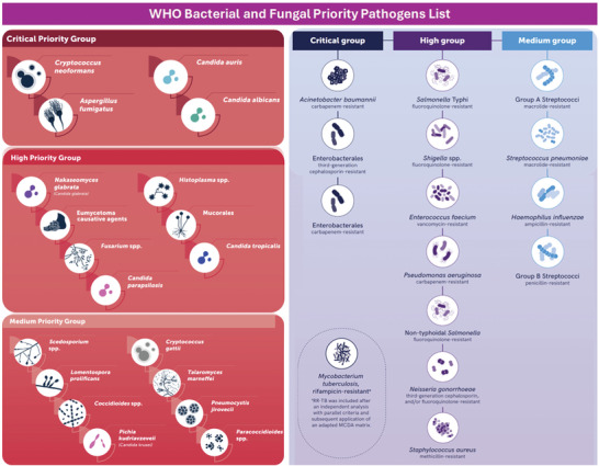
    1. WHO , WHO Fungal Priority Pathogens List to Guide Research, Development and Public Health Action, World Health Organization, Geneva: 2022, https://www.who.int/publications/i/item/9789240060241 (accessed: November 2024).
    1. WHO , WHO Bacterial Priority Pathogens List, 2024: Bacterial Pathogens of Public Health Importance to Guide Research, Development and Strategies to Prevent and Control Antimicrobial Resistance, World Health Organization, Geneva: 2024, https://www.who.int/publications/i/item/9789240093461 (accessed: November 2024).
    1. Reyes J., Komarow L., Chen L., Ge L., Hanson B. M., Cober E., Herc E., Alenazi T., Kaye K. S., Garcia‐Diaz J., Li L., Kanj S. S., Liu Z., Oñate J. M., Salata R. A., Marimuthu K., Gao H., Zong Z., Valderrama‐Beltrán S. L., Yu Y., Tambyah P., Weston G., Salcedo S., Abbo L. M., Xie Q., Ordoñez K., Wang M., Stryjewski M. E., Munita J. M., Paterson D. L., et al., Lancet Microbe 2023, 4, e159. - PMC - PubMed
    1. Krawczyk B., Wityk P., Gałęcka M., Michalik M., Microorganisms 2021, 9, 1900. - PMC - PubMed
    1. Venkateswaran P., Vasudevan S., David H., Shaktivel A., Shanmugam K., Neelakantan P., Solomon A. P., Front. Cell Infect. Microbiol. 2023, 13. - PMC - PubMed

Substances

LinkOut - more resources