. 2024 Dec 20;9(24):e185299.
doi: 10.1172/jci.insight.185299.

12-Lipoxygenase inhibition delays onset of autoimmune diabetes in human gene replacement mice

Affiliations

12-Lipoxygenase inhibition delays onset of autoimmune diabetes in human gene replacement mice

Titli Nargis et al. JCI Insight. .

Abstract

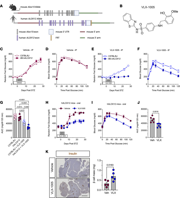
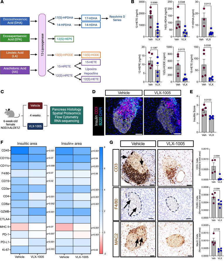
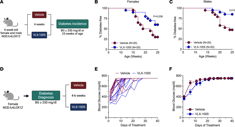
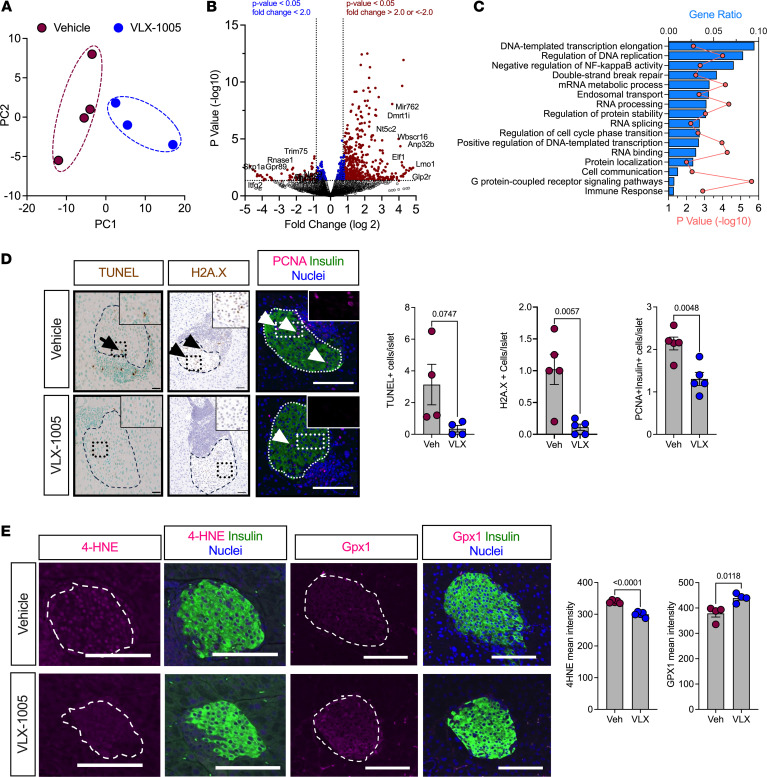
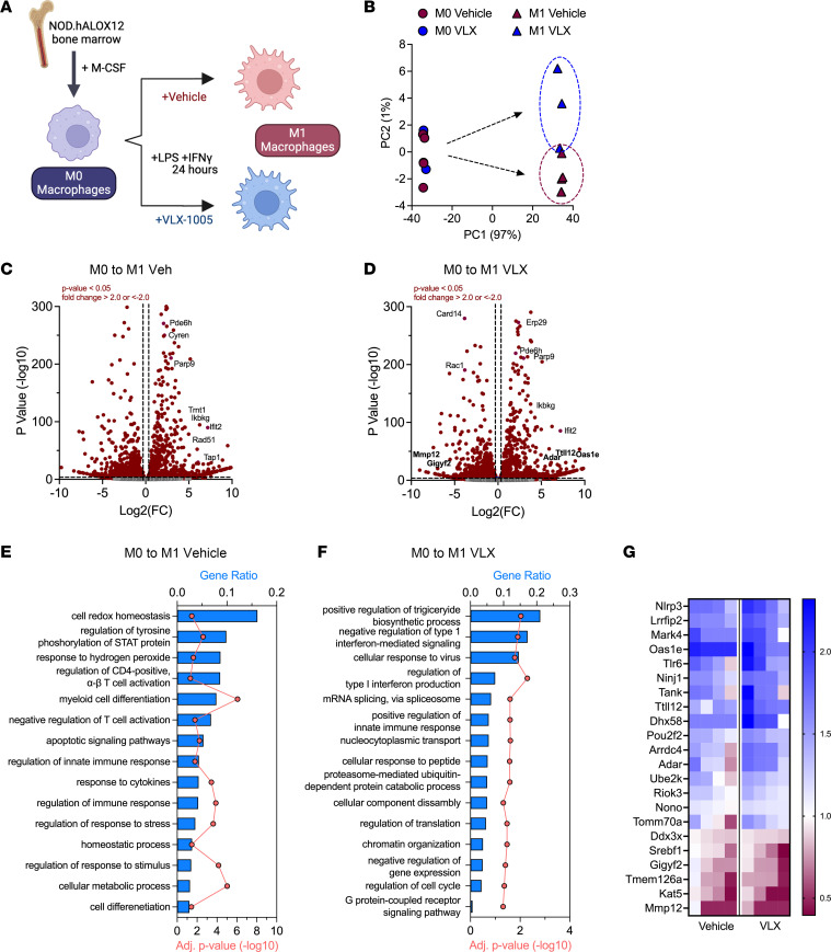
Type 1 diabetes (T1D) is characterized by the autoimmune destruction of insulin-producing β cells and involves an interplay between β cells and cells of the innate and adaptive immune systems. We investigated the therapeutic potential of targeting 12-lipoxygenase (12-LOX), an enzyme implicated in inflammatory pathways in β cells and macrophages, using a mouse model in which the endogenous mouse Alox15 gene is replaced by the human ALOX12 gene. Our finding demonstrated that VLX-1005, a potent 12-LOX inhibitor, effectively delayed the onset of autoimmune diabetes in human gene replacement non-obese diabetic mice. By spatial proteomics analysis, VLX-1005 treatment resulted in marked reductions in infiltrating T and B cells and macrophages, with accompanying increases in immune checkpoint molecule PD-L1, suggesting a shift toward an immunosuppressive microenvironment. RNA sequencing analysis of isolated islets and polarized proinflammatory macrophages revealed significant alteration of cytokine-responsive pathways and a reduction in IFN response after VLX-1005 treatment. Our studies demonstrated that the ALOX12 human replacement gene mouse provides a platform for the preclinical evaluation of LOX inhibitors and supports VLX-1005 as an inhibitor of human 12-LOX that engages the enzymatic target and alters the inflammatory phenotypes of islets and macrophages to promote the delay of autoimmune diabetes.

Keywords: Diabetes; Eicosanoids; Endocrinology; Islet cells; Therapeutics.


Conflict of interest statement

Conflict of interest: RGM and SAT received an investigator-initiated award from Veralox Therapeutics. RGM serves on the Scientific Advisory Board for Veralox Therapeutics. DJM and MBB are Veralox Therapeutics employees.

Figures

Figure 1
Figure 1. 12-LOX inhibition protects against streptozotocin-induced diabetes.
C57BL/6J and B6.hALOX12 male mice (n = 4–7 per group as indicated) were treated with 30 mg/kg i.p. or p.o. VLX-1005 and multiple low-dose streptozotocin (STZ). (A) Schematic of the generation of hALOX12 mice by replacing mouse Alox15 with human ALOX12. (B) Chemical structure of VLX-1005. (C) Random-fed blood glucose values in vehicle-treated male C57BL/6J and B6.hALOX12 mice after STZ. (D) GTT of vehicle-treated male C57BL/6J and B6.hALOX12 mice on day 4 after STZ treatment. (E) Random-fed blood glucose values in VLX-1005–treated male C57BL/6J and B6.hALOX12 mice after STZ. (F) GTT of VLX-1005–treated male C57BL/6J and B6.hALOX12 mice on day 4 after STZ treatment. (G) AUC of C57BL/6J and B6.hALOX12 on day 4 after STZ-treatment (1-way ANOVA). (H) Random-fed blood glucose values in male vehicle- or VLX-1005–treated (p.o.) B6.hALOX12 mice after STZ. (I) GTT of male vehicle- or VLX-1005–treated (p.o.) B6.hALOX12 mice on day 4 after STZ treatment. (J) AUC of B6.hALOX12 on day 4 after STZ treatment. (K) Pancreata stained for insulin (left panel) and β cell mass measurement (right panel) from male B6.hALOX12 mice on day 26 after STZ treatment. Scale bars: 500 μm. Data are presented as mean ± SEM and statistical significance was determined by a 2-tailed Student’s t test (all except G) or 1-way ANOVA with Tukey’s post hoc test (G).
Figure 2
Figure 2. VLX-1005 decreased islet inflammation in NOD.hALOX12 female mice.
Six-week-old female prediabetic NOD.hALOX12 mice were treated p.o. with 30 mg/kg VLX-1005 for 4 weeks prior to tissue analysis. (A) Schematic representation of 12-lipoxygenase products. (B) Serum lipidomics results of 12-lipoxygenase products as indicated (n = 4–5). (C) Schematic representation of mouse treatment paradigm. (D) Pancreata from mice stained for CD3 (magenta), B220 (teal), insulin (white), and nuclei (blue). Scale bars: 50 μm. (E) Average insulitis score; each dot represents an individual mouse (n = 4–5). (F) Heatmap of identified proteins in the insulitic area (left) and insulin-positive area (right). (G) Pancreata of mice stained and quantified for CD3 (brown, top panels: arrows indicate positive CD3 staining within the islet), F4/80 (brown, middle panels: arrows indicate positive F4/80 staining within the islet), or MAC2 (brown, bottom panels: arrows indicate positive MAC2 staining within the islet) and nuclei (blue). Each dot represents an individual mouse (n = 4–5). Scale bars: 50 μm. Data are presented as mean ± SEM and statistical significance was determined by a 2-tailed Student’s t test in all cases.
Figure 3
Figure 3. VLX-1005 treatment delays autoimmune diabetes onset in female and male NOD.hALOX12 mice.
NOD.hALOX12 mice (n = 20 per group) were treated during the prediabetic stage from 6 to 10 weeks of age or at the time of diabetes development (n = 11–12 per group). (A) Schematic representation of diabetes prevention experimental design. (B) Diabetes incidence in female NOD.hALOX12 mice. (C) Diabetes incidence in male NOD.hALOX12 mice. (D) Schematic representation of diabetes reversal experimental design. (E) Random-fed blood glucose levels in each female mouse. (F) Average random-fed blood glucose levels of female mice. Data are presented as mean ± SEM and statistical significance was determined by a Mantel-Cox log-rank test.
Figure 4
Figure 4. VLX-1005 decreased β cell death, proliferation, and oxidative stress in female NOD.hALOX12 mice.
Pancreata or islets were harvested from 10-week-old prediabetic female NOD.hALOX12 mice after 4 weeks of treatment with vehicle or VLX-1005 (n = 3–4 per group). (A) Principal component analysis plot of RNA sequencing results from isolated islets of vehicle- or VLX-1005–treated mice. (B) Volcano plot of differentially expressed genes. (C) Gene ontology pathway analysis of differentially expressed genes. (D) Pancreata from mice stained and quantified for TUNEL (brown, left panels: black arrow indicates positive TUNEL staining within the islet), H2A.X (brown, middle panels: black arrowheads indicate positive H2A.X staining within the islet), or PCNA (magenta, right panels: white arrowheads indicate positive PCNA staining within the islet), insulin (green) and nuclei (blue). Each dot represents an individual mouse (n = 4–5). Scale bars: 50 μm. (E) Pancreata from mice stained and quantified for 4-HNE (magenta, left panels), or GPx1 (magenta, right panels), and insulin (green) and nuclei (blue). Each dot represents an individual mouse (n = 4). Scale bars: 50 μm. Data are presented as mean ± SEM and statistical significance was determined by a 2-tailed Student’s t test.
Figure 5
Figure 5. RNA sequencing analysis of M1-like BMDMs reveals a reduction in the inflammatory response upon VLX-1005 treatment.
Bone marrow–derived macrophages (BMDMs) were isolated and polarized to the M1-like state and treated with vehicle or VLX-1005 (10 μM) during polarization. RNA was isolated and sequenced (n = 4 per group). (A) Schematic of experimental design. (B) Principal component analysis plot. (C) Volcano plot of differentially expressed genes in M0 and M1-like vehicle-treated macrophages. (D) Volcano plot of differentially expressed genes in M0 and M1-like VLX-1005–treated macrophages. (E) Gene ontology pathway analysis of differentially expressed genes in M0 vs. M1-like vehicle-treated macrophages. (F) Gene ontology pathway analysis of differentially expressed genes in M0 vs. M1-like VLX-1005–treated macrophages. (G) Heatmap of significantly altered IFN-related genes. Columns represent sequencing results from each sample (n = 4 per group). Numbers on the heatmap scale indicate fold change (FC) compared with M0 macrophages.

Update of

References

    1. Atkinson MA, Mirmira RG. The pathogenic “symphony” in type 1 diabetes: a disorder of the immune system, β cells, and exocrine pancreas. Cell Metab. 2023;35(9):1500–1518. doi: 10.1016/j.cmet.2023.06.018. - DOI - PMC - PubMed
    1. Kulkarni A, et al. 12-Lipoxygenase governs the innate immune pathogenesis of islet inflammation and autoimmune diabetes. JCI Insight. 2021;6(14):e147812. doi: 10.1172/jci.insight.147812. - DOI - PMC - PubMed
    1. Lee H, et al. Beta cell dedifferentiation induced by IRE1α deletion prevents type 1 diabetes. Cell Metab. 2020;31(4):822–836. doi: 10.1016/j.cmet.2020.03.002. - DOI - PMC - PubMed
    1. Tersey SA, et al. Islet β-cell endoplasmic reticulum stress precedes the onset of type 1 diabetes in the nonobese diabetic mouse model. Diabetes. 2012;61(4):818–827. doi: 10.2337/db11-1293. - DOI - PMC - PubMed
    1. Gonzalez-Duque S, et al. Conventional and neo-antigenic peptides presented by β cells are targeted by circulating naïve CD8+ T cells in type 1 diabetic and healthy donors. Cell Metab. 2018;28(6):946–960. doi: 10.1016/j.cmet.2018.07.007. - DOI - PubMed

MeSH terms