. 2024 Nov 12;135(2):e183656.
doi: 10.1172/JCI183656.

MAP3K1 mutations confer tumor immune heterogeneity in hormone receptor-positive HER2-negative breast cancer

Affiliations

MAP3K1 mutations confer tumor immune heterogeneity in hormone receptor-positive HER2-negative breast cancer

Yu-Wen Cai et al. J Clin Invest. .

Abstract

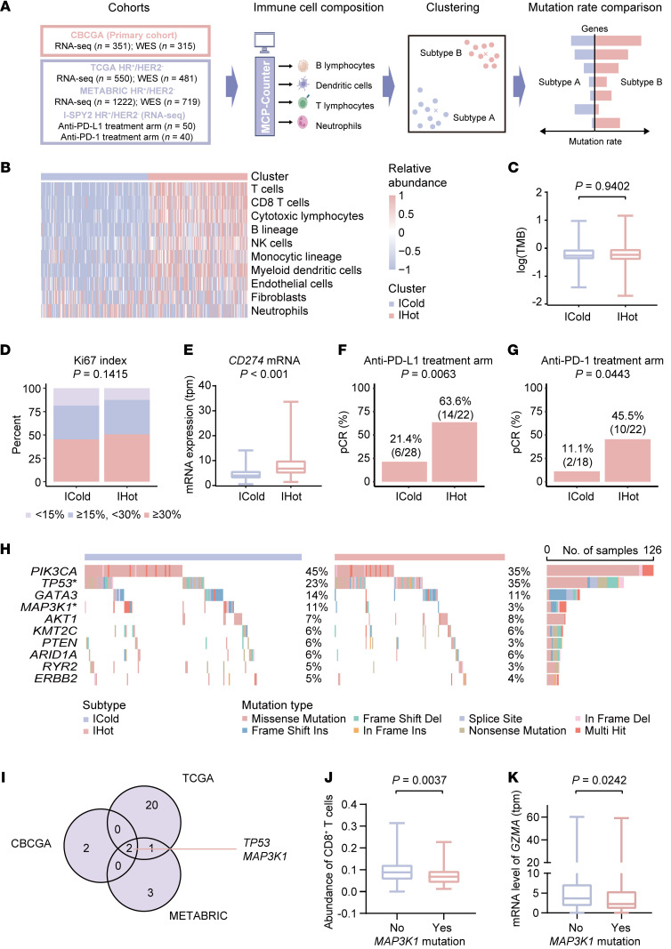
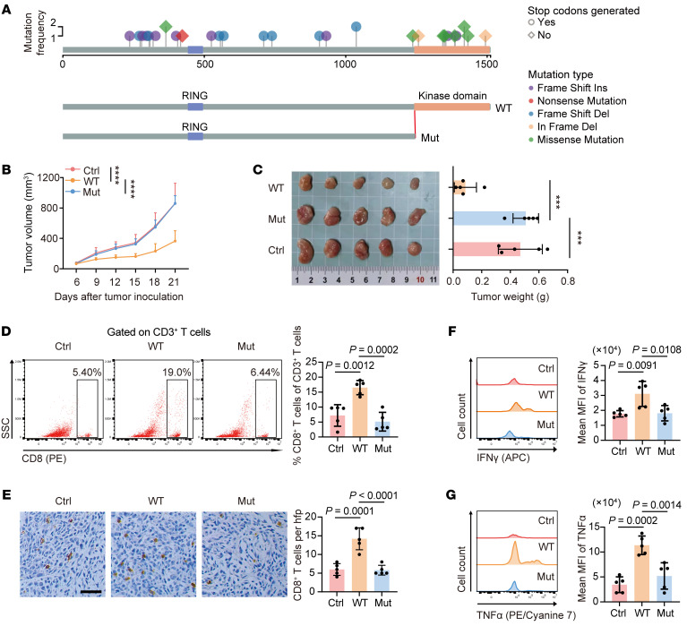
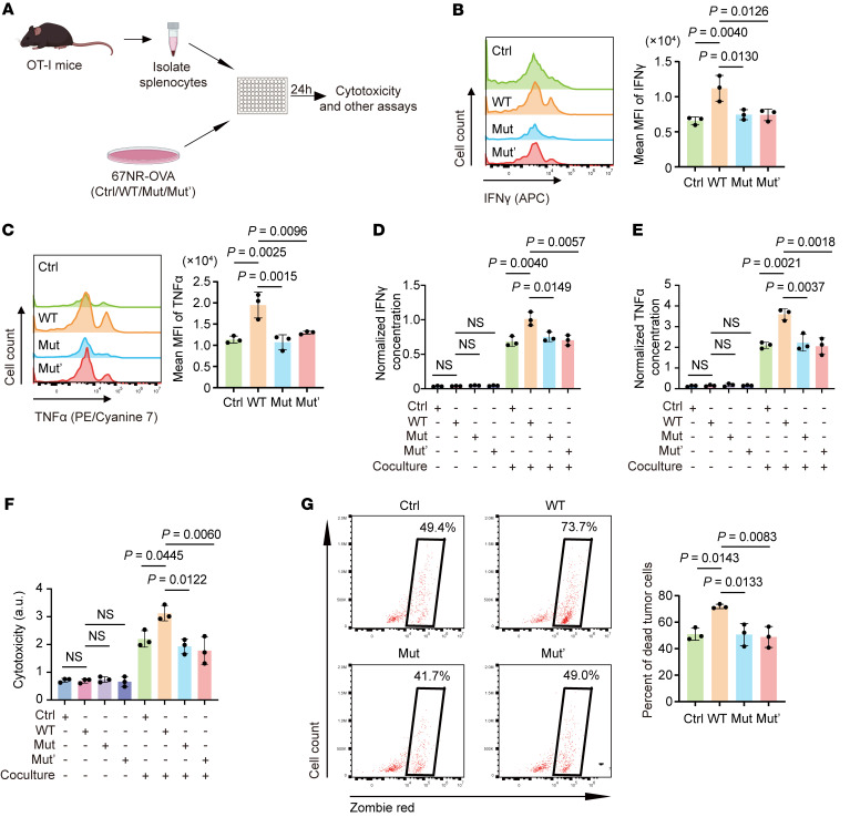
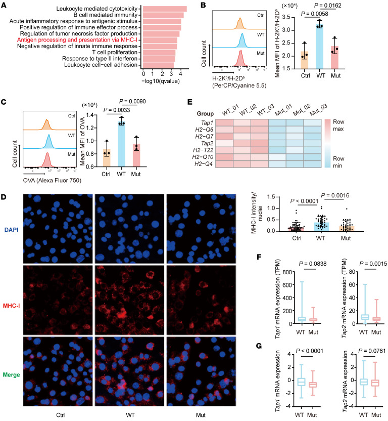
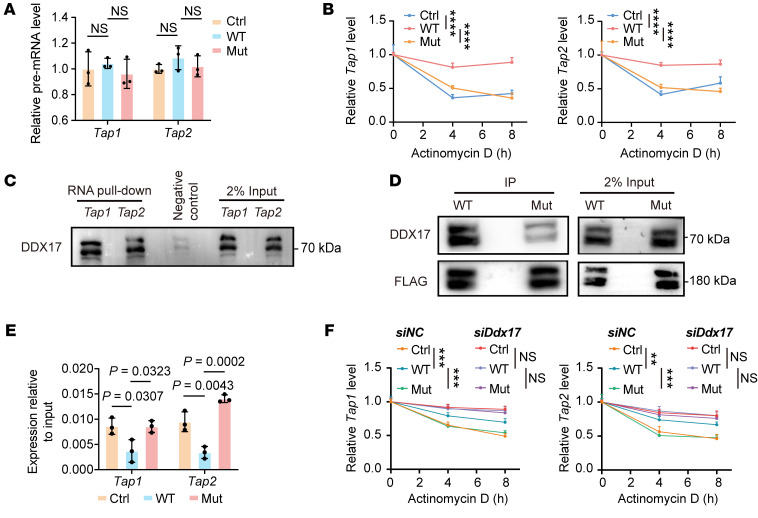
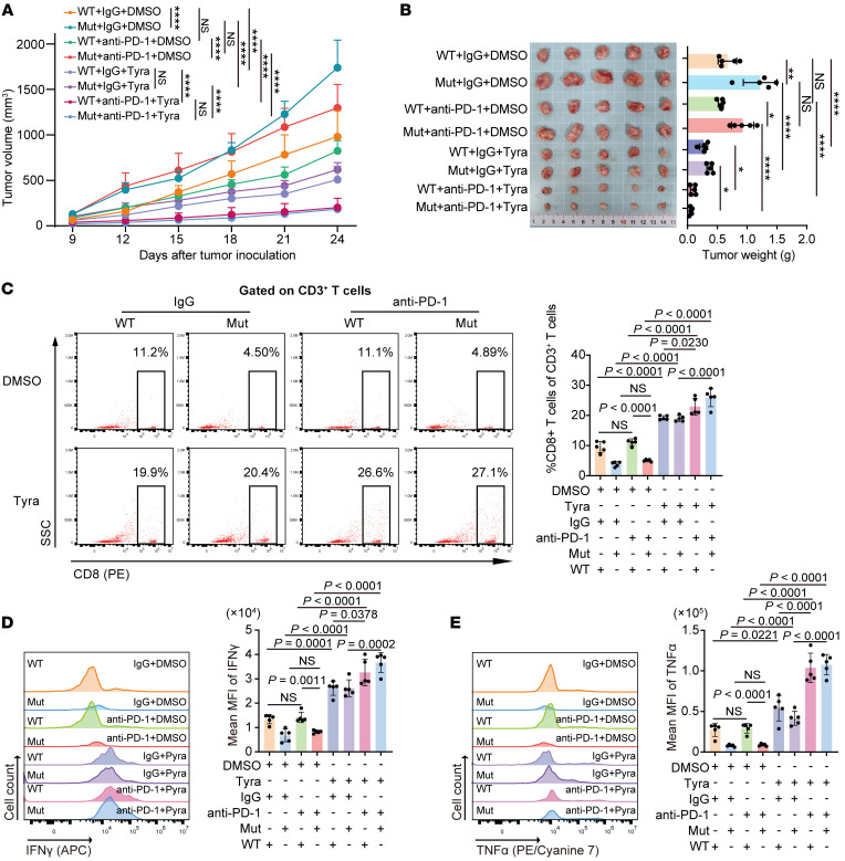
Treatment for hormone receptor-positive/human epidermal growth factor receptor 2-negative (HR+/HER2-) breast cancer, the most common type of breast cancer, has faced challenges such as endocrine therapy resistance and distant relapse. Immunotherapy has shown progress in treating triple-negative breast cancer, but immunological research on HR+/HER2- breast cancer is still in its early stages. Here, we performed a multi-omics analysis of a large cohort of patients with HR+/HER2- breast cancer (n = 351) and revealed that HR+/HER2- breast cancer possessed a highly heterogeneous tumor immune microenvironment. Notably, the immunological heterogeneity of HR+/HER2- breast cancer was related to mitogen-activated protein kinase kinase kinase 1 (MAP3K1) mutation and we validated experimentally that a MAP3K1 mutation could attenuate CD8+ T cell-mediated antitumor immunity. Mechanistically, MAP3K1 mutation suppressed MHC-I-mediated tumor antigen presentation through promoting the degradation of antigen peptide transporter 1/2 (TAP1/2) mRNA, thereby driving tumor immune escape. In preclinical models, the postbiotic tyramine could reverse the MAP3K1 mutation-induced MHC-I reduction, thereby augmenting the efficacy of immunotherapy. Collectively, our study identified the vital biomarker driving the immunological heterogeneity of HR+/HER2- breast cancer and elucidated the underlying molecular mechanisms, which provided the promise of tyramine as what we believe to be a novel therapeutic strategy to enhance the efficacy of immunotherapy.

Keywords: Cancer; Immunology; Immunotherapy; Oncology.


Figures

Figure 1
Figure 1. MAP3K1 mutation is closely correlated with immune microenvironment heterogeneity in HR+/HER2 breast cancer.
(A) Flowchart of bioinformatics analyses performed in the study. (B) Heatmap showing the relative abundance of immune and stromal cells calculated by MCP-Counter in each sample in CBCGA cohort (n = 351). The 2 immunological subtypes were annotated. (C) Comparison of tumor mutation burden (TMB) of tumors with the ICold and IHot subtypes in CBCGA cohort (n = 314). The center line represents the median. (D and E) Comparison of ki67 index (D) and CD274 mRNA expression level (E) of tumors with the ICold and IHot subtypes in CBCGA cohort (n = 350). The center line represents the median. (F and G) Pathological complete response (pCR) rate of patients with the ICold and IHot subtypes in the anti-PD-L1 (F) and anti-PD-1 (G) treatment arm of the I-SPY2 clinical trial. (H) The somatic mutations identified in tumors with the ICold and IHot subtypes in CBCGA cohort (n = 314). *P < 0.05. (I) Venn diagram showing genes with significantly different mutation prevalence between the ICold and IHot subtypes in CBCGA (n = 314), TCGA HR+/HER2 (n = 475), and METABRIC HR+/HER2 (n = 611) breast cancer cohorts. (J and K) Abundance of CD8+ T cells calculated by CIBERSORT (J) and GZMA mRNA expression (K) in HR+/HER2 breast tumors with or without MAP3K1 mutation in the TCGA cohort (n = 481). Statistical analysis: (C, E, J, and K) Wilcoxon signed-rank test; (D) χ2 test for trend; (FH) Fisher’s exact test. ICold, immune cold subtype; IHot, immune hot subtype; TMB, tumor mutation burden; HR, hormone receptor; HER2, human epidermal growth factor receptor 2.
Figure 2
Figure 2. Map3k1-mutant tumors evade CD8+ T cell–mediated immunity in vivo.
(A) MAP3K1 mutation atlas of tumors in the CBCGA cohort and schematic diagram of full-length (WT) and kinase domain-truncated (1–1,222 aa) Map3k1 (Mut) overexpression plasmids generated for the following experiments. The mutation type and whether a stop codon was generated are annotated. (B and C) 67NR mouse breast cancer cells with varying Map3k1 status (overexpression based on Map3k1-KO cell lines) were orthotopically injected into BALB/c mice (n = 5 per group). Tumor growth curves (B) and tumor weights with the images (C) are shown. (D) Representative flow cytometry data of CD8+ T cell infiltration gated on CD3+ T cells in tumor tissues. (E) Representative IHC images of tumor tissues are shown and the numbers of CD8+ T cell are quantified. Scale bar, 50 μm. (F and G) MFI of IFN-γ (F) and TNF-α (G) in CD8+ T cells in the tumor tissues. Data are mean ± SD (BG) (n = 5 per group). Statistical analysis: (B) 2-way ANOVA with Tukey’s test; (CG) 1-way ANOVA with Tukey’s test. Significance in tumor growth (B) and tumor weight (C). *P < 0.05, **P < 0.01, ***P < 0.001, ****P < 0.0001.
Figure 3
Figure 3. MAP3K1 mutation inhibits CD8+ T cell activation in vitro.
(A) Schematic diagram of the in vitro coculture assay. OVA-expressing 67NR tumor cells with varying Map3k1 status were cocultured with splenocytes isolated from OT-I mice at a ratio of 1:10. At 24 hours after coculture, tumor cells, immune cells, and the culture medium were collected for the following analyses. (B and C) MFI of IFN-γ (B) and TNF-α (C) in CD8+ T cells are shown. (D and E) Concentration of cytokines IFN-γ (D) and TNF-α (E) in the culture medium was measured by ELISA. (F) T cell cytotoxicity was measured by lactate dehydrogenase (LDH) assay. (G) T cell cytotoxicity was assessed by the percentage of dead cells in EpCAM+ tumor cells, which are indicated by black boxes. Data are mean ± SD (BG) (n = 3 per group). Statistical analysis: (BG) 1-way ANOVA with Tukey’s test.
Figure 4
Figure 4. MAP3K1 mutation suppresses tumor antigen presentation.
(A) Map3k1-WT and Map3k1-mut tumor tissues were collected for RNA-seq. GO enrichment analysis was performed and immune-related pathways that are significantly downregulated in Map3k1-mut compared with Map3k1-WT tumors are shown here. (n = 3 per group). (B and C) Surface expression of H-2Kb/H-2Db (B) and OVA (C) on 67NR tumor cells with varying Map3k1 status after coculture with OT-I splenocytes for 24 hours was determined by flow cytometry. (D) Surface expression of MHC-I on tumor cells in B and C was also measured by immunofluorescence. Scale bar: 50 μm. (E) Heatmap showing the significantly downregulated genes in Map3k1-mut compared with Map3k1-WT tumors in the pathway of antigen processing and presentation via MHC-I. Significance determined as P value less than 0.05 and fold change less than 0.8. (F and G) Comparison of mRNA expression of Tap1/2 in MAP3K1-WT and MAP3K1-mut tumors in TCGA (n = 481) (F) and METABRIC (n = 719) (G) cohorts. The center line represents the median. Data are mean ± SD (B and C) (n = 3 per group). Statistical analysis: (B-D) 1-way ANOVA with Tukey’s test. (F and G) Wilcoxon signed-rank test.
Figure 5
Figure 5. Map3k1 mutation promotes Tap1/2 RNA degradation.
(A) The premature mRNA level of Tap1/2 in 67NR-OVA cells with varying Map3k1 status in coculture was measured by RT-qPCR. (B) 67NR-OVA cells with varying Map3k1 status were cocultured with OT-I splenocytes for 24 hours and then treated with actinomycin D at a dose of 10 μg/mL. Tumor cells were collected at the indicated time points to perform RT-qPCR to test the RNA level of Tap1/2. (C) RNA pull-down assay was performed to examine the binding of Tap1/2 mRNAs to DDX17 in 67NR-OVA cells in coculture. (D) IP assay was performed to examine and compare the binding of Map3k1-WT and Map3k1-mut to DDX17 in 67NR-OVA cells in coculture. (E) 67NR-OVA cells with varying Map3k1 status in coculture were collected and RNA immunoprecipitation (RIP) assay was performed to extract the RNA binding to DDX17. RT-qPCR was then performed to measure the RNA levels of Tap1/2. (F) 67NR-OVA cells with varying Map3k1 status were transiently transfected with small interfering RNA targeting Ddx17 (siDdx17) or its control RNA (siNC). A day after transfection, tumor cells were cocultured with OT-I splenocytes for 24 hours and then treated with actinomycin D at a dose of 10 μg/mL. Tumor cells were collected at the indicated time points and RT-qPCR was performed to test the RNA level of Tap1/2. Data are mean ± SD (A, B, E, and F) (n = 3 per group). Statistical analysis: (A and E) 1-way ANOVA with Tukey’s test. (B and F) 2-way ANOVA with Tukey’s test. Significance in the RNA degradation experiments (B and F) is annotated as *P < 0.05, **P < 0.01, ***P < 0.001, ****P < 0.0001.
Figure 6
Figure 6. Tyramine augments the efficacy of immunotherapy in HR+/HER2 breast cancer.
(A and B) 67NR cells expressing Map3k1-WT or Map3k1-mut were orthotopically injected into BALB/c mice. Once tumors formed, mice were randomly assigned to receive tyramine (Tyra) or DMSO in combination with anti–PD-1 or its isotype control (IgG). Tumor growth curves (A) and tumor weights with the images (B) are shown here. (C) Representative flow cytometry data of CD8+ T cell infiltration in tumor tissues. (D and E) MFI of IFN-γ (D) and TNF-α (E) in CD8+ T cells in the tumor tissues. Data are mean ± SD (AE) (n = 5 per group). Statistical analysis: (A) 2-way ANOVA with Tukey’s test; (BE) 1-way ANOVA with Tukey’s test. Tyra, tyramine. HR, hormone receptor; HER2, human epidermal growth factor receptor 2.

References

    1. Huppert LA, et al. Systemic therapy for hormone receptor-positive/human epidermal growth factor receptor 2-negative early stage and metastatic breast cancer. CA Cancer J Clin. 2023;73(5):480–515. doi: 10.3322/caac.21777. - DOI - PubMed
    1. Ma CX, et al. Mechanisms of aromatase inhibitor resistance. Nat Rev Cancer. 2015;15(5):261–275. doi: 10.1038/nrc3920. - DOI - PubMed
    1. Pan H, et al. 20-year risks of breast-cancer recurrence after stopping endocrine therapy at 5 years. N Engl J Med. 2017;377(19):1836–1846. doi: 10.1056/NEJMoa1701830. - DOI - PMC - PubMed
    1. Dieci MV, et al. Neoadjuvant chemotherapy and immunotherapy in luminal B-like breast cancer: results of the Phase II GIADA Trial. Clin Cancer Res. 2022;28(2):308–317. doi: 10.1158/1078-0432.CCR-21-2260. - DOI - PMC - PubMed
    1. Rugo HS, et al. Safety and antitumor activity of pembrolizumab in patients with estrogen receptor-positive/human epidermal growth factor receptor 2-negative advanced breast cancer. Clin Cancer Res. 2018;24(12):2804–2811. doi: 10.1158/1078-0432.CCR-17-3452. - DOI - PubMed

MeSH terms