. 2025 Jan;12(2):e2404083.
doi: 10.1002/advs.202404083. Epub 2024 Nov 12.

NSUN5 Facilitates Hepatocellular Carcinoma Progression by Increasing SMAD3 Expression

Affiliations

NSUN5 Facilitates Hepatocellular Carcinoma Progression by Increasing SMAD3 Expression

Hexu Han et al. Adv Sci (Weinh). 2025 Jan.

Abstract

Hepatocellular carcinoma (HCC) is characterized by frequent intrahepatic and distant metastases, resulting in a poor prognosis for patients. Epithelial-mesenchymal transition (EMT) plays a pivotal role in this process. However, the expression of NOP2/Sun RNA methyltransferase 5 (NSUN5) in HCC and its role in mediating EMT remain poorly understood. In this study, clinicopathological analyses are conducted across multiple independent HCC cohorts and induced tumor formation in Nsun5-knockout mice. The findings reveal an upregulation of NSUN5 expression in tumor tissues; conversely, the absence of Nsun5 hinders the malignant progression of HCC, indicating that NSUN5 may serve as a significant oncogene in HCC. Furthermore, elevated levels of NSUN5 enhance EMT processes within HCC cells. NSUN5-knockout cells exhibit reduced invasion and migration capabilities under both in vivo and in vitro conditions, while overexpression of NSUN5 yields opposing effects. Mechanistically, high levels of NSUN5 promote the enrichment of trimethylated histone H3 at lysine 4 (H3K4me3) at the promoter region of SMAD3 through recruitment of the WDR5, thereby facilitating HCC metastasis via SMAD3-mediated EMT pathways. Collectively, this study identifies NSUN5 as a novel driver of metastasis in HCC and provides a theoretical foundation for potential therapeutic strategies against this malignancy.

Keywords: EMT; NSUN5; SMAD3; hepatocellular carcinoma (HCC); metastasis.


Conflict of interest statement

The authors declare no conflict of interest.

Figures
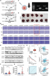
Figure 1
Figure 1
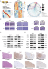
NSUN5 is highly expressed in hepatocellular carcinoma (HCC) tumor tissues. A) Western blotting was used to investigate the relationship between overexpression of several proteins potentially mediating RNA 5‐methylcytosinylation and epithelial–mesenchymal transition (EMT) in HepG2 and Hep3B cells. B) Results from wound healing assays indicated a significantly enhanced migration rate of HCC cells only upon overexpression of NSUN5. C) Homozygous Nsun5‐deficient mice were identified following genotyping. D) Modeling using CCL4 and DEN. After 16 weeks, analysis of liver tissues from wild‐type (n = 5) and Nsun5‐knockout (KO, n = 6) mice revealed a significant reduction in both the number and volume of tumor tissues in the KO group. E) Western blot analysis of NSUN5 levels in paired tumor and nontumor tissues, with GAPDH used as the loading control. F) Statistical analysis of western blot results from clinical samples demonstrated a significant increase in NSUN5 expression in HCC tumor tissues. G) qPCR assays of NSUN5 expression in paired tumors and corresponding paracancerous tissues showed significantly elevated NSUN5 mRNA levels in HCC tumor tissues, with β‐actin used as the control. H) Analysis of data from patients with HCC in TCGA database revealed the following results: NSUN5 was highly upregulated in tumor tissues compared to paracancerous tissues (upper). High NSUN5 expression was negatively correlated with patient prognosis (middle). The area under the receiver operating characteristic (ROC) curve (AUC > 0.9) indicated high predictive accuracy of NSUN5 for patient prognosis (lower). I) Analysis of TCGA database revealed that NSUN5 expression in HCC tumor tissues was inversely correlated with the expression of the epithelial marker E‐cadherin and positively correlated with that of the mesenchymal marker vimentin. Moreover, increased NSUN5 expression was significantly correlated with elevated SMAD3 expression in HCC. Results in (F) and (G) are shown as mean ± SD. P‐values are indicated by * < 0.05; ** < 0.01; *** < 0.001.
Figure 2
Figure 2
Upregulation of NSUN5‐induced EMT in HCC cells. A) Transcriptome analysis showed that Gene Ontology enrichment confirmed the involvement of NSUN5 in various biological behaviors, including mediating EMT. Kyoto Encyclopedia of Genes and Genomes signaling pathway and differential gene expression analyses suggested that NSUN5 could influence the SMAD3 signaling pathway by regulating SMAD3 expression. B) Results from wound healing and transwell assays demonstrated that higher NSUN5 expression enhanced the invasiveness and metastatic ability of HCC cells and vice versa. C) Western blot analysis of epithelial (E‐cadherin) and mesenchymal (vimentin and N‐cadherin) markers in Huh‐7‐sg‐vector/NSUN5 and HepG2‐ov‐vector/NSUN5 cells. D) Western blot analysis of several major EMT transcription factors (ZEB1/2, Snail1/2, and Twist) in Huh‐7‐sg‐vector/NSUN5 and HepG2‐ov‐vector/NSUN5 cells. E) Immunohistochemical (IHC) staining showed that Nsun5 knockout was accompanied by increased tumor epithelial marker (E‐cadherin) and decreased mesenchymal marker (vimentin) levels in spontaneous mouse HCC tissues.
Figure 3
Figure 3
Upregulation of NSUN5 increases SMAD3 expression to induce EMT. A) Dual‐luciferase reporter gene assays revealed significant downregulation of the SMAD3 signaling pathway in Huh‐7 cells after NSUN5 knockout, whereas it was markedly upregulated after NSUN5 overexpression in HepG2 cells (n = 3, ***P < 0.001). B) qPCR analyses demonstrated a significant increase in SMAD3 mRNA content in HepG2 cells following NSUN5 overexpression, whereas the opposite trend was observed in Huh‐7 cells (n = 3, ***P < 0.001). C) Western blot analysis of key molecules involved in signaling pathways that induce major EMT transcription factors revealed that NSUN5 mainly affected the SMAD3 signaling pathway without affecting the expression of other key transcription factors. D) Western blot analysis of key molecules in the SMAD3 signaling pathway showed that NSUN5 predominantly affected SMAD3 expression and the levels of its active form, p‐SMAD3, without significantly altering the levels of other important molecules. E) Western blot analysis demonstrated that decreased epithelial marker (E‐cadherin) and increased mesenchymal marker (vimentin and N‐cadherin) levels could be rescued by SMAD3 knockdown or inhibition in HepG2‐ov‐NSUN5 cells. F) Wound healing and transwell assays confirmed that the enhanced invasive and migration ability of HepG2 cells due to NSUN5 overexpression was dependent on the activation of the SMAD3 signaling pathway. G) IHC staining of SMAD3 and EMT markers in subcutaneous tumors showed that NSUN5 knockout suppressed the activity of the SMAD3 signaling pathway, accompanied by significant progression of MET in the Huh‐7 cells group (n = 5 per group). H) IHC staining of SMAD3 and EMT markers in subcutaneous tumors demonstrated that NSUN5 overexpression upregulated the SMAD3 signaling pathway, accompanied by significant progression of EMT in HepG2 cells (n = 5 per group). Results in (A) and (B) are shown as mean ± SD. P‐values are indicated by * < 0.05; ** < 0.01; *** < 0.001.
Figure 4
Figure 4
NSUN5 binds to the DNA promoter region of SMAD3 and enhances its accessibility to increase the expression of SMAD3. A) RNA immunoprecipitation (RIP) sequencing was applied to investigate the mRNA bound to NSUN5 in HepG2‐ov‐NSUN5 cells. B) Anti‐m5C methylated RNA immunoprecipitation (m5C meRIP) sequencing was used to detect the distribution and composition of m5C modifications of mRNA in HepG2‐ov‐NSUN5 and corresponding control cells before and after NSUN5 overexpression. C) Integration of mRNA‐seq, RIP‐seq, and meRIP‐seq data showed that NSUN5 exerts regulatory control over mRNA content in HCC cells by interacting with numerous mRNAs (n = 875) and influencing their m5C modification. D) NSUN5, an m5C writer, may not regulate all mRNAs. The results also revealed that the expression of mRNAs interacting with NSUN5 and their m5C abundance can be positively or negatively regulated. E) Results showed direct binding between NSUN5 and HDAC1 mRNA, significantly increasing the m5C modification of HDAC1 mRNA upon NSUN5 overexpression in HCC cells, followed by an increase in HDAC1 mRNA abundance. However, despite NSUN5 overexpression enhancing SMAD3 content in HCC cells, no direct interaction was observed between these two factors. F) ChIP‐seq was applied to investigate the genomic DNA distribution of NSUN5 in HepG2‐ov‐NSUN5 cells and analyze its localization pattern in HCC. G) ATAC‐seq was employed to examine alterations in chromatin accessibility in HCC cells following NSUN5 overexpression in HepG2‐ov‐NSUN5 and control cells. H) Integrated analysis using ChIP‐seq, ATAC‐seq, and mRNA‐seq revealed that NSUN5 exerts regulatory control over chromatin accessibility by directly binding to genomic DNA, thereby influencing the transcriptional regulation of specific genes, including SMAD3. I) IGV visualization results further validated the direct binding of NSUN5 to the transcription start site (TSS) of SMAD3 DNA, facilitating accessibility to the SMAD3 DNA region and promoting increased SMAD3 mRNA expression in HCC.
Figure 5
Figure 5
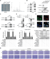
NSUN5 recruits WDR5 to increase SMAD3 expression. A) Cellular extracts expressing Flag‐NSUN5 were immunopurified using anti‐Flag, separated via SDS‐PAGE, and subjected to Coomassie brilliant blue staining to verify successful immunoprecipitation (IP) (left). Cellular extracts were then analyzed via LC–MS/MS to explore potential interacting partners of NSUN5 (right). B) Through co‐immunoprecipitation (Co‐IP) and pulldown assays, we speculated that NSUN5 directly interacts with WDR5 to facilitate recruitment of the lysine methyltransferase complex. C) Endogenous Co‐IP assays confirmed that NSUN5 interacts with WDR5 in HCC cells, consistent with previous mass spectrometry findings. D) GST‐pulldown and His‐pulldown assays demonstrated that NSUN5 can directly bind to WDR5 to form heterodimers. E) Endogenous immunofluorescence staining showed that NSUN5 was distributed in the nucleus and cytoplasm, significantly colocalizing with WDR5 in the nucleus. F) Rescue experiments revealed that downregulating WDR5 expression or inhibiting its function significantly attenuated the upregulation of the SMAD3 signaling pathway and elevated levels of SMAD3 mRNA induced by NSUN5 overexpression. Additionally, the aberrant EMT indices induced by NSUN5 overexpression returned to normal. G) Wound healing assays revealed that the enhanced migration ability of HCC cells due to NSUN5 overexpression was dependent on its interaction with WDR5. Results in (F) are shown as mean ± SD. P‐values are indicated by * < 0.05; ** < 0.01; *** < 0.001.
Figure 6
Figure 6
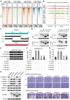
The 155–311 amino acid (aa) sequence of NSUN5 is indispensable for the function of the NSUN5‐WDR5 interaction. A) We employed a cut‐tag assay to assess alterations in WDR5 and H3K4me1/2/3 enrichment at the TSS of SMAD3 before and after NSUN5 overexpression in HCC cells. B) Visualization results demonstrated significant enhancement in the enrichment of WDR5 and H3K4me1/2/3 within the DNA promoter region of SMAD3 following NSUN5 overexpression in HCC cells. C) To explore the interaction domain between NSUN5 and WDR5, we constructed various truncated forms of NSUN5 using the pcDNA3.1‐Puro‐C‐3Flag plasmid. D) Using Co‐IP assays, we identified that the middle segment of NSUN5 (155–311 aa region, IS domain) is crucial for its interaction with WDR5 (right). E) ChIP‐qPCR analysis revealed that the binding of NSUN5 to the SMAD3 DNA region is dependent on its IS domain. The enrichment of WDR5 and H3K4me3 in the SMAD3 promoter region was not significant in the absence of this domain. F) Dual‐luciferase reporter gene assay showed that overexpression of the NSUN5‐JD2 truncated form did not upregulate the SMAD3 signaling pathway. G) qPCR assays demonstrated that the IS domain is a key domain on NSUN5 that promotes the increase in SMAD3 mRNA content in HCC cells. H) Western blot analysis demonstrated that when only the JD2 truncation (deletion of the 155–311 amino acid sequence) was overexpressed, the EMT indicators of HCC cells did not change significantly, revealing that the middle segment of NSUN5 is crucial for promoting the EMT process in HCC cells. I) Wound healing and transwell assays demonstrated that the IS domain of NSUN5 is required to enhance the invasion and migration abilities of HCC cells. Results in (F) are shown as mean ± SD. P‐values are indicated by * < 0.05; ** < 0.01; *** < 0.001.
Figure 7
Figure 7
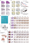
Overexpression of NSUN5 in vivo promotes the course of HCC. A) Hematoxylin and eosin (H&E) staining and IHC staining were used in spontaneous mouse HCC tissues. Experimental results revealed significant downregulation of the SMAD3 signaling pathway (indicated by p‐SMAD3) upon NSUN5 knockout. B) IHC staining results from the tissue microarray confirmed the increased expression of NSUN5 in HCC tumor tissues compared with that in adjacent tissues, and this high expression was often accompanied by poor prognosis in these patients. C) Using the same set of tissue microarrays, we found that p‐SMAD3 expression was higher in the NSUN5‐high group, validating our aforementioned findings and confirming a clinically significant correlation between NSUN5 and SMAD3 activation. D) Subcutaneous tumorigenic experiments in nude mice showed that NSUN5 overexpression resulted in increased HCC proliferation, which was significantly reduced by decreasing WDR5 content in HepG2‐ov‐NSUN5 cells (n = 5 per group). E) Corresponding IHC staining revealed increased SMAD3 expression in NSUN5‐overexpressing tumors, leading to enhanced SMAD3 signaling pathway (indicated by p‐SMAD3). This promoted EMT in HCC, which could be reversed upon knocking down WDR5. F) Tail vein metastasis mouse model (n = 5 per group). G) Lung metastases in mice injected with HepG2‐ov‐NSUN5 cells were more numerous and larger than those in the corresponding negative control group in the gross specimens, which was further confirmed through H&E and IHC staining. Results in (D) are shown as mean ± SD. P‐values are indicated by * < 0.05; ** < 0.01; *** < 0.001.
Figure 8
Figure 8
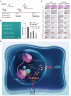
NSUN5 facilitates HCC progression by increasing SMAD3 expression. Schematic diagram demonstrating further experiments to explore the potential applicability of NSUN5 knockout for treating HCC. A) Compared with the corresponding control cells, Huh‐7‐sg‐NSUN5 cells showed higher drug sensitivity when Sorafenib (30 mg kg−1) was added, as confirmed by the statistical results (n = 5 per group). B) Corresponding IHC staining of EMT markers (E‐cadherin and vimentin) and related proliferation indexes (Ki‐67) in different groups. C) A diagram summarizing the findings: NSUN5, overexpressed in HCC, uses its IS domain to bind to SMAD3 DNA and recruit WDR5, thereby synergistically promoting the enrichment of H3K4me3 at the SMAD3 promoter. This enhances the accessibility of this DNA region and increases the expression of SMAD3 in HCC cells, facilitating EMT mediated by the SMAD3 signaling pathway to accelerate the progression of HCC. Results in (B) are shown as mean ± SD. P‐values are indicated by * < 0.05; ** < 0.01; *** < 0.001.

References

    1. a) D'Avola D., Granito A., Torre‐Aláez M., Piscaglia F. J. J. o. h., Journal of hepatology 2022, 76, 1185; - PubMed
    2. b) Han H., Lin T., Wang Z., Song J., Fang Z., Zhang J., You X., Du Y., Ye J., Zhou G. J. P. r., Pharmacological research 2023, 187, 106593. - PubMed
    1. Serper M., Parikh N., Thiele G., Ovchinsky N., Mehta S., Kuo A., Ho C., Kanwal F., Volk M., Asrani S., Ghabril M., Lake J., Merriman R., Morgan T., Tapper E. J. H., Hepatology 2022, 76, 251. - PMC - PubMed
    1. Rich N., Singal A. J. H., Hepatology 2022, 75, 740. - PMC - PubMed
    1. Lambert A., Weinberg R., Nature reviews. Cancer 2021, 21, 325. - PubMed
    1. Nieto M., Nature reviews. Molecular cell biology 2020, 21, 563. - PubMed

MeSH terms

LinkOut - more resources