

Physical inactivity exacerbates pathologic inflammatory signalling at the single cell level in patients with systemic lupus
- PMID: 39531917
- PMCID: PMC11603005
- DOI: 10.1016/j.ebiom.2024.105432
Physical inactivity exacerbates pathologic inflammatory signalling at the single cell level in patients with systemic lupus
Abstract
Background: Physical activity is an adjunctive therapy that improves symptoms in people living with systemic lupus erythematosus (SLE), yet the mechanisms underlying this benefit remain unclear.
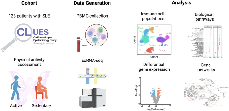
Methods: We carried out a cohort study of 123 patients with SLE enrolled in the California Lupus Epidemiology Study (CLUES). The primary predictor variable was self-reported physical activity, which was measured using a previously validated instrument. We analyzed peripheral blood mononuclear cell (PBMC) single-cell RNA sequencing (scRNA-seq) data available from the cohort. From the scRNA-seq data, we compared immune cell frequencies, cell-specific gene expression, biological signalling pathways, and upstream cytokine activation states between physically active and inactive patients, adjusting for age, sex and race.
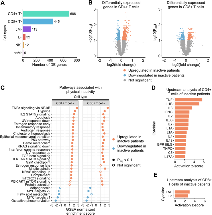
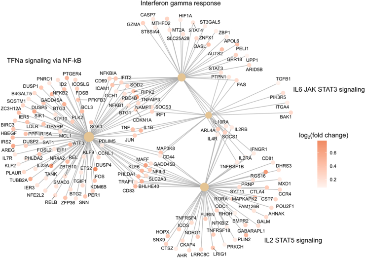
Findings: We found that physical activity influenced immune cell frequencies, with sedentary patients most notably demonstrating greater CD4+ T cell lymphopenia (Padj = 0.028). Differential gene expression analysis identified a transcriptional signature of physical inactivity across five cell types. In CD4+ and CD8+ T cells, this signature was characterized by 686 and 445 differentially expressed genes (Padj < 0.1). Gene set enrichment analysis demonstrated enrichment of proinflammatory genes in the TNF-α signalling through NF-kB, interferon-γ (IFN-γ), IL2/STAT5, and IL6/JAK/STAT3 signalling pathways. Computational prediction of upstream cytokine activation states suggested CD4+ T cells from physically inactive patients exhibited increased activation of TNF-α, IFN-γ, IL1Β, and other proinflammatory cytokines. Network analysis demonstrated interconnectivity of genes driving the proinflammatory state of sedentary patients. Findings were consistent in sensitivity analyses adjusting for corticosteroid treatment and physical function.
Interpretation: Taken together, our findings suggest a mechanistic explanation for the observed benefits of physical activity in patients with SLE. Specifically, we find that physical inactivity is associated with altered frequencies and transcriptional profiles of immune cell populations and may exacerbate pathologic inflammatory signalling via CD4+ and CD8+ T cells.
Funding: This work was supported by the US National Institutes of Health (NIH) (R01 AR069616, K23HL138461-01A1, K23AT011768) the US CDC (U01DP0670), and the CZ Biohub.
Keywords: Lifestyle behaviors; Physical activity; Rheumatoid arthritis; Single cell transcriptomics.
Copyright © 2024 The Authors. Published by Elsevier B.V. All rights reserved.
Conflict of interest statement
Declaration of interests JY declares grants from Aurinia, Astra Zeneca and the BMS Foundation, and consulting fees from Astra Zeneca, Aurinia, Pfizer and ImmPact Bio and Adelphi Values and Amgen grand rounds lecture to company employees and Research Lead for the RISE registry- ACR. C.J.Y. declares funding from NIH R01AI171184 and P01AI172523, and Lupus Research Alliance P0559324, consulting fees from Maze Therapeutics, HiBio, Santa Ana, TRex Bio, ImYoo and DeciBio, payment or honoraria from Renji Hospital School of Medicine, American College of Medical Genetics and Genomics, St Jude's Children Research Hospital, Yonsei University College of Medicine, Lupus Research Alliance, NIAMS, ACMG and MERCK, support from Icahn School of Medicine at Mount Sinai, Parker Institute for Cancer Immunotherapy, Renji Hospital School of Medicine, Northwestern University, St. Jude's Children Research Hospital and American Transplant Congress, NIAMS and Lupus 21st Century, participation on a Data Safety Monitoring Board for ImmunAI and Related Sciences, an advisory role in Survey Genomics, ImmunAI, and Arc Institute, stock options from Survey Genomics, Related Sciences, ImmunAI and Maze Therapeutics, receipt of materials from Illumina, and other interests with Chan Zuckerberg Biohub, Chan Zuckerberg Initiative, Genentech, ScaleBio, Parker Institute for Cancer Immunotherapy (PICI), Biolegend and Illumina. C.L. declares funding from the NIH Intramural Research Program. All other authors declare that there are no competing interests.
Figures

References
-
- Kang I., Park S.H. Infectious complications in SLE after immunosuppressive therapies. Curr Opin Rheumatol. 2003;15:528–534. - PubMed
-
- Blaess J., Goepfert T., Geneton S., et al. Benefits & risks of physical activity in patients with systemic lupus erythematosus: a systematic review of the literature. Semin Arthritis Rheum. 2023;58 - PubMed
-
- Vordenbäumen S., Kleefisch M., Sokolowski A., et al. Beneficial effects associated to a healthy lifestyle in systemic lupus erythematosus: a cross-sectional study. Lupus. 2023;32:855–863. - PubMed
MeSH terms
Substances
Grants and funding
LinkOut - more resources
Full Text Sources
Medical
Research Materials
Miscellaneous
