Detection challenges of temporal encephaloceles in epilepsy: A retrospective analysis
- PMID: 39532244
- PMCID: PMC11614672
- DOI: 10.1016/j.mri.2024.110272
Detection challenges of temporal encephaloceles in epilepsy: A retrospective analysis
Abstract
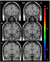
Temporal encephaloceles (TEs) are herniations of cerebral parenchyma through structural defects in the floor of the middle cranial fossa. They are a relatively common, but only relatively recently identified potential cause of drug-resistant epilepsy. Uncontrolled epilepsy is associated with many negative long term health consequences including a heightened risk of death. The most effective treatment for drug-resistant epilepsy is surgery. One of the most predictive factors associated with successful surgery is identification of an abnormality on imaging. However, TEs can be difficult to detect and are often overlooked on neuroimaging studies. Improving our ability to accurately detect TEs by MRI is an important step in improving surgical outcomes in patients with drug-resistant epilepsy. We performed a review on existing imaging modalities for detecting TEs and report on our attempt to use a voxel-based morphometry (VBM) algorithm to detect TEs in T1-weighted MRIs of 81 patients from a database comprised of 25 patients with confirmed encephaloceles and 56 controls. Our program's sensitivity and specificity were compared to those of two neuroradiologists and two epileptologists using visualization during surgery as the gold standard. On average, the neuroradiologists and epileptologists had sensitivities of 41 % and 58 % and specificities of 81 % and 60 % while our VBM-based approach had sensitivities and specificities ranging from 11 % to 50 % and 0.2 % to 17 %, respectively. This work provides an overview of the different imaging modalities utilized in the detection of TEs and highlights the difficulties associated with their detection for both experienced physicians and cutting-edge computational methods. Our findings suggest that VBM-based methods could potentially be used to enhance clinicians' ability to detect TEs thereby facilitating surgical planning, improving surgical outcomes by allowing for more specific targeting, and bettering the long-term health and well-being of patients with drug-resistant epilepsy secondary to TEs.
Keywords: Encephalocele; Epilepsy; Morphometry; Refractory epilepsy; Temporal encephalocele; Voxel-based.
Copyright © 2024. Published by Elsevier Inc.
Conflict of interest statement
Declaration of competing interest None of the authors has any conflict of interest to disclose.
Figures


References
-
- Fisher RS, Acevedo C, Arzimanoglou A, et al. ILAE official report: a practical clinical definition of epilepsy. Epilepsia 2014;55:475–82. - PubMed
-
- Kalilani L, Sun X, Pelgrims B, et al. The epidemiology of drug-resistant epilepsy: A systematic review and meta-analysis. Epilepsia 2018;59:2179–93. - PubMed
-
- Kwan P, Arzimanoglou A, Berg AT, et al. Definition of drug resistant epilepsy: consensus proposal by the ad hoc Task Force of the ILAE Commission on Therapeutic Strategies. Epilepsia 2010;51:1069–77. - PubMed
-
- Jobst BC, Cascino GD. Resective epilepsy surgery for drug-resistant focal epilepsy: a review. JAMA 2015;313:285–93. - PubMed
Publication types
MeSH terms
Grants and funding
LinkOut - more resources
Full Text Sources

