. 2024 Dec 17;13(12):1620-1626.
doi: 10.1021/acsmacrolett.4c00520. Epub 2024 Nov 12.

Generating Tooth Organoids Using Defined Bioorthogonally Cross-Linked Hydrogels

Affiliations

Generating Tooth Organoids Using Defined Bioorthogonally Cross-Linked Hydrogels

Xuechen Zhang et al. ACS Macro Lett. .

Abstract

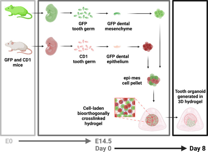
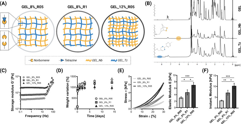
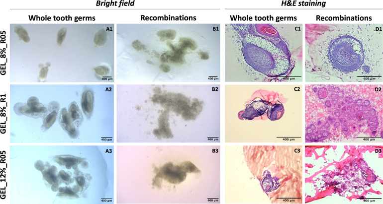
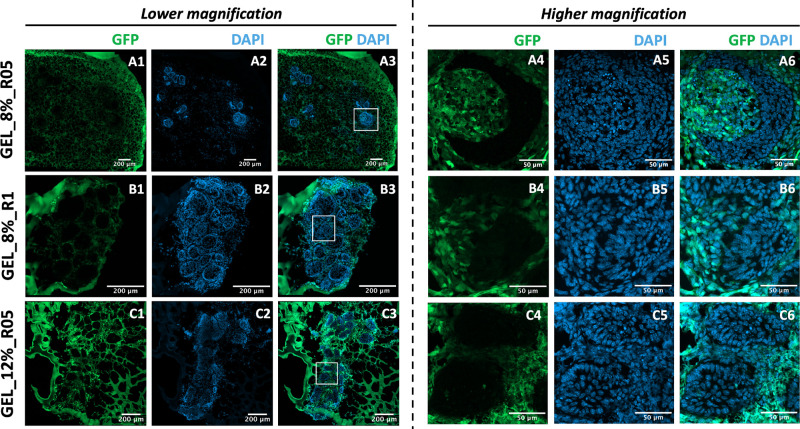
Generating teeth in vitro requires mimicking tooth developmental processes. Biomaterials are essential to support 3D tooth organoid formation, but their properties must be finely tuned to achieve the required biomimicry for tooth development. For the first time, we used bioorthogonally cross-linked hydrogels as defined 3D matrixes for tooth developmental engineering, and we highlighted how their properties play a pivotal role in enabling 3D tooth organoid formation in vitro. We prepared hydrogels by mixing gelatin precursors modified either with tetrazine (Tz) or norbornene (Nb) moieties. We tuned the hydrogel properties (E = 2-7 kPa; G' = 500-1500 Pa) by varying the gelatin concentration (8% vs 12% w/V) and stoichiometric ratio (Tz:Nb = 1 vs 0.5). We encapsulated dental epithelial-mesenchymal cell pellets in a library of hydrogels and identified a hydrogel formulation that enabled successful growth kinetics and morphogenesis of tooth germs, introducing a defined tunable platform for tooth organoid engineering and modeling.


Conflict of interest statement

The authors declare no competing financial interest.

Figures

Figure 1
Figure 1
Preparation of the 3D tooth organoids. Dental mesenchyme and epithelium were obtained in green fluorescent protein (GFP) and CD1 mouse embryos at embryonic day 14.5 (E14.5), separately. After digesting to single cell suspensions, the two cell populations were combined to obtain an epithelial-mesenchymal cell pellet, encapsulated in hydrogels (day 0), and cultured in vitro to generate 3D tooth organoids (day 8). Created with BioRender.com.
Figure 2
Figure 2
Bioorthogonally cross-linked click gelatin hydrogels. (A) Schematic of hydrogel structures prepared by varying the concentration (8% and 12% w/V) and ratio between gelatin modified with tetrazine (GEL_Tz, blue lines) and gelatin modified with norbornene (GEL_Nb, yellow lines) at 1:1 (R1) or 0.5:1 (R05). (B) Representative 1H NMR spectra of unmodified gelatin (GEL), gelatin modified with norbornene (GEL_Nb), and gelatin modified with tetrazine (GEL_Tz). (C) Representative rheological curves of the frequency response of the cross-linked hydrogels. (D) Swelling and weight variation in culture medium of the cross-linked hydrogels (n = 4). (E) Representative stress–strain curves (σ–ε) of cross-linked swollen hydrogels and elastic modulus (E). (F) Instantaneous modulus calculated from indentation tests (n = 3; * p < 0.05, ** p < 0.01, *** p < 0.001). Created with Biorender.com.
Figure 3
Figure 3
Whole tooth germs and recombination of dental mesenchymal and epithelial cells in bioorthogonally cross-linked hydrogels prepared by varying polymer concentrations and stoichiometric ratios. (A1–3) Tooth germs in GEL_8%_R05 (n = 6), GEL_8%_R1 (n = 6), or GEL_12%_R05 (n = 6). (B1–3) Recombination in GEL_8%_R05 (n = 13), GEL_8%_R1 (n = 9), or GEL_12%_R05 (n = 6). (C1–3) H&E staining images for A1–3. (D1–3) H&E staining images for B1–3. Scale bar: 400 μm.
Figure 4
Figure 4
Recombination of GFP dental mesenchymal cells and CD1 dental epithelial cells in bioorthogonally cross-linked hydrogels prepared by varying polymer concentrations and stoichiometric ratios. (A) GEL_8%_R05, (B) GEL_8%_R1, and (C) GEL_12%_R05 (1–3 are in low magnification; 4–6 are the small white squares in A3, B3 and C3 in high magnification.) Scale bar: 200 μm in lower magnification and 50 μm in higher magnification.

References

    1. Bernabe E.; Marcenes W.; Hernandez C. R.; Bailey J.; Abreu L. G.; Alipour V.; Amini S.; Arabloo J.; Arefi Z.; Arora A.; et al. Global, Regional, and National Levels and Trends in Burden of Oral Conditions from 1990 to 2017: A Systematic Analysis for the Global Burden of Disease 2017 Study. Journal of Dental Research 2020, 99 (4), 362–373. 10.1177/0022034520908533. - DOI - PMC - PubMed
    1. Zhang W.; Yelick P. C. Tooth repair and regeneration: potential of dental stem cells. Trends in molecular medicine 2021, 27 (5), 501–511. 10.1016/j.molmed.2021.02.005. - DOI - PMC - PubMed
    1. Mina M.; Kollar E. The induction of odontogenesis in non-dental mesenchyme combined with early murine mandibular arch epithelium. Archives of oral biology 1987, 32 (2), 123–127. 10.1016/0003-9969(87)90055-0. - DOI - PubMed
    1. Nakao K.; Morita R.; Saji Y.; Ishida K.; Tomita Y.; Ogawa M.; Saitoh M.; Tomooka Y.; Tsuji T. The development of a bioengineered organ germ method. Nat. Methods 2007, 4 (3), 227–230. 10.1038/nmeth1012. - DOI - PubMed
    1. Yamamoto H.; Kim E.-J.; Cho S.-W.; Jung H.-S. Analysis of tooth formation by reaggregated dental mesenchyme from mouse embryo. Microscopy 2003, 52 (6), 559–566. 10.1093/jmicro/52.6.559. - DOI - PubMed

Publication types

LinkOut - more resources