. 2025 Jan 1:191:189-204.
doi: 10.1016/j.actbio.2024.10.050. Epub 2024 Nov 12.

Harnessing synergistic effects of MMP-2 Inhibition and bFGF to simultaneously preserve and vascularize cardiac extracellular matrix after myocardial infarction

Affiliations

Harnessing synergistic effects of MMP-2 Inhibition and bFGF to simultaneously preserve and vascularize cardiac extracellular matrix after myocardial infarction

Hong Niu et al. Acta Biomater. .

Abstract

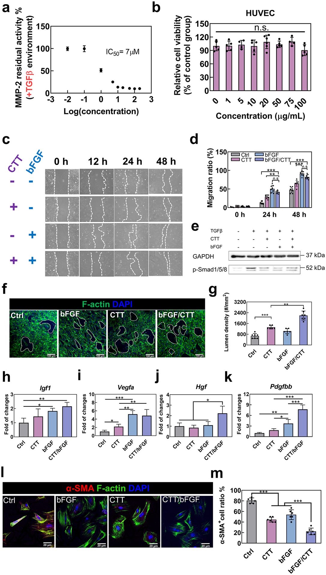
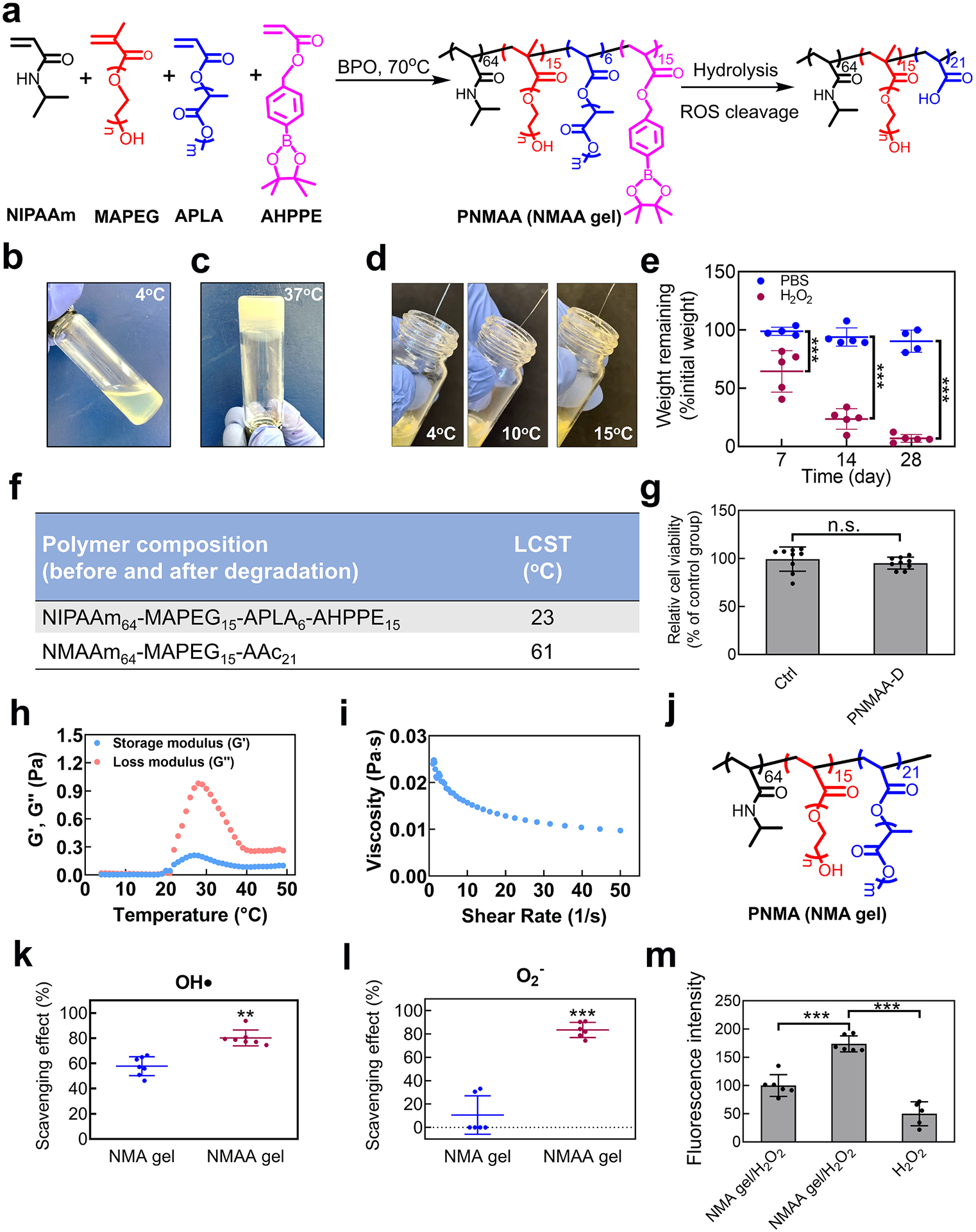
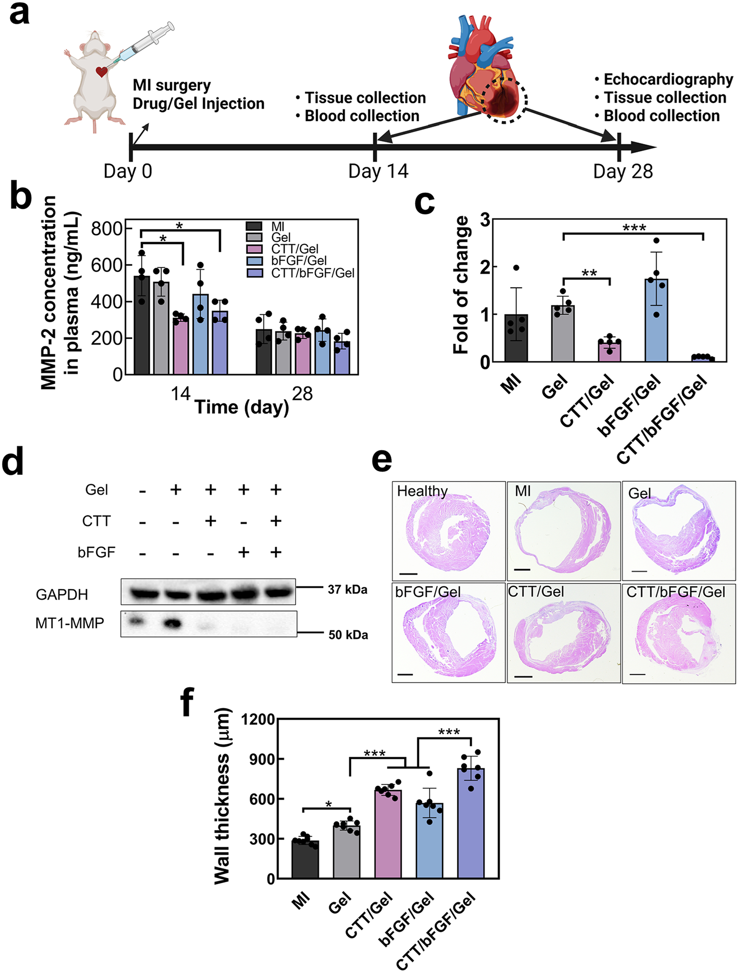
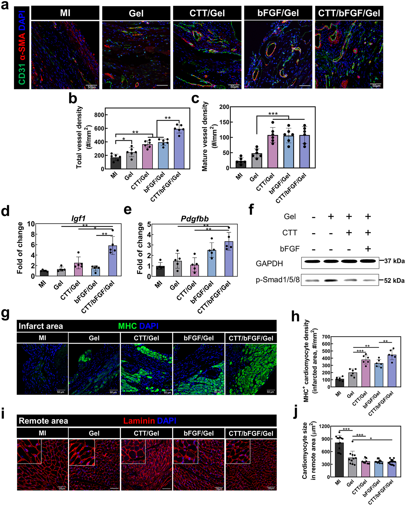
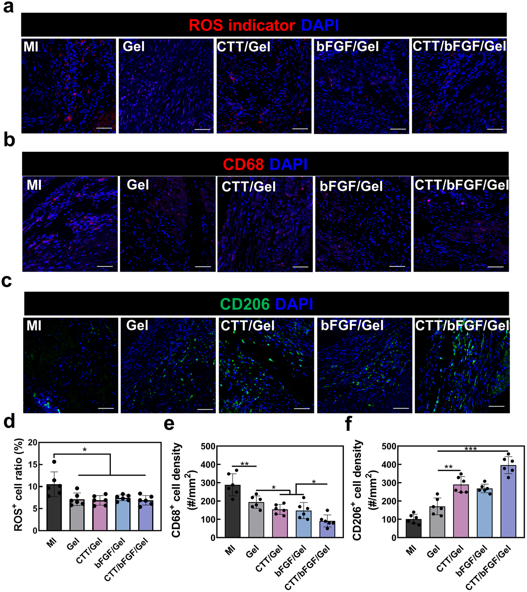
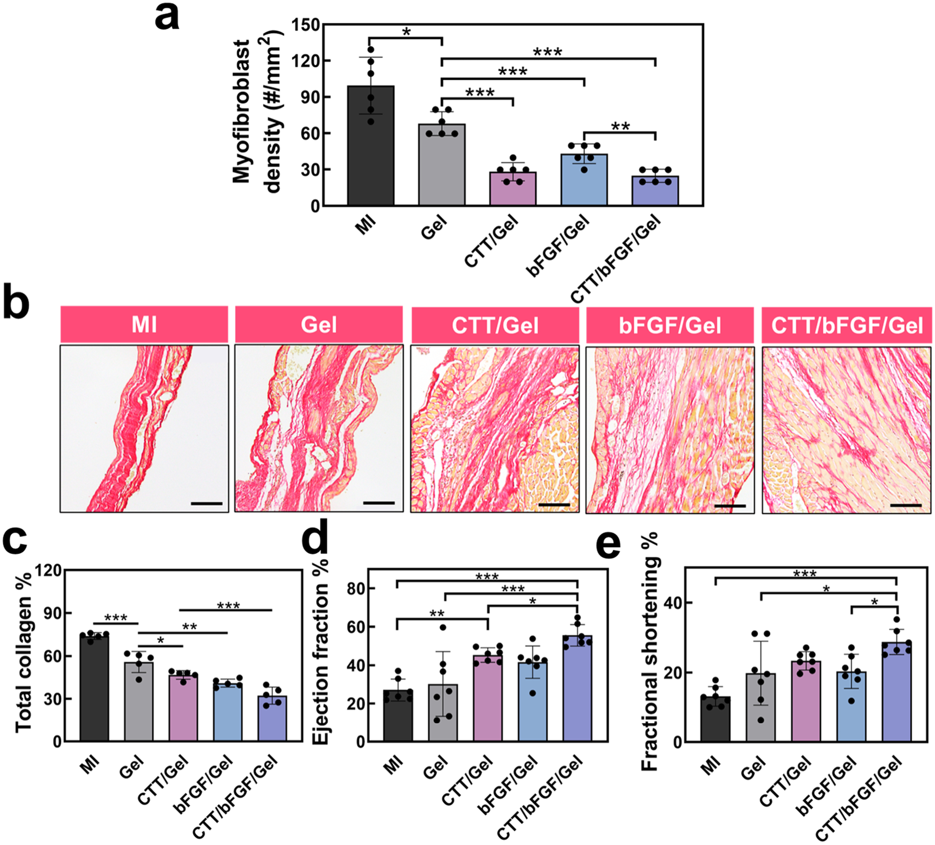
Myocardial infarction (MI) leads to cardiac extracellular matrix (ECM) degradation and fibrosis, reducing heart function. Consequently, simultaneously addressing ECM degradation and inhibiting cardiac fibrosis is essential for preserving heart function and mitigating adverse remodeling. However, the preserved ECM becomes unstable if not vascularized, as its structure and composition undergo changes over time. ECM vascularization is crucial to improve cardiac function. Presently, there is no clinically approved therapy that can simultaneously preserve and vascularize the ECM, and inhibit cardiac fibrosis. Our study develops a drug delivery system aiming to achieve these goals. It includes the peptide CTTHWGFTLC (CTT), a specific MMP-2 inhibitor, and basic fibroblast growth factor (bFGF), a potent factor with pro-angiogenic and anti-fibrotic properties. An injectable hydrogel serves as the carrier, featuring a rapid gelation that allows for the substantial retention of drugs. Additionally, the hydrogel has the capability to scavenge upregulated reactive oxygen species (ROS), thereby reducing tissue inflammation. Our findings indicate that CTT and bFGF synergistically enhance endothelial cell migration and tube formation while inhibiting the differentiation of fibroblasts into myofibroblasts. Upon delivery into hearts, the system significantly decreases MMP-2 level, promotes angiogenesis, attenuates cardiac fibrosis, and alleviates inflammation, resulting in a noteworthy cardiac function improvement. STATEMENT OF SIGNIFICANCE: 1) This work addresses key challenges in cardiac repair after myocardial infarction (MI), including extracellular matrix (ECM) degradation, vascularization, and fibrosis. 2) We combined an MMP-2/9 inhibitor (CTT) with bFGF to prevent ECM degradation, enhance vascularization, and inhibit fibrosis, providing a comprehensive strategy to improve cardiac function. 3) An injectable hydrogel was developed with rapid gelation and mechanical properties similar to heart tissue, ensuring efficient drug retention and reducing tissue stress. 4) The hydrogel enabled controlled, spatiotemporal release of CTT to dynamically reduce MMP-2/9 activity, and gradually released bFGF to promote angiogenesis and inhibit fibrosis.

Keywords: MMP-2 inhibitors; Myocardial infarction; ROS-scavenging; Vascularization; local delivery.


Conflict of interest statement

Declaration of competing interest The authors declare that they have no known competing financial interests or personal relationships that could have appeared to influence the work reported in this paper.

Figures

Fig. 1.
Fig. 1.
CTT and bFGF modulate the functions of HUVECs and reduce the activation of myofibroblasts. a. Concentration–dependence of CTT in inhibiting MMP-2 activity. b. Cell viability of HUVECs cultured with different concentrations of CTT tested by MTT assay. c. Representative images of HUVEC migration for 48 h in an environment where TGFβ was added (n = 8 images per condition). d. Quantification of migration ratio based on the images. e. Immunoblotting of p-Smad1/5/8 and GAPDH expression derived from HUVECs. HUVECs were treated with serum-free medium (control), TGFβ (10 ng/mL), CTT (10 μg/mL), and bFGF (1 μg/mL). f. Tube formation assay using HUVECs cultured on a 2D collagen model. g. Quantification of tube density. h-k. Gene expression of angiogenic factors by the cDNA derived from HUVECs with different treatment groups. (h). IGF1; (i). VEGFA; (j). HGF and (k). PDGFBB. l. Representative images of RCFs cultured on collagen gel with different treatments (n = 8 images per condition). m. Percentage of α-SMA+ cells in the different groups. n.s. p > 0.5, *p < 0.1, **p < 0.01, ***p < 0.001.
Fig. 2.
Fig. 2.
Development of NMAA hydrogel. a. Synthesis of poly (NIPAAm64-co-MAPEG15-co-APLA6-co-AHPPE15) by the copolymerization of NIPAAm, MAPEG, APLA, and AHPPE, and hydrolysis of the copolymer. b-d. Physical properties of the NMAA hydrogel, including (b) flowability at 4 °C, (c) gelation at 37 °C, and (d) injectability at 4 °C, 10 °C, and 15 °C. e. Biodegradation of NMAA gel with and without 10 mM H2O2 for 28 days. f. LCSTs of poly (NIPAAm65-co-MAPEG15-co-APLA5-co-AHPPE15) (NMAA) and its theoretical final degradation products poly (NIPAAm65-co-MAPEG15-co-AAc21) tested by rheometer. g. Cytotoxicity of degradation products collected at day 28 on RCFs. h. The storage modulus (G’) and the loss modulus (G”) of the hydrogel over the temperature range of 4 °C to 49 °C. i. The viscosity of the hydrogel was tested at 4 °C with a shear rate ranging from 1 to 50 s−1. j. Chemical structure of poly (NIPAAm64-co-MAPEG15-co-APLA21) that is non-ROS responsive and used as the control gel. k. The scavenging effect on hydroxyl radicals of NMAA hydrogel was determined by the Fenton reaction. Non-ROS responsive APLA gel was used as a control group. l. Scavenging effect on superoxide of NMAA gel tested by Pyrogallol assay. m. RCF proliferation under H2O2 (100 μM) cultured on NMAA film compared to the non-ROS scavenging film and plate well.
Fig. 3.
Fig. 3.
Release of CTT and bFGF encapsulated in NMAA hydrogel for 28 days. a. Release kinetics of CTT at different concentrations and CTT/bFGF in NMAA hydrogel (n = 6). b. Release kinetics of bFGF at different concentrations and bFGF/CTT in NMAA hydrogel (n = 6). c. Representative images of migration of HUVECs treated with the released medium at days 1, 14, 21, and 28. d. Quantification of migration ratio based on the images. e. Inhibition of MMP-2 by testing residual activity using the released medium collected from days 1, 7, 14, 21, and 28 (n = 3). f. Cell proliferation of RCFs treated with the release medium tested by dsDNA assay (n ≥ 3).
Fig. 4.
Fig. 4.
Delivery of CTT/bFGF reduced expression of MMPs 2 and 9 in plasma and tissues and effectively preserved ECM. a. Timeline of the mouse surgery (created with Biorender.com). b. Dynamic change of MMP2 in plasma at days 14 and 28 (n = 4). c. In vivo Mmp2 change in infarcted tissues tested by gene expression (n = 5). d. Immunoblotting of α-actinin and MT1-MMP from in vivo tissues. GAPDH was used as a loading control. e. Representative H&E staining images at day 28. f. Quantification of wall thickness based on H&E images (n = 7).
Fig. 5.
Fig. 5.
Delivery of CTT/bFGF via NMAA hydrogel effectively improved in vivo vessel formation without causing cardiac fibrosis. a. α-SMA/CD31/DAPI staining for injured heart tissues. Scale bar = 50 μm. b. Quantification of total blood vessel density (α-SMA/CD31+ vessels) (n = 6). c. Quantification of mature blood vessel (α-SMA+/CD31+ vessels) density (n = 6). d, e. Gene expression of in vivo angiogenic factors including (e) Igf1, and (f) Pdgfbb. f. Immunoblotting of p-Smad1/5/8 from in vivo tissues. GAPDH was used as a loading control. g. Representative images of MHC/DAPI staining in the infarcted area. Scale bar = 50 μm. h. Quantification of MHC+ cell density from IHC images (n = 6). i. Representative images of laminin/DAPI staining in remote areas. Scale bar = 50 μm. j. Quantification of remote cardiomyocytes’ size based on laminin signals in the images (n = 8–14 based on region size).
Fig. 6.
Fig. 6.
Local inflammation response after the treatment of CTT/bFGF/hydrogel. a. Representative images for CM-H2DCFDA staining. b. Representative images for CD68/DAPI staining. c. Representative images for CD206/DAPI staining. d. Quantification of CM-H2DCFDA+ cell density (n = 6). e. Quantification of CD68+ cell density. f. Quantification of CD206+ cell density (n = 6). Scale bar = 50 μm.
Fig. 7.
Fig. 7.
Cardiac regeneration and function repair by the treatment of CTT/bFGF/Gel. a. Myofibroblast density at the injured area based on the images in Fig. 5a (n = 6). b. Representative images for tissue samples stained with picrosirius red (PSR). Scale bar = 50 μm. c. Quantification of total collagen deposition calculated from PSR staining images (n = 5). d. Echocardiogram analysis of LV ejection fraction 28 days after surgery. e. Echocardiogram analysis of LV fractional shortening (n = 7).

Similar articles

Cited by

References

    1. Rienks M, Papageorgiou A-P, Frangogiannis NG, Heymans S, Myocardial extracellular matrix: an ever-changing and diverse entity, Circ. Res 114 (5) (2014) 872–888. - PubMed
    1. DeLeon-Pennell KY, Meschiari CA, Jung M, Lindsey ML, Matrix metalloproteinases in myocardial infarction and heart failure, Prog. Mol. Biol. Transl. Sci 147 (2017) 75–100. - PMC - PubMed
    1. Lindsey ML, Assigning matrix metalloproteinase roles in ischaemic cardiac remodelling, Nat. Rev. Cardiol 15 (8) (2018) 471–479. - PMC - PubMed
    1. Webb CS, Bonnema DD, Ahmed SH, Leonardi AH, McClure CD, Clark LL, Stroud RE, Corn WC, Finklea L, Zile MR, Specific temporal profile of matrix metalloproteinase release occurs in patients after myocardial infarction: relation to left ventricular remodeling, Circulation 114 (10) (2006) 1020–1027. - PubMed
    1. Herzog E, Gu A, Kohmoto T, Burkhoff D, Hochman JS, Early activation of metalloproteinases after experimental myocardial infarction occurs in infarct and non-infarct zones, Cardiovasc. Pathol 7 (6) (1998) 307–312. - PubMed

Publication types

MeSH terms

Substances