. 2024 Nov 12;7(1):1493.
doi: 10.1038/s42003-024-07201-6.

Optimisation of cell fate determination for cultivated muscle differentiation

Affiliations

Optimisation of cell fate determination for cultivated muscle differentiation

Lea Melzener et al. Commun Biol. .

Abstract

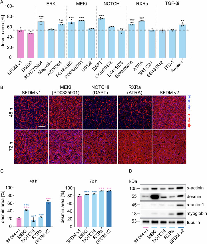
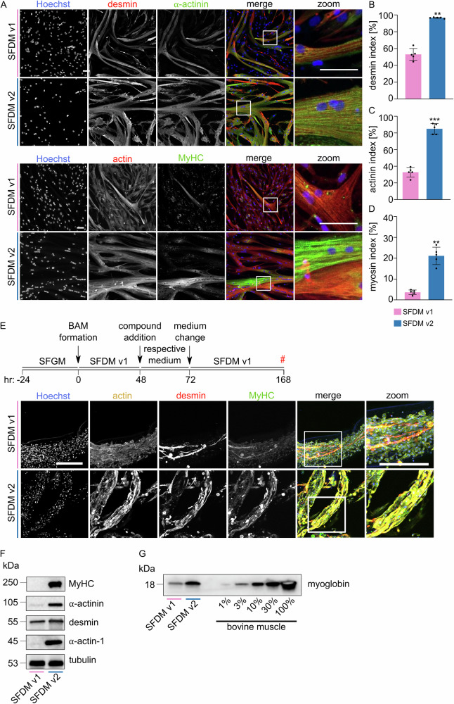
Production of cultivated meat requires defined medium formulations for the robust differentiation of myogenic cells into mature skeletal muscle fibres in vitro. Although these formulations can drive myogenic differentiation levels comparable to serum-starvation-based protocols, the resulting cultures are often heterogeneous, with a significant proportion of cells not participating in myofusion, limiting maturation of the muscle. To address this problem, we employed RNA sequencing to analyse heterogeneity in differentiating bovine satellite cells at single-nucleus resolution, identifying distinct cellular subpopulations including proliferative cells that fail to exit the cell cycle and quiescent 'reserve cells' that do not commit to myogenic differentiation. Our findings indicate that the MEK/ERK, NOTCH, and RXR pathways are active during the initial stages of myogenic cell fate determination, and by targeting these pathways, we can promote cell cycle exit while reducing reserve cell formation. This optimised medium formulation consistently yields fusion indices close to 100% in 2D culture. Furthermore, we demonstrate that these conditions enhance myotube formation and actomyosin accumulation in 3D bovine skeletal muscle constructs, providing proof of principle for the generation of highly differentiated cultivated muscle with excellent mimicry to traditional muscle.


Conflict of interest statement

Competing interests L.M., L.S., M.F., T.M., D.R., A.K., T.v.H., S.S., A.D. and J.E.F. were all employees of Mosa Meat B.V., a company aiming to commercialise cultivated meat technologies, at the time of writing. M.J.P. is co-founder and stakeholder of Mosa Meat B.V. Study was funded by Mosa Meat B.V. Mosa Meat B.V. has patent applications pending on serum-free proliferation and differentiation media (WO2021158103, WO2022114955). All authors declare no other competing interests.

Figures

Fig. 1
Fig. 1. Only a subset of SCs fuse during myogenic differentiation.
A Fluorescence images of differentiating SCs to visualise myogenic fusion at time points indicated (#). Blue, Hoechst; red, desmin. Scale bar, 500 µm. B Mean quantified fusion indices (proportion of nuclei within desmin-stained areas) for images in (A). Error bars indicate s.d., n = 10. C Mean quantified desmin area (proportion of total image field that is desmin-stained) for images in (A). Error bars indicate s.d., n = 10. D Mean nuclei counts for images in (A). Error bars indicate s.d., n = 10. E Representative brightfield images of differentiating SC cultures prior to nuclei harvest for snRNA-seq at 0, 24, 48, 72 and 96 h. Scale bar, 500 µm. F Combined UMAP plots showing single nuclei from all five snRNA-seq time points (shown in E). Nuclei from each time point are coloured in the respective plot, all other nuclei are shown in grey. Nuclei clusters are annotated for clarity of explanation.
Fig. 2
Fig. 2. snRNAseq identifies differentiating, proliferating and reserve SC subpopulations.
A UMAP of single nuclei from all five time points, where nuclei are coloured and assigned according to identified clusters. B UMAP as for (A), coloured according to expression of indicated canonical marker gene(s) for each subpopulation. C Significantly enriched GO terms corresponding to differentially expressed genes between the subpopulations identified in (A). Numerals indicate the proportion of genes for each GO term that are differentially expressed. D Dotplot showing normalised expression of selected genes in each subpopulation (averaged across all nuclei); size of points indicates percentage of nuclei expressing the respective gene, colour indicates average expression level. E Timecourse of fluorescence images for analysis of SC subpopulations (identified in A) during differentiation. Blue, Hoechst; green, Pax7; pink, Ki-67; yellow, myogenin; red, desmin. Scale bar, 100 µm. F Quantification of SC subpopulations for images in E. Ki-67+, Proliferating; Pax7+/Ki-67, Reserve; myogenin+/desmin, Committed myoblast; myogenin+/desmin+, Myotube. Error bars indicate s.d., n = 3.
Fig. 3
Fig. 3. MEK/ERKi, NOTCHi and RXRa promote myogenic differentiation.
A Mean desmin areas after 72 h differentiation with indicated compounds (targeting MEK/ERK, RXR, NOTCH and TGF-β pathways). Error bars indicate s.d., n = 3. B Representative fluorescence images of differentiation after indicated timeframe with three treatments showing significant effect in (A) (MEKi, PD0325901; NOTCHi, DAPT; RXRa, ATRA), and their combination (SFDM v2). Blue, Hoechst; red, desmin; Scale bar, 500 µm. C Quantification of (B), showing mean desmin areas after 48 (left) and 72 h (right) of myogenic differentiation with indicated compound treatment. Error bars show s.d., n = 3. D Muscle-related protein expression, analysed by western blot after 72 h differentiation with the indicated treatment. *p < 0.01, **p < 0.001, ***p < 0.0001.
Fig. 4
Fig. 4. MEKi, NOTCHi and RXRa act during early commitment to myogenic differentiation.
A Representative fluorescence images after 48 h of differentiation for analysis of SC subpopulations during differentiation with indicated treatments. Blue, Hoechst; green, Pax7; pink, Ki-67; yellow, myogenin; red, desmin. Scale bar, 100 µm. B Quantification of SC subpopulation images in (A). Ki-67+, Proliferating; Pax7+/Ki-67, Reserve; myogenin+/desmin, Committed myoblast; myogenin+/desmin+, Myotube. Error bars indicate s.d., n = 3. C Mean desmin areas at time point indicated (#) after compound treatment during only low seeding density (SD, 5 × 103 cm2) phase, only high seeding density (1 × 105 cm2) phase, or both. Error bars show s.d., n = 3. D Quantification of Ki-67, MyoD and myogenin positivity after 48 h of indicated treatment at low seeding density. Error bars show s.d., n = 8. E Mean desmin areas after compound treatment during the ‘second wave’ of differentiation. Error bars show s.d., n = 9. F Model proposing mechanistic basis for improved myogenic differentiation in SFDM v2. *p < 0.01, **p < 0.001, ***p < 0.0001.
Fig. 5
Fig. 5. Inducers promote differentiation in animal-free bioartificial muscles.
A Representative fluorescence images after 72 h myogenic differentiation in SFDM v1 and SFDM v2 (indicated respectively) for two fluorescent staining panels. Blue, Hoechst; red, desmin/actin; green, α-actinin/myosin heavy chain. Scale bar, 250 µm. B Mean quantified desmin indices for images in (A). Error bars indicate s.d., n = 5. C As (B), but α-actinin indices. Error bars indicate s.d., n = 5. D As (B), but myosin indices. Error bars indicate s.d., n = 5. E Representative maximum-intensity projection fluorescence images after 7 days of 3D culture in an RGD-alginate-based animal-free bioartificial muscle system. Blue, Hoechst; yellow, actin; red, desmin; green, myosin heavy chain. Lower panels show a zoom of the merged image. Scale bars, 200 µm. F Muscle-related protein expression during 3D myogenic differentiation, analysed by western blot after 168 h differentiation with the indicated media. G As (F), for myoglobin expression and compared to a dilution series of bovine muscle tissue lysate. *p < 0.01, **p < 0.001, ***p < 0.0001.

References

    1. Post, M. J. Cultured meat from stem cells: challenges and prospects. Meat Sci.92, 297–301 (2012). - PubMed
    1. Post, M. J. et al. Scientific, sustainability and regulatory challenges of cultured meat. Nat. Food1, 403–415 (2020).
    1. Parodi, A. et al. The potential of future foods for sustainable and healthy diets. Nat. Sustain.1, 782–789 (2018).
    1. Rizou, M., Galanakis, I. M., Aldawoud, T. M. S. & Galanakis, C. M. Safety of foods, food supply chain and environment within the COVID-19 pandemic. Trends Food Sci. Technol.102, 293–299 (2020). - PMC - PubMed
    1. Humbird, D. Scale‐up economics for cultured meat. Biotechnol. Bioeng.118, 3239–3250 (2021). - PMC - PubMed

MeSH terms

LinkOut - more resources