. 2024 Dec;43(24):6555-6572.
doi: 10.1038/s44318-024-00294-z. Epub 2024 Nov 12.

LARP1 binds ribosomes and TOP mRNAs in repressed complexes

Affiliations

LARP1 binds ribosomes and TOP mRNAs in repressed complexes

James A Saba et al. EMBO J. 2024 Dec.

Abstract

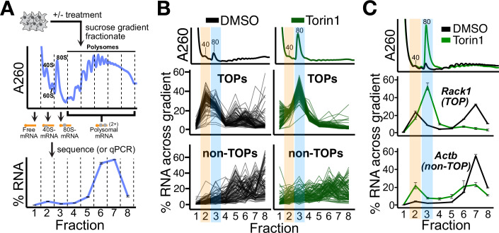
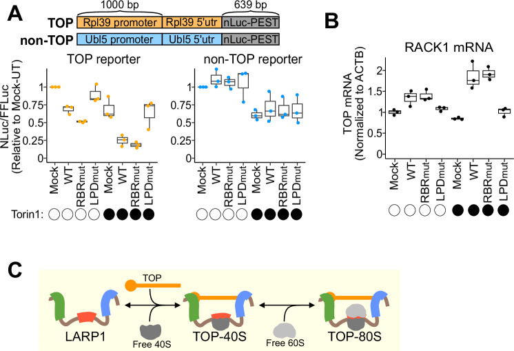
Terminal oligopyrimidine motif-containing mRNAs (TOPs) encode all ribosomal proteins in mammals and are regulated to tune ribosome synthesis to cell state. Previous studies have implicated LARP1 in 40S- or 80S-ribosome complexes that are thought to repress and stabilize TOPs. However, a molecular understanding of how LARP1 and TOPs interact with these ribosome complexes is lacking. Here, we show that LARP1 directly binds non-translating ribosomal subunits. Cryo-EM structures reveal a previously uncharacterized domain of LARP1 bound to and occluding the mRNA channel of the 40S subunit. Increased availability of free ribosomal subunits downstream of various stresses promote 60S joining at the same interface to form LARP1-80S complexes. Simultaneously, LARP1 engages the TOP via its previously characterized La/PAM2 and DM15 domains. Contrary to expectations, ribosome binding within these complexes is not required for LARP1-mediated TOP repression or stabilization, two canonical LARP1 functions. Together, this work provides molecular insight into how LARP1 directly binds ribosomal subunits and challenges existing models describing the function of repressed LARP1-40S/80S-TOP complexes.

Keywords: Cryo-EM; LARP1; Ribosome; TOP mRNA; Translation.


Conflict of interest statement

Disclosure and competing interests statement. RG is a member of the Advisory Editorial Board of The EMBO Journal. This has no bearing on the editorial consideration of this article for publication. RG is on the scientific advisory board of Alltrna, Initial Therapeutics, and Arrakis Pharmaceuticals and serves as a consultant for Vertex Pharmaceuticals, Bristol-Myers Squibb (Celgene), Monta Rosa Therapeutics, and Flagship Pioneering. RG previously served on the scientific advisory board at Moderna. WT has two patents (8,748,091 and 8,394,584) licensed to ONT and received reimbursement for travel, accommodation, and/or conference fees to speak at events organized by ONT.

Figures

Figure 1
Figure 1. System to study TOP association with 40S and 80S ribosomes.
(A) Schematic of experimental setup. Cell lysates were separated by ultracentrifugation through a sucrose gradient and fractionated. The amount of each RNA in each fraction is presented as a percentage of the whole across the gradient, as shown for a representative mRNA. (B) U266B1 cells were treated with DMSO or 300 nM Torin1 for 1 h, lysed, and processed as shown in (A), followed by PCR-cDNA nanopore sequencing. A260 traces for each treatment are shown in the upper panels. Each line represents the fractional distribution of a single mRNA across the gradient. Top: 96 annotated TOPs are plotted ±Torin1. Bottom: A random sample of 96 non-TOPs are plotted ±Torin1. Orange and blue highlights correspond to the TOP-40S and TOP-80S, respectively. (C) HEK293T cells were treated with DMSO or 300 nM Torin1 for 1 h and processed as shown in (A), followed by qPCR against genes of interest. Orange and blue highlights correspond to the TOP-40S and TOP-80S, respectively. For (AC), “40”, “60”, and “80” designations correspond to the canonical 40S, 60S, and 80S peaks by A260. For qPCR plots, error bars are centered at the average and represent the SD from two to four technical replicates. Source data are available online for this figure.
Figure 2
Figure 2. Characterization of the TOP-80S complex.
(A) HEK293T cells were treated with DMSO or 300 nM Torin1 for 1 h, and lysates fractionated on 15–35% sucrose gradients containing 100 mM KOAc followed by qPCR against genes of interest. Gray highlight corresponds to the TOP-80S. (B) HEK293T cells were treated with 300 nM Torin1 for 1 h, and lysates fractionated along 15–35% sucrose gradients containing either 100 mM KOAc (Norm-K+) or 200 mM KOAc (High-K+) followed by qPCR against genes of interest. Gray highlight corresponds to the TOP-80S. Western blots against LARP1 and EtBr gel for 18S rRNA from the same samples are presented below qPCR traces. (C) HEK293T cells were treated with siRNAs targeting scrambled (si-Scr), Rps24, Rpl31, or eIF5B followed by 300 nM Torin1 for 1 h (western blots demonstrating knockdown efficiency are shown). Lysates were fractionated along 15–35% sucrose gradients containing 200 mM KOAc (High-K+) followed by qPCR against genes of interest. Green, orange, and blue highlights correspond to free mRNA, TOP-40S, and TOP-80S, respectively. For (AC), “40”, “60”, and “80” designations correspond to the canonical 40S, 60S, and 80S peaks by A260. For qPCR plots, error bars are centered at the average and represent the SD from two to four technical replicates. For (AC), biological replicate experiments are shown in Appendix Fig. S2A, C, D, respectively. Source data are available online for this figure.
Figure 3
Figure 3. Increases in free ribosomes drive TOP-80S formation.
(A) HEK293T cells were treated with siRNAs targeting scrambled (si-Scr), eIF4E, or eIF4G and lysates fractionated along 10-50% sucrose gradients followed by qPCR against genes of interest. Western blots for knockdown efficiency and proteins of interest are shown. (B) Identical to (A) except with siRNAs targeting scrambled (si-Scr), eIF4A1/2, eIF3B, or eIF2S1. (C) Identical to (A) except cells were treated with Torin1 (300 nM, 1 h), Silvestrol (30 nM, 1 h), Sodium arsenite (Arsenite; 100 µM, 1 h), or Puromycin (250 µM, 30 min). (D) Phos-tag gel for LARP1 following identical treatments to those described throughout Fig. 3. For (AD), gray highlights correspond to the TOP-80S; biological replicate experiments are shown in Appendix Figs. S3B, D, F, S4F, respectively. For qPCR plots, error bars are centered at the average and represent the SD of two to four technical replicates. Source data are available online for this figure.
Figure 4
Figure 4. Cryo-EM analysis of the interaction between LARP1 and the 40S ribosome.
(A) Schematic showing the domain arrangement of LARP1 protein. (B) Three different views of the cryo-EM structure of the human LARP1-40S ribosome complex (from the PYM1 sample). (C) Back view of cryo-EM structure of the LARP1-40S ribosome complex (from the LARP1 sample). (D) Zoom-in of the RBR of LARP1 bound within the mRNA channel of the 40S ribosomal subunit. References to specific figure panels are annotated with the corresponding letter circled in red. (E) LARP1 residues 660-665 bind to the decoding center of the 40S ribosome. Key residues interacting with RPS3 (blue) and 18S rRNA (yellow) are shown. (FI) Detailed schematics of LARP1 residues 666-694; key residues interacting with 18S rRNA helices 1 and 18 (yellow, F), RPS3 (blue, G), RPS2 (green, H) and RPS30 (pink, I) are shown. (J) LARP1 residues 708–724 interacting with 18S rRNA (yellow), RPS3 (blue), and RPS17 (light green) are shown. For (DJ), residues and nucleobases are annotated, and stacking interactions are indicated by black bidirectional arrows.
Figure 5
Figure 5. LARP1 simultaneously binds ribosomes and TOPs via multimodal interactions.
(A) Schematic showing the amino acid substitutions for the RBRmut- and LPDmut-LARP1 constructs. Amino acid numbering corresponds to the ENSEMBL LARP1-204 (“long isoform”) annotation. (B) Western blot for LARP1 levels from WT or LARP1-KO cells transfected with the indicated constructs. Two independent samples are shown for WT cells (lanes 1 and 2). (C) LARP1-KO cells were transfected with the indicated constructs, followed by treatment with 300 nM Torin1 for 1 h. Lysates were fractionated along 15–35% high-K+ (200 mM KOAc) sucrose gradients followed by qPCR against genes of interest. Error bars are centered at the average and represent the SD of two to four technical replicates. Gray highlight corresponds to the TOP-80S. Western blots against LARP1 from the same samples are presented below qPCR traces. (D) Schematic of the complexes formed by the indicated LARP1 proteins. TOP mRNA (orange), ribosomes (gray), LARP1 (brown), La/PAM2 (blue), RBR (red), and DM15 (green) domains are depicted. WT-LARP1 binds both ribosomes and TOPs to form the TOP-80S. RBRmut-LARP1 fails to bind ribosomes. LPDmut-LARP1 fails to bind TOPs. For (B, C), biological replicate experiments for RBRmut-LARP1 are shown in Appendix Fig. S9A, B. Source data are available online for this figure.
Figure 6
Figure 6. LARP1-ribosome binding is not required for TOP repression or stabilization.
(A) Top: Schematic showing the reporter constructs. FFluc expressed from a CMV promoter on the same plasmid served as a transfection control. Bottom: NLuc-PEST expression (normalized to FFLuc) from three biological replicates for the indicated reporters and conditions are shown. Values were normalized to the mock-transfected, untreated sample. Torin1 treatment was 300 nM for 90 min. (B) Steady-state mRNA levels (qPCR) from three biological replicates for RACK1 normalized to ACTB mRNA. Values were normalized to the mock-transfected, untreated sample. Torin1 treatment was 300 nM for 24 h. (C) Model figure: Repressed LARP1-TOP complexes form by sequential binding of the 40S and 60S subunits to the RBR of LARP1 while the DM15 and La/PAM2 domains bind the TOP. TOP mRNA (orange), ribosomal subunits (gray), La/PAM2 (blue), RBR (red), and DM15 (green) domains of LARP1 (brown) are depicted. For box plots in (A-B): whiskers represent minima and maxima; bounds of box represent the quartiles (25th and 75th percentile); center line represents the median. Source data are available online for this figure.

References

    1. Adams PD, Afonine PV, Bunkóczi G, Chen VB, Davis IW, Echols N, Headd JJ, Hung L-W, Kapral GJ, Grosse-Kunstleve RW et al (2010) PHENIX: a comprehensive Python-based system for macromolecular structure solution. Acta Crystallogr D Biol Crystallogr 66:213–221 - PMC - PubMed
    1. Al-Ashtal HA, Rubottom CM, Leeper TC, Berman AJ (2021) The LARP1 La-module recognizes both ends of TOP mRNAs. RNA Biol 18:248–258 - PMC - PubMed
    1. Ameismeier M, Cheng J, Berninghausen O, Beckmann R (2018) Visualizing late states of human 40S ribosomal subunit maturation. Nature 558:249–253 - PubMed
    1. Aoki K, Adachi S, Homoto M, Kusano H, Koike K, Natsume T (2013) LARP1 specifically recognizes the 3’ terminus of poly(A) mRNA. FEBS Lett 587:2173–2178 - PubMed
    1. Azzam ME, Algranati ID (1973) Mechanism of puromycin action: fate of ribosomes after release of nascent protein chains from polysomes. Proc Natl Acad Sci USA 70:3866–3869 - PMC - PubMed