. 2024 Nov 12;26(1):199.
doi: 10.1186/s13075-024-03417-3.

Small heterodimer partner-interacting leucine zipper protein suppresses pain and cartilage destruction in an osteoarthritis model by modulating the AMPK/STAT3 signaling pathway

Affiliations

Small heterodimer partner-interacting leucine zipper protein suppresses pain and cartilage destruction in an osteoarthritis model by modulating the AMPK/STAT3 signaling pathway

Jeonghyeon Moon et al. Arthritis Res Ther. .

Abstract

Objective: Osteoarthritis (OA) is a degenerative joint disease caused by the breakdown of joint cartilage and adjacent bone. Joint injury, being overweight, differences in leg length, high levels of joint stress, abnormal joint or limb development, and inherited factors have been implicated in the etiology of OA. In addition to physical damage to the joint, a role for inflammatory processes has been identified as well. Small heterodimer partner-interacting leucine zipper protein (SMILE) regulates transcription and many cellular functions. Among the proteins activated by SMILE is the peroxisome proliferator-activated receptor (PPAR) γ, which mediates the activities of CD4 + T helper cells, including Th1, Th2, and Th17, as well as Treg cells. PPAR-γ binds to STAT3 to inhibit its transcription, thereby suppressing the expression of the NF-κB pathway, and in turn, the expression of the inflammatory cytokines interferon (IFN), interleukin (IL)-1β, IL-6, and tumor necrosis factor (TNF)-α, which are sub-signals of STAT3 and NF-κB.

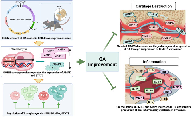
Methods: OA was induced in control C57BL/6 mice and in C57BL/6-derived SMILE-overexpressing transgenic (SMILE Tg) mice. The protein expression levels in the joint and spleen tissues were analyzed by immunohistochemistry and immunofluorescence images. In addition, flow cytometry was performed for detecting changes of the changes of immune cells.

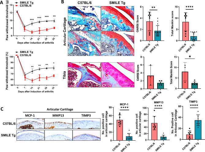
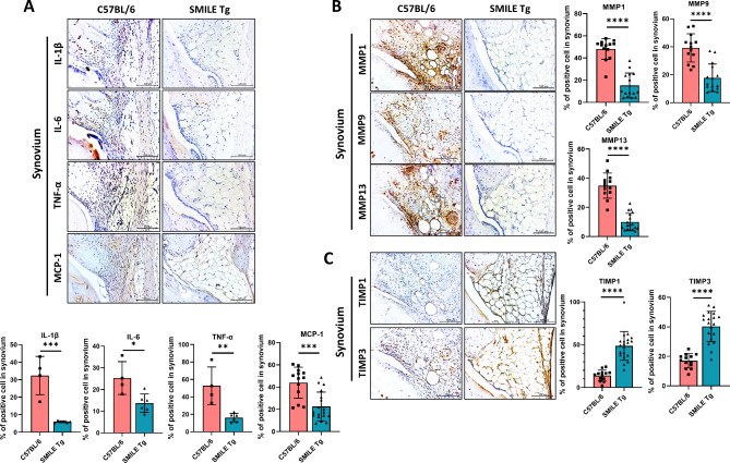
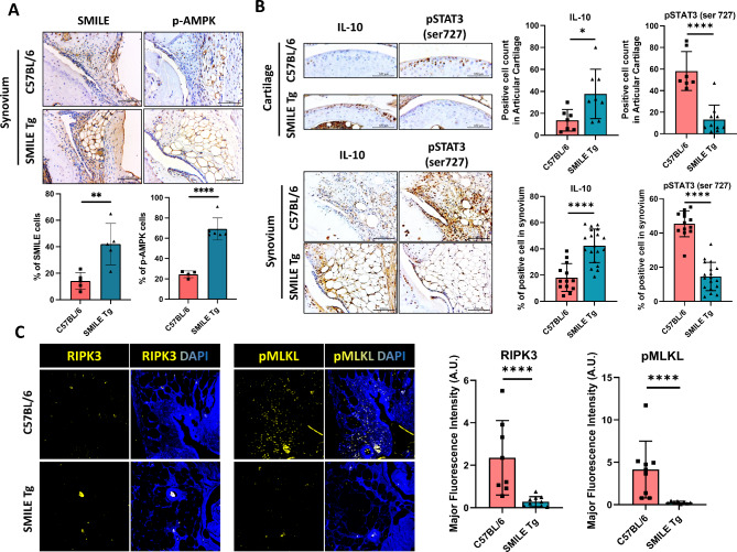
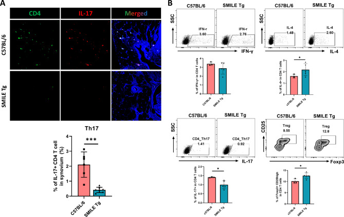
Results: Less cartilage damage and significantly reduced levels of OA biomarkers (MMP13, TIMP3 and MCP-1) were observed in SMILE Tg mice. Immunohistochemistry performed to identify the signaling pathway involved in the link between SMILE expression and OA revealed decreased levels of IL-1β, IL-6, TNF-α, and phosphorylated AMPK in synovial tissues as well as a significant decrease in phosphorylated STAT3 in both cartilage and synovium. Changes in systemic immune cells were investigated via flow cytometry to analyze splenocytes isolated from control and SMILE Tg mice. SMILE Tg mice had elevated proportions of CD4 + IL-4 + cells (Th2) and CD4 + CD25 + Foxp3 + cells (Treg) and a notable decrease in CD4 + IL-17 + cells (Th17).

Conclusion: Our results show that overexpressed SMILE attenuates the symptoms of OA, by increasing AMPK signaling and decreasing STAT3, thus reducing the levels of inflammatory immune cells.

Keywords: Cyclic AMP-response element binding protein Zhangfei (CREBZF); Helper T cell (th cell); Osteoarthritis (OA); Signal transducer and activator of transcription 3 (STAT3); Small heterodimer partner-interacting leucine zipper protein (SMILE).


Conflict of interest statement

Declarations Ethics approval and consent to participate The study protocol involving human participants was approved by the Institutional Review Board of Seoul St. Mary’s Hospital. All patients/participants provided written informed consent to participate in this study. The animal study protocol was approved by the Animal Research Ethics Committee of the Catholic University of Korea (IACUC approval numbers: 2020-0306-02 and 2021-0010-01). Consent for publication Not applicable. Competing interests The authors declare no competing interests.

Figures

Fig. 1
Fig. 1
Upregulation of SMILE inhibits the progression of OA and cartilage damage in MIA-induced OA in mice. (A) Joint pain was analyzed as the difference between the PWL (left) and PWT (right) in control C57BL/6 MIA-induced OA mice (C57BL/6) and SMILE overexpression vector injected transgenic mice (SMILE Tg). The mice were evaluated for 5 weeks. Nociceptive testing was performed using a dynamic plantar esthesiometer, which is an automated version of the von Frey hair assessment tool. The threshold for pain was shown as weight (g) and relative weight load (%) based on Day 0 as 100%. (B) Safranin O staining of the articular cartilage and tibia tissues of C57BL/6 mice and SMILE Tg. Bar graphs show the average OARSI and Mankin scores. (C) Immunohistochemistry was performed to assess the expression of MCP-1, MMP13 and TIMP3 in articular cartilage of C57BL/6 and SMILE Tg mice. The data are presented as the mean ± standard deviation. Scale bars = 100 μm (B and C). *p < 0.05, **p < 0.01, ***p < 0.001, and ****p < 0.0001
Fig. 2
Fig. 2
Levels of proinflammatory cytokines and OA-catabolic factors are decreased and those of OA-anabolic factors are increased in the synovium of SMILE Tg mice. Representative immunohistochemistry images of the synovium of control C57BL/6 and SMILE Tg mice showing the levels of (A) pro-inflammatory cytokines such as IL-1β, IL-6, TNF-α, and MCP-1, (B) OA-catabolic (MMP1, MMP9, and MMP13) and (C) OA-anabolic factors (TIMP1 and TIMP3). The data are presented as the mean ± standard deviation. Scale bars = 100 μm (A, B and C). *p < 0.05, **p < 0.01, ***p < 0.001, and ****p < 0.0001
Fig. 3
Fig. 3
AMPK and STAT3 signaling pathways and necroptosis are significantly altered by SMILE overexpression. (A) Immunohistochemistry images show AMPK expression in the synovial tissues of C57BL/6 and SMILE Tg mice. (B) The expressions of IL-10 and phosphorylated STAT3 (pSTAT3) in the synovial and cartilage tissues of C57BL/6 and SMILE Tg mice. (C) Immunofluorescence shows the cell nuclei (DAPI; blue) and the expressions of RIPK3 (yellow) and phosphorylated MLKL (pMLKL; yellow) in synovial tissues of C57BL/6 and SMILE Tg. The data are presented as the mean ± standard deviation. Scale bars = 100 μm (A and B). *p < 0.05 and ****p < 0.0001
Fig. 4
Fig. 4
Frequency of changes in systemic helper T (Th) cells in SMILE Tg mice. (A) Representative immunofluorescence images show the spleen tissues of C57BL/6 and SMILE Tg stained with anti-CD4 antibody (green) and anti-IL-17 antibody (red). Nuclei were stained by DAPI (blue). (B) The splenocytes of C57BL/6 and SMILE Tg were analyzed by flow cytometry. Th1 (CD4+ IFNγ+), Th2 (CD4+ IL-4+), Th17 (CD4+ IL-17+) and regulatory T cell (Treg; CD4+ CD25high Foxp3+) populations are shown. The data are presented as the mean ± standard deviation. *p < 0.05 and ***p < 0.001
Fig. 5
Fig. 5
Schematic diagram of therapeutic effect of SMILE on OA through the regulation of AMPK and cytokine production. SMILE overexpression leads to an increase in AMPK and a decrease in STAT3 in joint tissue, in addition to changing the proportion of immune cell subsets. SMILE overexpression also suppresses both the expression of OA-catabolic factors and inflammatory cytokine production

References

    1. Glyn-Jones S, Palmer AJ, Agricola R, Price AJ, Vincent TL, Weinans H, Carr AJ. Osteoarthritis. Lancet. 2015;386(9991):376–87. - PubMed
    1. Loeser RF, Goldring SR, Scanzello CR, Goldring MB. Osteoarthritis: a disease of the joint as an organ. Arthritis Rheum. 2012;64(6):1697–707. - PMC - PubMed
    1. Martel-Pelletier J, Barr AJ, Cicuttini FM, Conaghan PG, Cooper C, Goldring MB, Goldring SR, Jones G, Teichtahl AJ, Pelletier JP. Osteoarthritis. Nat Rev Dis Primers. 2016;2:16072. - PubMed
    1. Barnett R. Osteoarthr Lancet. 2018;391(10134):1985. - PubMed
    1. Loeser RF, Collins JA, Diekman BO. Ageing and the pathogenesis of osteoarthritis. Nat Rev Rheumatol. 2016;12(7):412–20. - PMC - PubMed

Substances