. 2024 Aug 23;9(11):3301-3317.
doi: 10.1016/j.ekir.2024.08.019. eCollection 2024 Nov.

TRPC6-Calpain-1 Axis Promotes Tubulointerstitial Inflammation by Inhibiting Mitophagy in Diabetic Kidney Disease

Affiliations

TRPC6-Calpain-1 Axis Promotes Tubulointerstitial Inflammation by Inhibiting Mitophagy in Diabetic Kidney Disease

Cong-Cong Liu et al. Kidney Int Rep. .

Abstract

Introduction: Renal tubulointerstitial inflammation represents an effective indicator for predicting the progression of diabetic kidney disease (DKD). Mitophagy abnormality is 1 of the most important factors involved in tubule injury. However, the exact molecular mechanism underlying mitophagy abnormality-mediated tubulointerstitial inflammation in DKD remains poorly understood.

Methods: In this study, a streptozotocin-induced DKD mouse model was established and HK-2 cells treated with high glucose (HG) served as an in vitro model. Tubular mitophagy was regulated through pharmacological urolithin A (UA) administration. The functional effect of the transient receptor potential cation channel, subfamily C, member 6 (TRPC6) was explored using genetic interventions in vivo and in vitro.

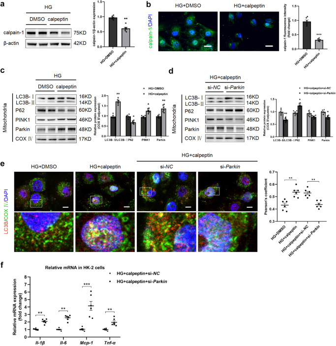
Results: We found that renal tubulointerstitial inflammation in DKD was closely associated with mitophagy inhibition, which was mediated by disturbance of PINK1/Parkin pathway. Mitophagy activation significantly attenuated tubular injury and tubulointerstitial inflammation. Further, it was found that TRPC6 was markedly increased in DKD and played an essential role in mitophagy inhibition by activating calpain-1. Knockdown of Trpc6 partially reversed mitophagy abnormality and consequently attenuated tubular injury and tubulointerstitial inflammation in vivo and in vitro. Finally, we found that tubular TRPC6-mediated mitophagy inhibition was blocked with BAPTA (a specific Ca2+ chelator) or calpeptin (a specific calpain-1 inhibitor).

Conclusion: Our study reveals that TRPC6-calpain-1 axis promotes tubulointerstitial inflammation in DKD by inhibiting mitophagy.

Keywords: TRPC6; calpain-1; diabetic kidney disease; mitophagy; tubulointerstitial inflammation.


Figures

None
Graphical abstract
Figure 1
Figure 1
Renal tubular injury and tubulointerstitial inflammation were observed in DKD. (a) The levels of fasting blood glucose (FBG). (b) Urinary ACR at week 8 and week 12 after successful construction of DKD mice. (c) Kidney weight-to-body weight ratio (KW/BW). (d) The blood urea nitrogen (BUN) levels. (e) Representative images of Periodic acid-Schiff (PAS) staining of kidney sections. Scale bars, 20 μm. (f) Representative western blotting images and densitometric analysis of KIM-1 in kidney. (g) Representative images of Masson’s trichrome staining and the quantitative analysis of fibrotic area. Scale bar, 20 μm. (h) Representative images of F4/80 immunohistochemical staining. Scale bars, 20 μm. (i) Real-time polymerase chain reaction analysis of the mRNA expression levels of Il-1β,Il-6, Mcp-1, and Tnf-α in kidney. (j) Real-time polymerase chain reaction analysis of the mRNA expression levels of Il-1β, Il-6, Mcp-1 and Tnf-α in HK-2 cells. All data are represented as means ± SEM. n = 6, P < 0.05, ∗∗P < 0.01, ∗∗∗P < 0.001. ACR, albumin-to-creatinine ratio; Ctrl, control; DKD, diabetic kidney disease; IL, interleukin; MCP-1, monocyte chemoattractant protein-1; TNF-α, tumor necrosis factor-α.
Figure 2
Figure 2
Mitophagy inhibition was associated with tubulointerstitial inflammation in DKD. (a) Representative transmission electron microscopy (TEM) images of intracellular mitophagosomes (arrows) in renal tubular epithelial cells from the 2 groups of mice. Scale bars, 400 nm. (b) Representative images of PINK1 immunohistochemical staining. Scale bars, 20 μm. (c) Representative images of Parkin immunohistochemical staining. Scale bars, 20 μm. (d) Representative western blotting images and densitometric analysis of LC3B, P62, PINK1, and Parkin in kidney. (e) Representative western blotting images and densitometric analysis of LC3B, P62, PINK1, and Parkin in HK-2 cells. (f) Representative confocal microscopic images and the Pearson correlation coefficient showing the colocalization of LC3B and COX Ⅳ in HK-2 cells. Nuclei were revealed using 4′,6-diamidino-2-phenylindole (DAPI) staining. Scale bars, 10 μm. All data are represented as means ± SEM. n = 6, P < 0.05, ∗∗P < 0.01, ∗∗∗P < 0.001. DKD, diabetic kidney disease.
Figure 3
Figure 3
Mitophagy activation attenuated renal tubular injury and tubulointerstitial inflammation in DKD. (a) Representative transmission electron microscopy (TEM) images of intracellular mitophagosomes (arrows) in renal tubular epithelial cells from the 2 groups of mice. Scale bars, 400 nm. (b) Representative images of PINK1 immunohistochemical staining. Scale bars, 20 μm. (c) Representative images of Parkin immunohistochemical staining. Scale bars, 20 μm. (d) Representative western blotting images and densitometric analysis of LC3B, P62, PINK1, and Parkin in kidney. (e) Representative western blotting images and densitometric analysis of LC3B, P62, PINK1, and Parkin in HK-2 cells. (f) Representative confocal microscopic images and the Pearson correlation coefficient showing the colocalization of LC3B and COX Ⅳ in HK-2 cells. Nuclei were revealed using DAPI staining. Scale bars, 10 μm. (g) The levels of fasting blood glucose (FBG). (h) Kidney weight-to-body weight ratio (KW/BW). (i) Urinary ACR. (j) The blood urea nitrogen (BUN) levels. (k) Representative images of Periodic acid-Schiff (PAS) staining of kidney sections. Scale bars, 20 μm. (l) Representative western blotting images and densitometric analysis of KIM-1 in kidney. (m) Representative images of Masson’s trichrome staining and the quantitative analysis of fibrotic area. Scale bar, 20 μm. (n) Representative images of F4/80 immunohistochemical staining. Scale bars, 20 μm. (o) Real-time polymerase chain reaction analysis of the mRNA expression levels of Il-1β, Il-6, Mcp-1, and Tnf-α in kidney. (p) Real-time polymerase chain reaction analysis of the mRNA expression levels of Il-1β, Il-6, Mcp-1, and Tnf-α in HK-2 cells. All data above are represented as means ± SEM. n = 6, P < 0.05, ∗∗P < 0.01, ∗∗∗P < 0.001. ACR, albumin-to-creatinine ratio; DAPI, 4′,6-diamidino-2-phenylindole; DKD, diabetic kidney disease; DMSO, dimethyl sulfoxide; HG, high glucose; MCP-1, monocyte chemoattractant protein-1; PAS, Periodic acid-Schiff staining; TNF-α, tumor necrosis factor-α; UA, urolithin A; Veh, vehicle.
Figure 3
Figure 3
Mitophagy activation attenuated renal tubular injury and tubulointerstitial inflammation in DKD. (a) Representative transmission electron microscopy (TEM) images of intracellular mitophagosomes (arrows) in renal tubular epithelial cells from the 2 groups of mice. Scale bars, 400 nm. (b) Representative images of PINK1 immunohistochemical staining. Scale bars, 20 μm. (c) Representative images of Parkin immunohistochemical staining. Scale bars, 20 μm. (d) Representative western blotting images and densitometric analysis of LC3B, P62, PINK1, and Parkin in kidney. (e) Representative western blotting images and densitometric analysis of LC3B, P62, PINK1, and Parkin in HK-2 cells. (f) Representative confocal microscopic images and the Pearson correlation coefficient showing the colocalization of LC3B and COX Ⅳ in HK-2 cells. Nuclei were revealed using DAPI staining. Scale bars, 10 μm. (g) The levels of fasting blood glucose (FBG). (h) Kidney weight-to-body weight ratio (KW/BW). (i) Urinary ACR. (j) The blood urea nitrogen (BUN) levels. (k) Representative images of Periodic acid-Schiff (PAS) staining of kidney sections. Scale bars, 20 μm. (l) Representative western blotting images and densitometric analysis of KIM-1 in kidney. (m) Representative images of Masson’s trichrome staining and the quantitative analysis of fibrotic area. Scale bar, 20 μm. (n) Representative images of F4/80 immunohistochemical staining. Scale bars, 20 μm. (o) Real-time polymerase chain reaction analysis of the mRNA expression levels of Il-1β, Il-6, Mcp-1, and Tnf-α in kidney. (p) Real-time polymerase chain reaction analysis of the mRNA expression levels of Il-1β, Il-6, Mcp-1, and Tnf-α in HK-2 cells. All data above are represented as means ± SEM. n = 6, P < 0.05, ∗∗P < 0.01, ∗∗∗P < 0.001. ACR, albumin-to-creatinine ratio; DAPI, 4′,6-diamidino-2-phenylindole; DKD, diabetic kidney disease; DMSO, dimethyl sulfoxide; HG, high glucose; MCP-1, monocyte chemoattractant protein-1; PAS, Periodic acid-Schiff staining; TNF-α, tumor necrosis factor-α; UA, urolithin A; Veh, vehicle.
Figure 4
Figure 4
TRPC6 inhibited mitophagy in DKD. (a) Representative western blotting images and densitometric analysis of TRPC6 in kidney. (b) Representative western blotting images and densitometric analysis TRPC6 and in HK-2 cells. (c) Real-time polymerase chain reaction analysis of the mRNA expression level of Trpc6 in kidney. (d) Representative western blotting images and densitometric analysis of TRPC6 in HK-2 cells. (e) Representative images of PINK1 immunohistochemical staining. Scale bars, 20 μm. (f) Representative images of Parkin immunohistochemical staining. Scale bars, 20 μm. (g) Representative western blotting images and densitometric analysis of LC3B, P62, PINK1, and Parkin in kidney. (h) Representative western blotting images and densitometric analysis of LC3B, P62, PINK1, and Parkin in HK-2 cells. (i) Representative western blotting images and densitometric analysis of LC3B, P62, PINK1, and Parkin in HK-2 cells. (j) Representative confocal microscopic images and the Pearson correlation coefficient showing the colocalization of LC3B and COX Ⅳ in HK-2 cells. Nuclei were revealed using DAPI staining. Scale bars, 10 μm. (k) Real-time polymerase chain reaction analysis of the mRNA expression levels of Il-1β, Il-6, Mcp-1, and Tnf-α in HK-2 cells. All data above are represented as means ± SEM. n = 6, P < 0.05, ∗∗P < 0.01, ∗∗∗P < 0.001. DAPI, 4′,6-diamidino-2-phenylindole; DKD, diabetic kidney disease; HG, high glucose; IL, interleukin; LV, lentivirus; MCP-1, monocyte chemoattractant protein-1; NC, negative control; TNF-α, tumor necrosis factor-α; TRPC6, transient receptor potential cation channel subfamily C member 6.
Figure 5
Figure 5
Trpc6 knockdown attenuated renal tubular injury and tubulointerstitial inflammation. (a) The levels of fasting blood glucose (FBG). (b) Kidney weight-to-body weight ratio (KW/BW). (c) Urinary ACR. (d) The blood urea nitrogen (BUN) levels. (e) Representative images of Periodic acid-Schiff (PAS) staining of kidney sections. Scale bars, 20 μm. (f) Representative western blotting images and densitometric analysis of KIM-1 in kidney. (g) Representative images of Masson’s trichrome staining and the quantitative analysis of fibrotic area. Scale bar, 20 μm. (h) Representative transmission electron microscopy (TEM) images of intracellular mitophagosomes (arrows) in renal tubular epithelial cells from the 2 groups of mice. Scale bars, 400 nm. (i) Representative images of F4/80 immunohistochemical staining. Scale bars, 20 μm. (j) Real-time polymerase chain reaction analysis of the mRNA expression levels of Il-1β, Il-6, Mcp-1, and Tnf-α in kidney. (k) Real-time polymerase chain reaction analysis of the mRNA expression levels of Il-1β, Il-6, Mcp-1 and Tnf-α in HK-2 cells. All data above are represented as means ± SEM. n = 6, P < 0.05, ∗∗P < 0.01, ∗∗∗P < 0.001. ACR, albumin-to-creatinine ratio. IL, interleukin; MCP-1, monocyte chemoattractant protein-1; TNF-α, tumor necrosis factor-α; Trpc6, transient receptor potential cation channel subfamily C member 6.
Figure 6
Figure 6
Calpain-1 activation was associated with TRPC6-mediated intracellular Ca2+ influx. (a) Representative western blotting images and densitometric analysis of calpain-1 in kidney. (b) Representative western blotting images and densitometric analysis of calpain-1 in HK-2 cells. (c) Representative western blotting images and densitometric analysis of calpain-1 in kidney. (d) Representative western blotting images and densitometric analysis of calpain-1 in HK-2 cells. (e) Representative western blotting images and densitometric analysis of calpain-1 in HK-2 cells. (f) Representative confocal microscopic images and quantitative analysis showing the expressions of Ca2+ and calpain-1 in HK-2 cells. Nuclei were revealed using DAPI staining. Scale bars, 20 μm. All data above are represented as means ± SEM. n = 6, P < 0.05, ∗∗P < 0.01, ∗∗∗P < 0.001. DAPI, 4′,6-diamidino-2-phenylindole; TRPC6, transient receptor potential cation channel subfamily C member 6.
Figure 7
Figure 7
Calpain-1 inhibition activated mitophagy. (a) Representative western blotting images and densitometric analysis of calpain-1 in HK-2 cells. (b) Representative confocal microscopic images and quantitative analysis showing the expression of calpain-1 in HK-2 cells. Nuclei were revealed using DAPI staining. Scale bars, 20 μm. (c) Representative western blotting images and densitometric analysis of LC3B, P62, PINK1, and Parkin in HK-2 cells. (d) Representative western blotting images and densitometric analysis of LC3B, P62, PINK1, and Parkin in HK-2 cells. (e) Representative confocal microscopic images and the Pearson correlation coefficient showing the colocalization of LC3B and COX Ⅳ in HK-2 cells. Nuclei were revealed using DAPI staining. Scale bars, 10 μm. (f) Real-time polymerase chain reaction analysis of the mRNA expression levels of Il-1β, Il-6, Mcp-1, and Tnf-α in HK-2 cells. All data above are represented as means ± SEM. n = 6, P < 0.05, ∗∗P < 0.01, ∗∗∗P < 0.001. DAPI, 4′,6-diamidino-2-phenylindole; IL, interleukin; MCP-1, monocyte chemoattractant protein-1; TNF-α, tumor necrosis factor-α.
Figure 8
Figure 8
Schematic illustration of the mechanism of upregulated TRPC6 affecting renal tubulointerstitial inflammation in DKD. In tubular epithelial cells under the condition of DKD, the increased inflammatory cytokines release is associated with increased TRPC6 expression. Increased TRPC6 activates calpain-1 by Ca2+ influx, leading to the inhibition of mitophagy. Damaged or dysfunctional mitochondria cannot be removed. Then, the release of increased inflammatory cytokines results in renal tubular injury. DKD, diabetic kidney disease; TRPC6, transient receptor potential cation channel subfamily C member 6.

Similar articles

Cited by

References

    1. Tuttle K.R., Agarwal R., Alpers C.E., et al. Molecular mechanisms and therapeutic targets for diabetic kidney disease. Kidney Int. 2022;102:248–260. doi: 10.1016/j.kint.2022.05.012. - DOI - PubMed
    1. Tang S.C.W., Yiu W.H. Innate immunity in diabetic kidney disease. Nat Rev Nephrol. 2020;16:206–222. doi: 10.1038/s41581-019-0234-4. - DOI - PubMed
    1. Jiang W.J., Xu C.T., Du C.L., et al. Tubular epithelial cell-to-macrophage communication forms a negative feedback loop via extracellular vesicle transfer to promote renal inflammation and apoptosis in diabetic nephropathy. Theranostics. 2022;12:324–339. doi: 10.7150/thno.63735. - DOI - PMC - PubMed
    1. Yao L., Liang X., Qiao Y., Chen B., Wang P., Liu Z. Mitochondrial dysfunction in diabetic tubulopathy. Metabolism. 2022;131 doi: 10.1016/j.metabol.2022.155195. - DOI - PubMed
    1. Wang S., Long H., Hou L., et al. The mitophagy pathway and its implications in human diseases. Signal Transduct Target Ther. 2023;8:304. doi: 10.1038/s41392-023-01503-7. - DOI - PMC - PubMed

LinkOut - more resources