

Image2SSM: Reimagining Statistical Shape Models from Images with Radial Basis Functions
- PMID: 39534229
- PMCID: PMC11555643
- DOI: 10.1007/978-3-031-43907-0_49
Image2SSM: Reimagining Statistical Shape Models from Images with Radial Basis Functions
Abstract
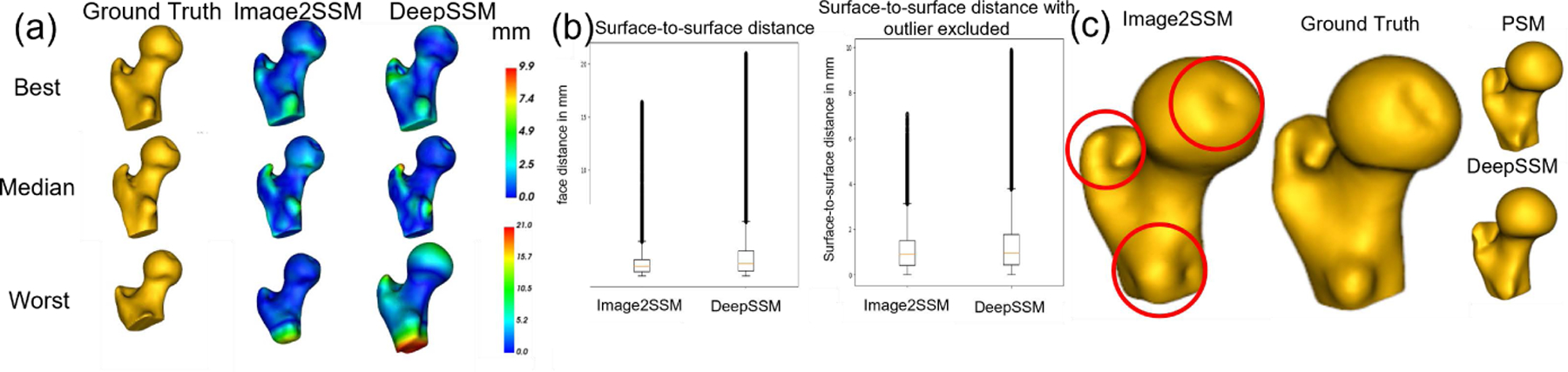
Statistical shape modeling (SSM) is an essential tool for analyzing variations in anatomical morphology. In a typical SSM pipeline, 3D anatomical images, gone through segmentation and rigid registration, are represented using lower-dimensional shape features, on which statistical analysis can be performed. Various methods for constructing compact shape representations have been proposed, but they involve laborious and costly steps. We propose Image2SSM, a novel deep-learning-based approach for SSM that leverages image-segmentation pairs to learn a radial-basis-function (RBF)-based representation of shapes directly from images. This RBF-based shape representation offers a rich self-supervised signal for the network to estimate a continuous, yet compact representation of the underlying surface that can adapt to complex geometries in a data-driven manner. Image2SSM can characterize populations of biological structures of interest by constructing statistical landmark-based shape models of ensembles of anatomical shapes while requiring minimal parameter tuning and no user assistance. Once trained, Image2SSM can be used to infer low-dimensional shape representations from new unsegmented images, paving the way toward scalable approaches for SSM, especially when dealing with large cohorts. Experiments on synthetic and real datasets show the efficacy of the proposed method compared to the state-of-art correspondence-based method for SSM.
Keywords: Deep Learning; Polyharmonic Splines; Radial Basis Function Interpolation; Statistical Shape Modeling.
Figures


Similar articles
-
DeepSSM: A blueprint for image-to-shape deep learning models.Med Image Anal. 2024 Jan;91:103034. doi: 10.1016/j.media.2023.103034. Epub 2023 Nov 17. Med Image Anal. 2024. PMID: 37984127 Free PMC article.
-
DeepSSM: A Deep Learning Framework for Statistical Shape Modeling from Raw Images.Shape Med Imaging (2018). 2018 Sep;11167:244-257. doi: 10.1007/978-3-030-04747-4_23. Epub 2018 Nov 23. Shape Med Imaging (2018). 2018. PMID: 30805572 Free PMC article.
-
Progressive DeepSSM: Training Methodology for Image-To-Shape Deep Models.Shape Med Imaging (2023). 2023 Oct;14350:157-172. doi: 10.1007/978-3-031-46914-5_13. Epub 2023 Oct 31. Shape Med Imaging (2023). 2023. PMID: 38745942 Free PMC article.
-
MASSM: An End-to-End Deep Learning Framework for Multi-Anatomy Statistical Shape Modeling Directly From Images.Shape Med Imaging (2024). 2025;15275:149-163. doi: 10.1007/978-3-031-75291-9_12. Epub 2024 Oct 26. Shape Med Imaging (2024). 2025. PMID: 39649703
-
Statistical shape models for 3D medical image segmentation: a review.Med Image Anal. 2009 Aug;13(4):543-63. doi: 10.1016/j.media.2009.05.004. Epub 2009 May 27. Med Image Anal. 2009. PMID: 19525140 Review.
References
-
- Adams J, Elhabian S: From images to probabilistic anatomical shapes: A deep variational bottleneck approach. In: Medical Image Computing and Computer Assisted Intervention–MICCAI 2022: 25th International Conference, Singapore, September 18–22, 2022, Proceedings, Part II. pp. 474–484. Springer; (2022) - PMC - PubMed
-
- Atkins PR, Elhabian SY, Agrawal P, Harris MD, Whitaker RT, Weiss JA, Peters CL, Anderson AE: Quantitative comparison of cortical bone thickness using correspondence-based shape modeling in patients with cam femoroacetabular impingement. Journal of Orthopaedic Research 35(8), 1743–1753 (2017) - PMC - PubMed
Grants and funding
LinkOut - more resources
Full Text Sources
Other Literature Sources