. 2024 Jul 2:21:140-167.
doi: 10.1016/j.xjon.2024.06.013. eCollection 2024 Oct.

Using machine learning to predict neurologic injury in venovenous extracorporeal membrane oxygenation recipients: An ELSO Registry analysis

Collaborators, Affiliations

Using machine learning to predict neurologic injury in venovenous extracorporeal membrane oxygenation recipients: An ELSO Registry analysis

Andrew Kalra et al. JTCVS Open. .

Abstract

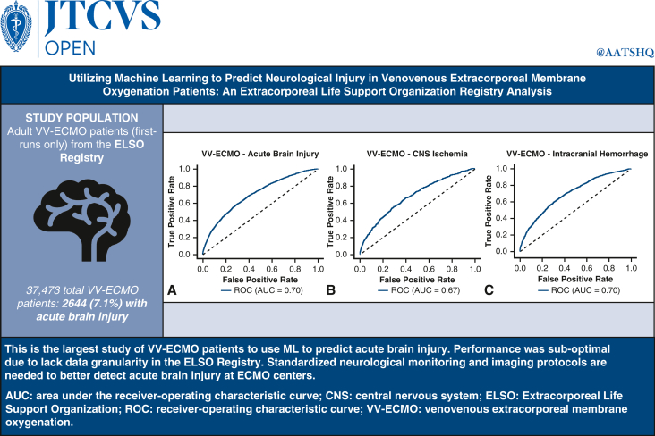
Background: Venovenous extracorporeal membrane oxygenation (VV-ECMO) is associated with acute brain injury (ABI), including central nervous system (CNS) ischemia (defined as ischemic stroke or hypoxic-ischemic brain injury [HIBI]) and intracranial hemorrhage (ICH). Data on prediction models for neurologic outcomes in VV-ECMO are limited.

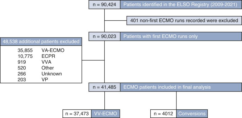
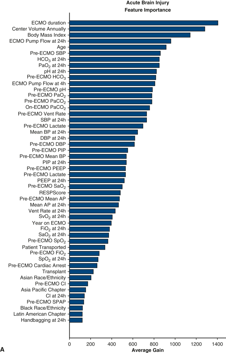
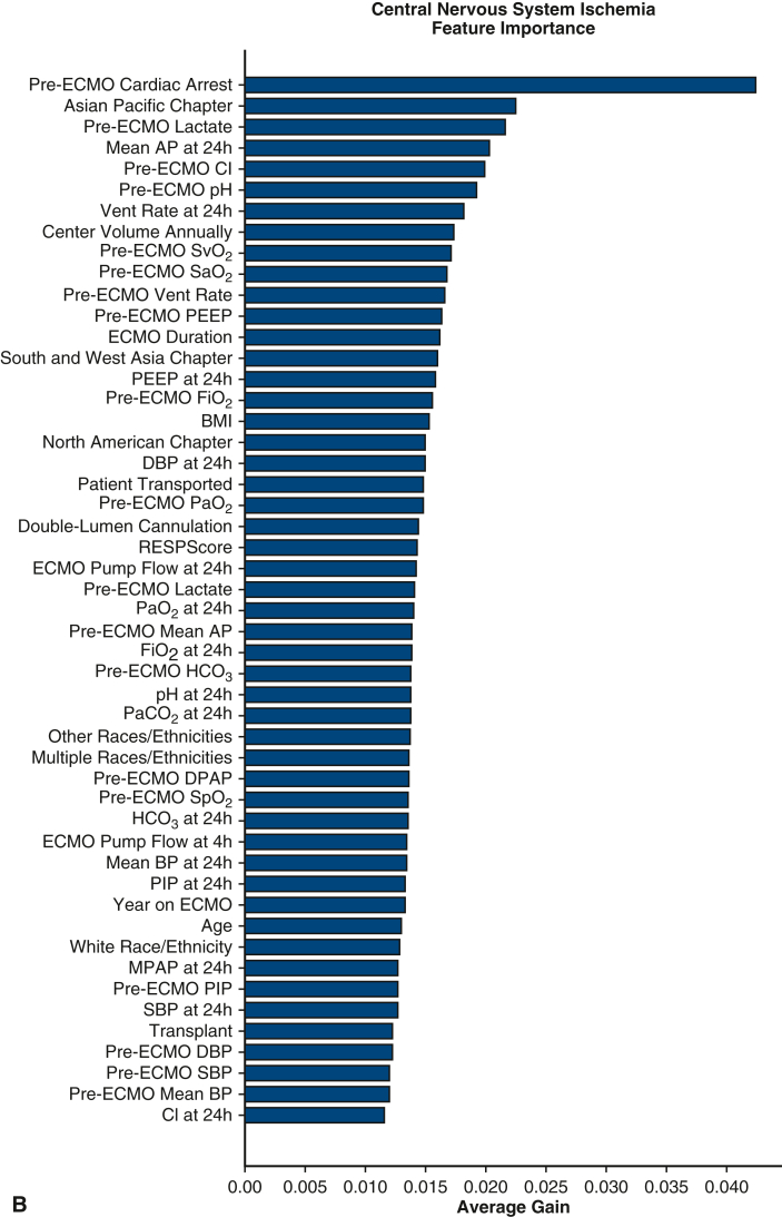
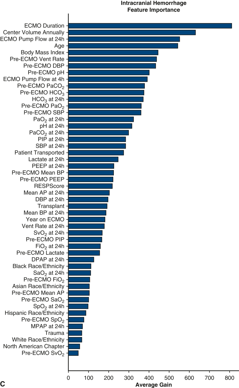
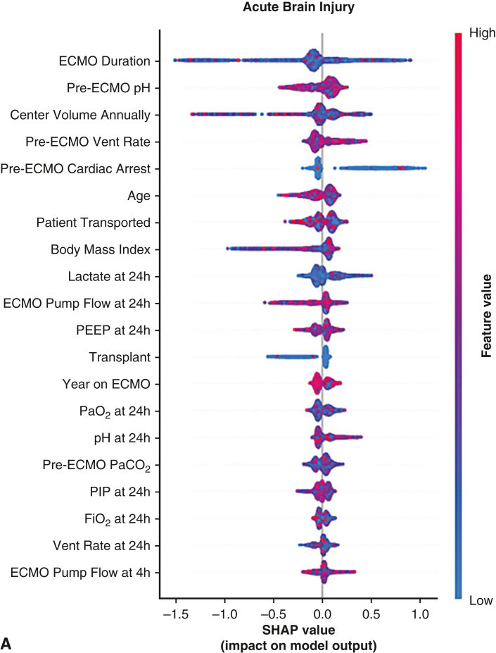
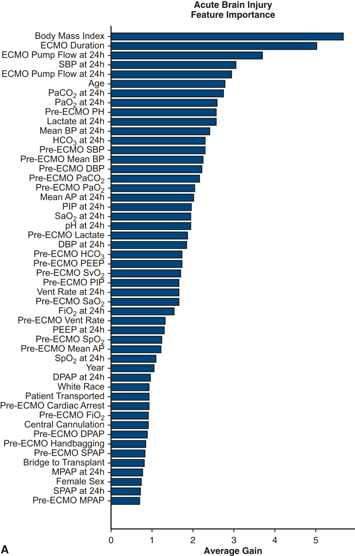
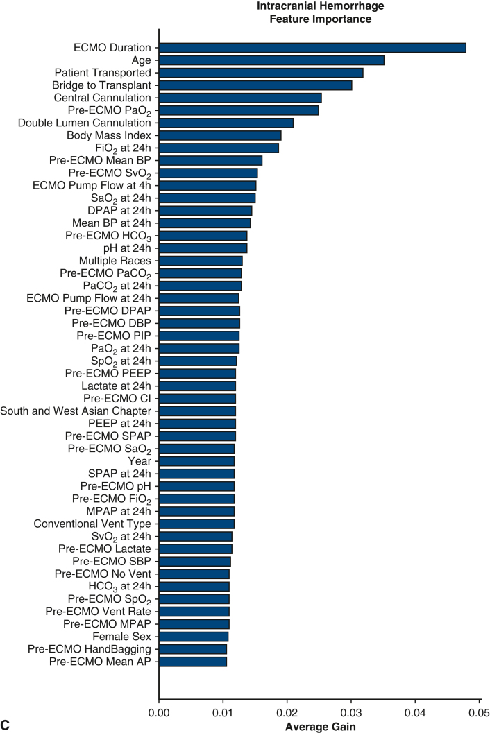
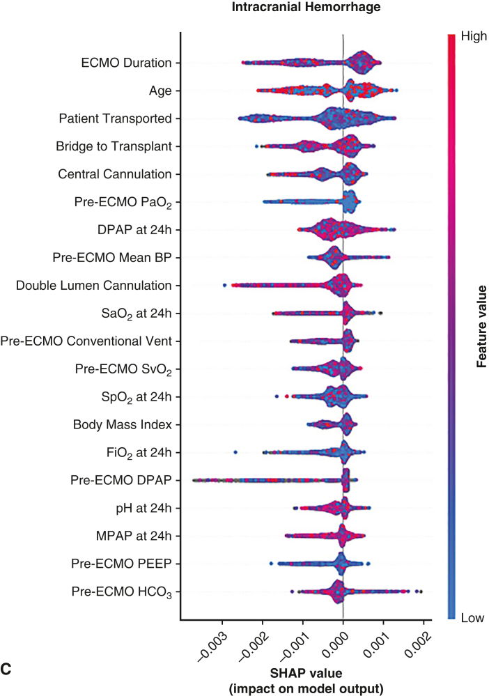
Methods: We analyzed adult (age ≥18 years) VV-ECMO patients in the Extracorporeal Life Support Organization (ELSO) Registry (2009-2021) from 676 centers. ABI was defined as CNS ischemia, ICH, brain death, and seizures. Data on 67 variables were extracted, including clinical characteristics and pre-ECMO/on-ECMO variables. Random forest, CatBoost, LightGBM, and XGBoost machine learning (ML) algorithms (10-fold leave-one-out cross-validation) were used to predict ABI. Feature importance scores were used to pinpoint the most important variables for predicting ABI.

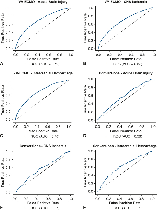
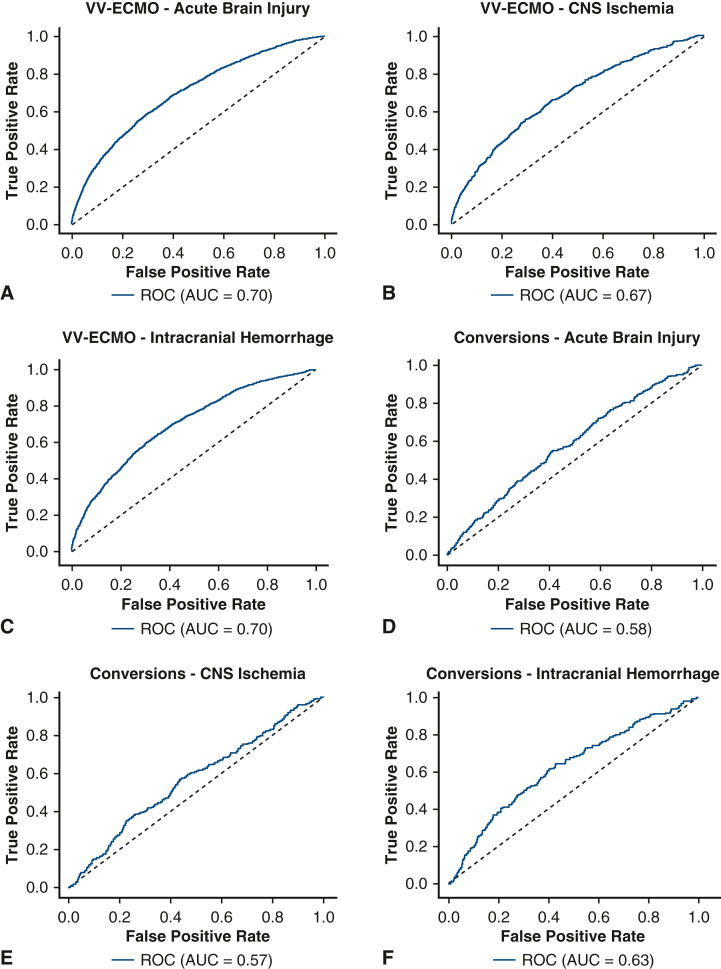
Results: Of 37,473 VV-ECMO patients (median age, 48.1 years; 63% male), 2644 (7.1%) experienced ABI, including 610 (2%) with CNS ischemia and 1591 (4%) with ICH. The areas under the receiver operating characteristic curve for predicting ABI, CNS ischemia, and ICH were 0.70, 0.68, and 0.70, respectively. The accuracy, positive predictive value, and negative predictive value for ABI were 85%, 19%, and 95%, respectively. ML identified higher center volume, pre-ECMO cardiac arrest, higher ECMO pump flow, and elevated on-ECMO serum lactate level as the most important risk factors for ABI and its subtypes.

Conclusions: This is the largest study of VV-ECMO patients to use ML to predict ABI reported to date. Performance was suboptimal, likely due to lack of standardization of neuromonitoring/imaging protocols and data granularity in the ELSO Registry. Standardized neurologic monitoring and imaging are needed across ELSO centers to detect the true prevalence of ABI.

Keywords: acute brain injury; machine learning; neurologic complications; venovenous extracorporeal membrane oxygenation.


Conflict of interest statement

Dr Brodie receives research support from and consults for LivaNova. He has been on the medical advisory boards for Xenios, Medtronic, Inspira and Cellenkos. He is the President-elect of ELSO and the Chair of the Executive Committee of the International ECMO Network (ECMONet), and he writes for UpToDate. Dr Ventetuolo has been a consultant or served on advisory boards for Merck, Janssen, and Regeneron outside of the submitted work. Sung-Min Cho is supported by the National Heart, Lung and Blood Institute (1K23HL157610) and Hyperfine (SAFE MRI ECMO study). All other authors reported no conflicts of interest. The Journal policy requires editors and reviewers to disclose conflicts of interest and to decline handling or reviewing manuscripts for which they may have a conflict of interest. The editors and reviewers of this article have no conflicts of interest.

Figures

None
Graphical abstract
None
Machine learning performance to predict ABI in VV-ECMO patients using the ELSO Registry.
Figure 1
Figure 1
Variables used in our machine learning pipeline to predict ABI, ranging from demographics to on-ECMO laboratory values. ECMO, Extracorporeal membrane oxygenation; BP, blood pressure; PaCO2, partial pressure of carbon dioxide; PaO2, partial pressure of oxygen; PCWP, pulmonary capillary wedge pressure; DPAP, diastolic pulmonary arterial pressure; SaO2, arterial blood gas oxygen saturation; SpO2, peripheral oxygen saturation; SvO2, mixed venous oxygen saturation; MPAP, mean pulmonary arterial pressure; FiO2, fraction of inspired oxygen; PEEP, positive-end expiratory pressure; PIP, peak inspiratory pressure; EEG, electroencephalogram.
Figure 2
Figure 2
Flow diagram for creation of the cohort study. ECMO, Extracorporeal membrane oxygenation; VA, venoarterial; VV, venovenous; Conversions, VA→VV or VV→VA; ECPR, extracorporeal cardiopulmonary resuscitation; VVA, venovenoarterial; Other, mode not defined; VP, venopulmonary.
Figure 3
Figure 3
Receiver-operating characteristic curves (ROC) for predicting area under the receiver-operating characteristic curves (AUC) (A) acute brain injury, (B) central nervous system (CNS) ischemia, and (C) intracranial hemorrhage in venovenous extracorporeal membrane oxygenation (VV-ECMO) and for predicting (D) acute brain injury, (E) CNS ischemia, and (F) intracranial hemorrhage in “Conversion” patients.
Figure 4
Figure 4
Most important features for each neurological outcome: (A) acute brain injury, (B) central nervous system ischemia, and (C) intracranial hemorrhage in VV-ECMO patients. ECMO, Extracorporeal membrane oxygenation; SBP, systolic blood pressure; PaO2, partial pressure of oxygen; PaCO2, partial pressure of carbon dioxide; BP, blood pressure; DBP, diastolic blood pressure; PIP, peak inspiratory pressure; PEEP, positive-end expiratory pressure; SaO2, arterial blood gas oxygen saturation; SpO2, peripheral oxygen saturation; CI, cardiac index; SPAP, systolic pulmonary arterial pressure; AP, arterial pressure; Vent, ventilator; BMI, body mass index; DPAP, diastolic pulmonary arterial pressure.
Figure 4
Figure 4
Most important features for each neurological outcome: (A) acute brain injury, (B) central nervous system ischemia, and (C) intracranial hemorrhage in VV-ECMO patients. ECMO, Extracorporeal membrane oxygenation; SBP, systolic blood pressure; PaO2, partial pressure of oxygen; PaCO2, partial pressure of carbon dioxide; BP, blood pressure; DBP, diastolic blood pressure; PIP, peak inspiratory pressure; PEEP, positive-end expiratory pressure; SaO2, arterial blood gas oxygen saturation; SpO2, peripheral oxygen saturation; CI, cardiac index; SPAP, systolic pulmonary arterial pressure; AP, arterial pressure; Vent, ventilator; BMI, body mass index; DPAP, diastolic pulmonary arterial pressure.
Figure 5
Figure 5
Summary of our study's findings demonstrating the performance of machine learning to predict acute brain injury and its subtypes in patients receiving venovenous extracorporeal membrane oxygenation. Overall, the performance of the models was sub-optimal (area under the receiver-operating characteristic curve of 0.70, 0.67, and 0.70 for acute brain injury, central nervous system ischemia, and intracranial hemorrhage, respectively). Standardized neurological monitoring and imaging protocols are recommended to accurately diagnose acute brain injury across all Extracorporeal Life Support Organization centers.
Figure E1
Figure E1
Shapley additive explanations (SHAP) value plots for acute brain injury (A), central nervous system ischemia (B), and intracranial hemorrhage in venovenous extracorporeal membrane oxygenation patients (C). ECMO, Extracorporeal membrane oxygenation; PEEP, positive-end expiratory pressure; PaO2, partial pressure of oxygen; PIP, peak inspiratory pressure; FiO2, fraction of inspired oxygen; AP, Arterial pressure; SaO2, arterial blood gas oxygen saturation; DBP, diastolic blood pressure; Vent, ventilator.
Figure E1
Figure E1
Shapley additive explanations (SHAP) value plots for acute brain injury (A), central nervous system ischemia (B), and intracranial hemorrhage in venovenous extracorporeal membrane oxygenation patients (C). ECMO, Extracorporeal membrane oxygenation; PEEP, positive-end expiratory pressure; PaO2, partial pressure of oxygen; PIP, peak inspiratory pressure; FiO2, fraction of inspired oxygen; AP, Arterial pressure; SaO2, arterial blood gas oxygen saturation; DBP, diastolic blood pressure; Vent, ventilator.
Figure E2
Figure E2
The most important features of each neurologic outcome. A, Acute brain injury. B, Central nervous system ischemia. C, Intracranial hemorrhage in conversion patients. ECMO, Extracorporeal membrane oxygenation; PaCO2, partial pressure of carbon dioxide; PaO2, partial pressure of oxygen; BP, blood pressure; AP, Arterial pressure; PIP, peak inspiratory pressure; SaO2, arterial blood gas oxygen saturation; PEEP, positive-end expiratory pressure; SvO2, mixed venous oxygen saturation; Vent, ventilator; DPAP, diastolic pulmonary arterial pressure; FiO2, fraction of inspired oxygen; SpO2, peripheral oxygen saturation; SPAP, systolic pulmonary arterial pressure; MPAP, mean pulmonary arterial pressure.
Figure E2
Figure E2
The most important features of each neurologic outcome. A, Acute brain injury. B, Central nervous system ischemia. C, Intracranial hemorrhage in conversion patients. ECMO, Extracorporeal membrane oxygenation; PaCO2, partial pressure of carbon dioxide; PaO2, partial pressure of oxygen; BP, blood pressure; AP, Arterial pressure; PIP, peak inspiratory pressure; SaO2, arterial blood gas oxygen saturation; PEEP, positive-end expiratory pressure; SvO2, mixed venous oxygen saturation; Vent, ventilator; DPAP, diastolic pulmonary arterial pressure; FiO2, fraction of inspired oxygen; SpO2, peripheral oxygen saturation; SPAP, systolic pulmonary arterial pressure; MPAP, mean pulmonary arterial pressure.
Figure E3
Figure E3
Shapley additive explanations (SHAP) value plots for acute brain injury (A), central nervous system ischemia (B), and intracranial hemorrhage (C) in conversion patients. ECMO, Extracorporeal membrane oxygenation; PaO2, partial pressure of oxygen; BP, blood pressure; SaO2, arterial blood gas oxygen saturation; PaCO2, partial pressure of carbon dioxide; SvO2, mixed venous oxygen saturation; Vent, ventilator; DPAP, diastolic pulmonary arterial pressure; FiO2, fraction of inspired oxygen; MPAP, mean pulmonary arterial pressure.
Figure E3
Figure E3
Shapley additive explanations (SHAP) value plots for acute brain injury (A), central nervous system ischemia (B), and intracranial hemorrhage (C) in conversion patients. ECMO, Extracorporeal membrane oxygenation; PaO2, partial pressure of oxygen; BP, blood pressure; SaO2, arterial blood gas oxygen saturation; PaCO2, partial pressure of carbon dioxide; SvO2, mixed venous oxygen saturation; Vent, ventilator; DPAP, diastolic pulmonary arterial pressure; FiO2, fraction of inspired oxygen; MPAP, mean pulmonary arterial pressure.

References

    1. Thiagarajan R.R., Barbaro R.P., Rycus P.T., et al. Extracorporeal life support organization registry international report 2016. ASAIO J. 2017;63(1):60–67. doi: 10.1097/mat.0000000000000475. - DOI - PubMed
    1. Kalra A., Kang J.K., Khanduja S., et al. Long-term neuropsychiatric, neurocognitive, and functional outcomes of patients receiving ECMO: a systematic review and meta-analysis. Neurology. 2024;102 doi: 10.1212/wnl.0000000000208081. - DOI - PMC - PubMed
    1. Cho S.M., Canner J., Caturegli G., et al. Risk factors of ischemic and hemorrhagic strokes during venovenous extracorporeal membrane oxygenation: analysis of data from the extracorporeal life support organization registry. Crit Care Med. 2021;49(1):91–101. doi: 10.1097/ccm.0000000000004707. - DOI - PMC - PubMed
    1. Yoon J.H., Pinsky M.R., Clermont G. Artificial intelligence in critical care medicine. Crit Care. 2022;26(1):75. doi: 10.1186/s13054-022-03915-3. - DOI - PMC - PubMed
    1. Mamdani M., Slutsky A.S. Artificial intelligence in intensive care medicine. Intensive Care Med. 2021;47(2):147–149. doi: 10.1007/s00134-020-06203-2. - DOI - PubMed

LinkOut - more resources