Review
. 2024 Nov 12;14(48):35769-35970.
doi: 10.1039/d4ra06290f. eCollection 2024 Nov 4.

Exploring heterocyclic scaffolds in carbonic anhydrase inhibition: a decade of structural and therapeutic insights

Affiliations
Review

Exploring heterocyclic scaffolds in carbonic anhydrase inhibition: a decade of structural and therapeutic insights

Nafeesa Naeem et al. RSC Adv. .

Abstract

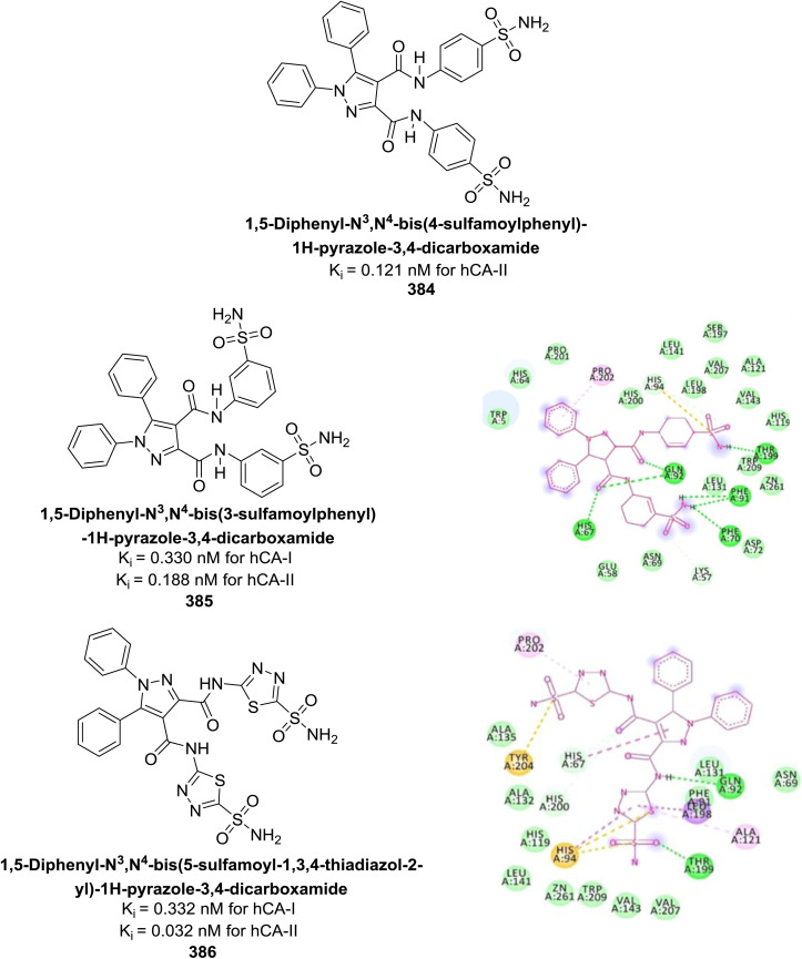
Heterocyclic compounds represent a prominent class of molecules with diverse pharmacological activities. Among their therapeutic applications, they have gained significant attention as carbonic anhydrase (CA) inhibitors, owing to their potential in the treatment of various diseases such as epilepsy, cancer and glaucoma. CA is a widely distributed zinc metalloenzyme that facilitates the reversible interconversion of carbon dioxide and bicarbonate. This reaction is essential for numerous physiological and pathological processes. In humans, CA exists in sixteen different isoforms, labeled hCA-I to hCA-XV, each distributed across various tissues and organs and involved in crucial physiological functions. Clinically utilized CA inhibitors, such as brinzolamide, dorzolamide and acetazolamide, exhibit poor selectivity, leading to undesirable side effects. A significant challenge in designing effective CA inhibitors is achieving balanced isoform selectivity, prompting the exploration of new chemotypes. This review compiles recent strategies employed by various researchers in developing CAIs across different structural classes, including pyrazoline, quinoline, imidazole, oxadiazole, pyrimidine, coumarin, chalcone, rhodanine, phthalazine, triazole, isatin, and indole. Additionally, the review summarizes structure-activity relationship (SAR) analyses, isoform selectivity evaluations, along with mechanistic and in silico investigations. Insights derived from SAR studies provide crucial directions for the rational design of next-generation heterocyclic CA inhibitors, with improved therapeutic efficacy and reduced side effects. To the best of our knowledge, for the first time, we have comprehensively summarized all known isoforms of CA in relation to various heterocyclic motifs. This review examines the use of different heterocycles as CA inhibitors, drawing on research published over the past 11 years. It offers a valuable resource for early-career researchers, encouraging further exploration of synthetic heterocycles in the development of CA inhibitors.


Conflict of interest statement

Authors have no conflict of interests to declare.

Figures

Fig. 1
Fig. 1. The role of CA in erythrocytes involves facilitating the hydration of CO2 in tissues and the dehydration of HCO3 in erythrocytes within the lungs.
Fig. 2
Fig. 2. Various isoforms of human carbonic anhydrase.
Fig. 3
Fig. 3. Catalytic mechanism of hCAs entails the reversible hydration and dehydration reactions (A–D) between CO2 with HCO3.
Fig. 4
Fig. 4. Therapeutic applications of CAIs.
Fig. 5
Fig. 5. Diuretic therapy with CAIs.
Fig. 6
Fig. 6. Role of CA-VA, VB and CA-II in the de novo lipogenesis.
Fig. 7
Fig. 7. Schematic representation illustrating the role of hCA-IX in regulating pH in hypoxic tumor cells.
Fig. 8
Fig. 8. Chemical structure of compound 11 and its hCA-I and II inhibition.
Fig. 9
Fig. 9. Chemical structures of compounds 12 and 13 and their IC50 and Ki values against hCA-I and II.
Fig. 10
Fig. 10. Chemical structures of compounds 14 and 15 and their IC50 values against hCA-II and IX.
Fig. 11
Fig. 11. Chemical structures of compounds 16 and 17 and their IC50 and Ki values against hCA-I and II.
Fig. 12
Fig. 12. Chemical structures of compounds 18 and 19 and their IC50 values against hCA-I and II.
Fig. 13
Fig. 13. Chemical structures of compounds 20 and 21 and their Ki values against hCA-I and II.
Fig. 14
Fig. 14. Chemical structure and Ki value of compound 22 and its docking image inside the active pocket of CA-IX enzyme.
Fig. 15
Fig. 15. Chemical structures of compounds 23, 24 and 25 and their IC50 values against hCA-I and II.
Fig. 16
Fig. 16. Chemical structures of compounds 26 and 27 and their IC50 values against hCA-IX and docking image of compound 26 against CA-IX (PDB ID: 3IAI).
Fig. 17
Fig. 17. Chemical structure of compound 28 and its Ki values against hCA-IX and XII.
Fig. 18
Fig. 18. Chemical structure of compound 45 and its Ki values against hCA-I, II, IX, XII.
Fig. 19
Fig. 19. Chemical structure of compounds 48 and 49 and their Ki values against hCA isoforms.
Fig. 20
Fig. 20. Chemical structure of compounds 50 and 51 and their Kd values against hCA isoforms.
Fig. 21
Fig. 21. Chemical structure of compounds 58 and 59 and their Kd values against hCA-I and II.
Fig. 22
Fig. 22. Chemical structure of compounds 60–67 and their Ki values against hCA-IX and XII.
Fig. 23
Fig. 23. Chemical structure of compounds 68 and 69 and their IC50 values against hCA-I and II.
Fig. 24
Fig. 24. Chemical structure of compounds 70 and 71 and their Ki values against hCA-XI and XII.
Fig. 25
Fig. 25. Chemical structure of compounds 72 and 73 and their IC50 values against hCA-I and II.
Fig. 26
Fig. 26. Chemical structure of compound 74 and its IC50 value against bCA-II along with its docking image.
Fig. 27
Fig. 27. Chemical structure of compounds 75 and 76 and their Ki values against hCA-I and II.
Fig. 28
Fig. 28. Chemical structure of compounds 77 and 78 and their Ki values against hCA-IX and XII.
Fig. 29
Fig. 29. Chemical structure of compound 79 its Ki values against hCA-IX and XII.
Fig. 30
Fig. 30. Chemical structure of compound 80 and its IC50 value against hCA-I.
Fig. 31
Fig. 31. Chemical structures of compounds 81 and 82 and their IC50 values against hCA-I and II.
Fig. 32
Fig. 32. Chemical structure of compounds 83 and 84 and their Ki values against hCA-I and II.
Fig. 33
Fig. 33. Chemical structure of compounds 85 and 86 and their IC50 values against bCA-II.
Fig. 34
Fig. 34. Chemical structure of compounds 87, 88 and 89 and their Ki values against hCA-I, II, IX and XII.
Fig. 35
Fig. 35. Chemical structure of compounds 90 and 91 and their Ki values against hCA-I, II, IX and XII.
Fig. 36
Fig. 36. Chemical structure of compound 92 and their Ki values against hCA-II and IX.
Fig. 37
Fig. 37. Chemical structure of compound 103 and its IC50 value against hCA-II along with its docking image.
Fig. 38
Fig. 38. Chemical structure of compounds 104–109 and their Ki values against hCA-IX.
Fig. 39
Fig. 39. Chemical structure of compounds 110–113 and their Ki values against hCA-XII.
Fig. 40
Fig. 40. Chemical structure of compound 114 and its IC50 values against hCA-I and II.
Fig. 41
Fig. 41. Chemical structure of compounds 123–126 and their IC50 values against bCA-II.
Fig. 42
Fig. 42. Chemical structure of compound 127 and its Ki value against hCA-XII along with its docking image.
Fig. 43
Fig. 43. Chemical structures of compounds 128–130 and their Ki values against hCA-I, II and VII.
Fig. 44
Fig. 44. Chemical structure of compound 131 and its Ki value against hCA-II.
Fig. 45
Fig. 45. Chemical structures of compounds 132 and 133, and their Ki value against hCA-I and II.
Fig. 46
Fig. 46. Chemical structures of compounds 134 and 135, and their Ki value against hCA-I and II.
Fig. 47
Fig. 47. Chemical structures of compounds 136 and 137, and their Ki value against hCA-I and II.
Fig. 48
Fig. 48. Chemical structure of compound 138 and its Ki value against hCA-I and II.
Fig. 49
Fig. 49. Chemical structures of compounds 139 and 140 and their Ki value against hCA-I and IX.
Fig. 50
Fig. 50. Chemical structures of compounds 147–152 and their Ki value against hCA-XII.
Fig. 51
Fig. 51. Chemical structure of compound 153 and its Ki value against bCA-II along with its docking image.
Fig. 52
Fig. 52. Chemical structures of compounds 154–157 and their IC50 value against bCA-II along with docking images of 154 and 157.
Fig. 53
Fig. 53. Chemical structures of compounds 158 and 159 and their Ki values against hCA-I and II.
Fig. 54
Fig. 54. Chemical structures of compounds 160 and 161 and their Ki values against hCA-IX.
Fig. 55
Fig. 55. Chemical structures of compounds 162, 163 and 164 and their Ki values against hCA-IX.
Fig. 56
Fig. 56. Chemical structures of compounds 165 and 166 and their Ki values against hCA-II.
Fig. 57
Fig. 57. Chemical structures of compounds 167 and 168 and their Ki values against hCA-I & II.
Fig. 58
Fig. 58. Chemical structures of compounds 169 and 170 and their Ki values against hCA-I & II.
Fig. 59
Fig. 59. Chemical structures of compounds 171 and 172 and their Ki values against hCA-I & II.
Fig. 60
Fig. 60. Chemical structure of compound 173 and its Ki value against hCA-IX along with its docking images in hydrolyzed and non-hydrolyzed forms.
Fig. 61
Fig. 61. Chemical structures of compounds 174, 175 and 176 and their Ki values against hCA-I & II.
Fig. 62
Fig. 62. Chemical structure of compound 177 and its Ki values against hCA-I & II.
Fig. 63
Fig. 63. Chemical structures of compounds 178 and 179 and their Ki values against hCA-I & II.
Fig. 64
Fig. 64. Chemical structures of compounds 180, 181 and 182 and their Ki values against hCA-I & bCA-II along with docking image of compound 180.
Fig. 65
Fig. 65. Chemical structures of compounds 183 and 184 and their Ki values against hCA-IX along with their docking images.
Fig. 66
Fig. 66. Chemical structures of compounds 185 and 186 and their Ki values against hCA-I and IX.
Fig. 67
Fig. 67. Chemical structure of compound 187 and its Ki value against hCA-IV.
Fig. 68
Fig. 68. Chemical structures of compounds 188, 189 and 190 and their Ki values against hCA-IX.
Fig. 69
Fig. 69. Chemical structure of compound 191 and its Ki values against hCA-I and II.
Fig. 70
Fig. 70. Chemical structure of compound 195 and its Ki value against bCA-II along with docking image of compound 195.
Fig. 71
Fig. 71. Chemical structures of compounds 196 and 197; their Ki values against hCA-I and II along with docking images of compounds 196 and 197.
Fig. 72
Fig. 72. Chemical structure of compound 205; its IC50 value against bCA-II along with docking image of compound 205.
Fig. 73
Fig. 73. Chemical structures of compounds 206 and 207; their Ki values against hCA-I and II along with docking images of compound 206 and 207.
Fig. 74
Fig. 74. Chemical structure of compound 208; its Ki value against hCA-IX and XII along with docking images of compound 208.
Fig. 75
Fig. 75. Chemical structures of compounds 209 and 210; their Ki values against hCA-IX and XII along with their docking images.
Fig. 76
Fig. 76. Chemical structure of compound 211; its Ki value against hCA-IX along with docking image of compound 211.
Fig. 77
Fig. 77. Chemical structures of compounds 212, 213 and 214; their Ki values against hCA-I and II along with docking image of compound 214.
Fig. 78
Fig. 78. Chemical structures of compounds 215, 216 and 217; their Ki values against hCA-IX and XII along with docking image of compound 217.
Fig. 79
Fig. 79. Chemical structure of compound 218; its Ki values against hCA-I and II along with docking images of compound 218.
Fig. 80
Fig. 80. Chemical structures of compounds 219, 220 and 221; their Ki values against hCA-IX along with docking image of compound 220.
Fig. 81
Fig. 81. Chemical structures of compounds 222 and 223; their Ki values against hCA-I and II along with their docking images.
Fig. 82
Fig. 82. Chemical structures of compounds 224–230; their Ki values against hCA-I and II along with docking images of compounds 224, 225 and 226.
Fig. 83
Fig. 83. Chemical structures of compounds 231–234; their Ki values against hCA-IX and XII along with docking images of compounds 233 and 234.
Fig. 84
Fig. 84. Chemical structures of compounds 235–237; their IC50 values against bCA-II and hCA-II along with docking image of compound 235.
Fig. 85
Fig. 85. Chemical structures of compounds 238 and 239; their IC50 values against hCA-II along with docking image of compound 238.
Fig. 86
Fig. 86. Chemical structures of compounds 240 and 241; their Ki values against hCA-XII along with docking image of compound 240.
Fig. 87
Fig. 87. Chemical structures of compounds 242–245; their Ki values against hCA-IX and XII along with docking images of compounds 242 and 243.
Fig. 88
Fig. 88. Chemical structure of compound 246; its Ki values against hCA-II and IX along with docking images of compound 246.
Fig. 89
Fig. 89. Chemical structures of compounds 247–250; their Ki values against hCA-II.
Fig. 90
Fig. 90. Chemical structures of compounds 251–254; their Ki values against hCA-IX and XII along with docking images of compounds 251–254.
Fig. 91
Fig. 91. Chemical structures of compounds 255–258; their Ki values against hCA-I & XIII.
Fig. 92
Fig. 92. Chemical structure of compound 259; its Ki values against hCA-VII.
Fig. 93
Fig. 93. Chemical structure of compound 260; its Ki values against bCA-II and its docking image.
Fig. 94
Fig. 94. Chemical structures of compounds 261 and 262; their Ki values against hCA-IX along with docking images.
Fig. 95
Fig. 95. Chemical structures of compounds 263–266; their Ki values against hCA-II.
Fig. 96
Fig. 96. Chemical structures of compounds 267–269; their IC50 values against hCA-IX along with docking images.
Fig. 97
Fig. 97. Chemical structures of compounds 270–273; their IC50 values against bCA-II along with docking images.
Fig. 98
Fig. 98. Chemical structures of compounds 274 and 275; their Ki values against hCA-II, XI and XII.
Fig. 99
Fig. 99. Chemical structure of compound 276; its IC50 value against bCA-II and its docking image.
Fig. 100
Fig. 100. Chemical structure of compound 277; its Ki value against hCA-IX along with docking image.
Fig. 101
Fig. 101. Chemical structures of compounds 278–280; their Ki values against hCA-IX and XII along with docking images.
Fig. 102
Fig. 102. Chemical structures of compounds 286–290; their Ki values against hCA-IX along with docking images of 286 and 290.
Fig. 103
Fig. 103. Chemical structures of compounds 291 and 292; their Ki values against hCA-I.
Fig. 104
Fig. 104. Chemical structure of compound 293; its Ki values against hCA-I, II, IX and XII along with its docking images.
Fig. 105
Fig. 105. Chemical structure of compound 294; its Ki values against hCA-IX and XII along with its docking image against hCA-XII.
Fig. 106
Fig. 106. Chemical structures of compounds 295 and 296; their Ki values against hCA-II, IX and XII along with docking images.
Fig. 107
Fig. 107. Chemical structure of compound 297; its Ki values against hCA-I, II, IV, VII, IX, and XII.
Fig. 108
Fig. 108. Chemical structures of compounds 298–303; their Ki values against hCA-I, II, IX and XII along with docking images of 302 and 303.
Fig. 109
Fig. 109. Chemical structures of compounds 304 and 305; their Ki values against hCA-I, II, IX and XII along with their docking images (predicted binding modes of compounds 304 (orange) and 305 (green)).
Fig. 110
Fig. 110. Chemical structures of compounds 306–309; their Ki values against hCA-I, IX and XII along with their docking images.
Fig. 111
Fig. 111. Chemical structure of compound 310; its Ki values against hCA-IX and XII along with its docking images.
Fig. 112
Fig. 112. Chemical structures of compounds 311 and 312; their Ki values against hCA-I and II.
Fig. 113
Fig. 113. Chemical structures of compounds 313 and 314; their Ki values against hCA-I and II along with their docking images.
Fig. 114
Fig. 114. Chemical structures of compounds 315 and 316; their Ki values against hCA-IX and XII along with their docking images.
Fig. 115
Fig. 115. Chemical structures of compounds 317–322; their Ki values against hCA-IX and XII along with docking images of 318 and 319.
Fig. 116
Fig. 116. Chemical structures of compounds 323–326; their Ki values against hCA-I and II.
Fig. 117
Fig. 117. Chemical structures of compounds 327 and 328; their Ki values against hCA-I and II along with their docking images.
Fig. 118
Fig. 118. Chemical structures of compounds 329–332; their IC50 values against hCA-I and II along with their docking images.
Fig. 119
Fig. 119. Chemical structures of compounds 333 and 334; their Ki values against hCA-IX and XII along with docking image of 333.
Fig. 120
Fig. 120. Chemical structures of compounds 335–337; their IC50 values against hCA-I and II.
Fig. 121
Fig. 121. Chemical structures of compounds 338–342; their Ki values against hCA-I, II and IV; docking image of compound 339.
Fig. 122
Fig. 122. Chemical structure of compound 343; its Ki values against hCA-IX and XII.
Fig. 123
Fig. 123. Chemical structures of compounds 344–347; their IC50 values against hCA-IX and XII along with docking images of 344 and 347.
Fig. 124
Fig. 124. Chemical structures of compounds 348–352; their IC50 values against hCA-II, IX and XII along with their docking images.
Fig. 125
Fig. 125. Chemical structure of compound 353; its IC50 values against hCA-I and II along with its docking images.
Fig. 126
Fig. 126. Chemical structure of compound 354; its Ki values against hCA-I and II along with its docking images.
Fig. 127
Fig. 127. Chemical structures of compounds 355 and 356; their Ki values against hCA-I and II along with their docking images.
Fig. 128
Fig. 128. Chemical structure of compound 357; its Ki value against hCA-XII.
Fig. 129
Fig. 129. Chemical structures of compounds 358–362; their Ki values against hCA-XI and XII along with docking images of 360 and 362.
Fig. 130
Fig. 130. Chemical structures of compounds 363–366; their Ki values against hCA-I, II, XI and XII along with their docking images.
Fig. 131
Fig. 131. Chemical structures of compounds 367 and 368; their Ki values against hCA-I and IX along with their docking images.
Fig. 132
Fig. 132. Chemical structures of compounds 369 and 370; their Ki values against mtCA-2 along with their docking images.
Fig. 133
Fig. 133. Chemical structures of compounds 371 and 372; their Ki values against hCA-IX and XIII.
Fig. 134
Fig. 134. Chemical structures of compounds 373–379; their Ki values against hCA-I, IX and XII.
Fig. 135
Fig. 135. Chemical structures of compounds 380 and 381; their Ki values against hCA-I and II along with their docking images.
Fig. 136
Fig. 136. Chemical structures of compounds 382 and 383; their Ki values against hCA-IX and XII along with their docking images.
Fig. 137
Fig. 137. Chemical structures of compounds 384–386; their Ki values against hCA-I and II along with docking images of 385 and 386.
Fig. 138
Fig. 138. Chemical structures of compounds 387–391; their Ki values against hCA-II and IX.
Fig. 139
Fig. 139. Chemical structures of compounds 392 and 393; their Ki values against hCA-I and II along with their docking images.
Fig. 140
Fig. 140. Chemical structures of compounds 394 and 395; their Ki values against hCA-II and IX along with their docking images.
Fig. 141
Fig. 141. Chemical structures of compounds 396 and 397; their Ki values against hCA-IX and XII.
Fig. 142
Fig. 142. Chemical structures of compounds 398–400; their IC50 values against hCA-IX and docking image of 398.
Fig. 143
Fig. 143. Chemical structure of compound 401; its IC50 values against hCA-I and II, and its docking images.
Fig. 144
Fig. 144. SAR of pyrazoline-based CAIs.
Fig. 145
Fig. 145. SAR of triazole and thiazole-based CAIs.
Fig. 146
Fig. 146. SAR of pyrimidine-based CAIs.
Fig. 147
Fig. 147. SAR of phthalazine-based CAIs.
Fig. 148
Fig. 148. SAR of imidazole and oxadiazole-based CAIs.
Fig. 149
Fig. 149. SAR of indole and isatin-based CAIs.
Fig. 150
Fig. 150. SAR of chalcone and coumarin-based CAIs.
Fig. 151
Fig. 151. SAR of rhodanine-based CAIs.
Fig. 152
Fig. 152. SAR of quinoline-based CAIs.
Fig. 153
Fig. 153. SAR of sulfonamide-based CAIs.
Fig. 154
Fig. 154. SAR of different heterocyclic scaffolds against hCA isoforms based on docking study.
None
Ehsan Ullah Mughal

Similar articles

Cited by

References

    1. Qadir T. Amin A. Sharma P. K. Jeelani I. Abe H. Open Med. Chem. J. 2022;16:1–34.
    1. Kabir E. Uzzaman M. Results Chem. 2022;4:100606. - PMC - PubMed
    1. Jassas R. S. Naeem N. Sadiq A. Mehmood R. Alenazi N. A. Al-Rooqi M. M. Mughal E. U. Alsantali R. I. Ahmed S. A. RSC Adv. 2023;13:16413–16452. - PMC - PubMed
    1. Obaid R. J. Naeem N. Mughal E. U. Al-Rooqi M. M. Sadiq A. Jassas R. S. Moussa Z. Ahmed S. A. RSC Adv. 2022;12:19764–19855. - PMC - PubMed
    1. Kalaria P. N. Karad S. C. Raval D. K. Eur. J. Med. Chem. 2018;158:917–936. - PubMed

LinkOut - more resources