. 2024 Dec 5;68(12):e0049624.
doi: 10.1128/aac.00496-24. Epub 2024 Nov 13.

LiaR-dependent gene expression contributes to antimicrobial responses in group A Streptococcus

Affiliations

LiaR-dependent gene expression contributes to antimicrobial responses in group A Streptococcus

Luis Alberto Vega et al. Antimicrob Agents Chemother. .

Abstract

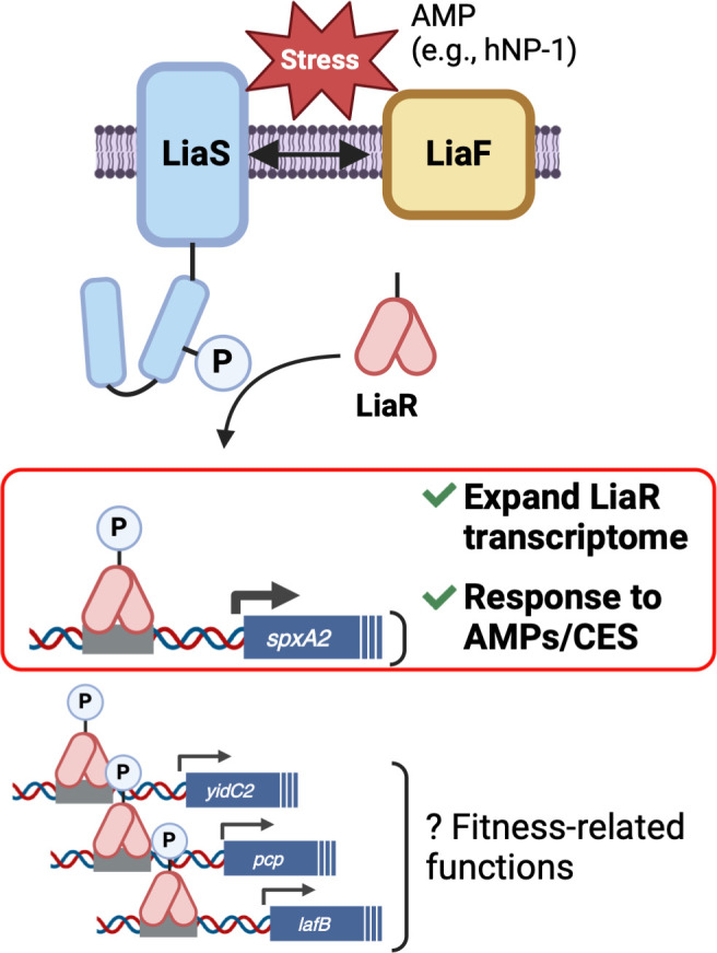
The ability to sense and respond to host defenses is essential for pathogen survival. Some mechanisms involve two-component systems (TCSs) that respond to host molecules, such as antimicrobial peptides (AMPs), and activate specific gene regulatory pathways to aid in survival. Alongside TCSs, bacteria coordinate cell division proteins, chaperones, cell wall sortases, and secretory translocons at discrete locations within the cytoplasmic membrane, referred to as functional membrane microdomains (FMMs). In group A Streptococcus (GAS), the FMM or "ExPortal" coordinates protein secretion, cell wall synthesis, and sensing of AMP-mediated cell envelope stress via the LiaFSR three-component system. Previously, we showed that GAS exposure to a subset of AMPs (α-defensins) activates the LiaFSR system by disrupting LiaF and LiaS co-localization in the ExPortal, leading to increased LiaR phosphorylation, expression of the transcriptional regulator SpxA2, and altered GAS virulence gene expression. The mechanisms by which LiaFSR integrates cell envelope stress with responses to AMP activity and virulence are not fully elucidated. Here, we show the LiaFSR regulon is comprised of genes encoding SpxA2 and three membrane-associated proteins: a PspC domain-containing protein (PCP), the lipoteichoic acid-modifying protein LafB, and the membrane protein insertase YidC2. Our data support that phosphorylated LiaR induces transcription of these genes via a conserved operator, whose disruption attenuates GAS virulence and increases susceptibility to AMPs in a manner primarily dependent on differential expression of SpxA2. Our work expands our understanding of the LiaFSR regulatory network in GAS and identifies targets for further investigation of mechanisms of cell envelope stress tolerance contributing to GAS pathogenesis.

Keywords: LiaFSR; Spx; antimicrobial peptides; gene regulation; global regulatory networks; group A streptococcus; two-component regulatory systems.


Conflict of interest statement

The authors declare no conflict of interest.

Figures

Fig 1
Fig 1
Opposing patterns of LiaR phosphorylation result in differential transcription of spxA2 and correlate with ∆liaF and ∆liaR transcriptomes. (A) Mutagenesis of amino acid residues of the LiaS kinase involved in phosphorylation/dephosphorylation of LiaR protein (Q146, D142, and R135) results in altered levels of phosphorylated LiaR in the presence/absence of exposure to bacitracin (BAC). Homology of the H-box harboring the invariant histidine residue (site of histidine phosphorylation) across LiaS proteins from GAS (MGAS10870 LiaS), Streptococcus agalactiae (Sagalac LiaS), Streptococcus mutans (Smutans LiaS), Streptococcus pneumoniae (Spneumo LiaS), Streptococcus thermophilus (Sthermo LiaS), and Bacillus subtilis DesK is shown, highlighting identical (black), similar (gray), and distinct (white) residues. Substitutions for Ala residues (Q146A, D142A, blue box) and the previously isolated R135G mutation (red box) associated with altered LiaFSR activity are shown. (B) Measurement of spxA2 transcript levels by quantitative real-time PCR (qRT-PCR) in WT MGAS10870 (emm3), Q146A, and D142A isogenic LiaS mutants exposed to BAC confirms spxA2 transcript levels correlate with LiaR phosphorylation. (C) Correlation plot of 41 significantly (P  <  0.05; Bonferroni correction) differentially expressed genes (≥1.5-fold relative to WT MGAS10870) shared between the Q146A (x-axis) and the ∆liaF (y-axis) transcriptome. (D) Correlation plot of 86 significantly (P  <  0.05, Bonferroni correction) differentially expressed genes (≥1.5-fold relative to WT MGAS10870) shared between the emergent R135G (x-axis) and the ΔliaR (y-axis) transcriptome. Log2 values are plotted, and colors correspond to operons or individual genes whose differential expression was inversely correlated in ∆liaF and ∆liaR transcriptomes (∆liaF/R DGE, previously published in reference [35]). Names of virulence genes of interest are listed. Bacitracin exposure = 1 µg/mL, 1 h, mid-log phase, 37°C. Transcripts assessed by qRT-PCR analysis were measured in technical triplicate of biological quadruplicates, with significance determined by a t-test (*P  <  0.05; **P  <  0.01). Data are presented as the mean transcript level with standard error of the mean (SEM). Transcriptomes were analyzed by RNA-seq in biological triplicate and DGE was determined for the core genome only, excluding phage and other mobile elements.
Fig 2
Fig 2
The regulon directly controlled by LiaR consists of spxA2, yidC2, lafB, and pcp transcripts. (A) Identified peaks and read mapping results of ChIP-seq analysis of the Q146A mutant relative to ∆liaR (negative control) reveal four transcripts directly regulated by phosphorylated LiaR. Quantitative real-time PCR (qRT-PCR) measurement of identified targets directly regulated by phosphorylated LiaR (spxA2, SpyM3_1799; pcp, SpyM3_0411; lafB, SpyM3_0363; yidC2, SpyM3_0256) in the bacitracin-exposed (1 µg/mL, 1 h, mid-log phase culture, 37°C) (B) wild-type (emm3) MGAS10870 strain, LiaR phosphorylation-lacking LiaR_D56A (D56A), and (C) LiaR operator scramble mutants (p*spxA2, p*pcp, p*lafB, p*yidC2). Transcripts were measured in technical triplicate of biological quadruplicates, with significance determined by t-test (*P  <  0.05). Data are presented as the mean transcript level with standard error of the mean (SEM) relative to untreated cultures (B) or untreated WT MGAS10870 (emm3) strain (C).
Fig 3
Fig 3
Disruption of LiaR phosphorylation and LiaR regulon control alter the GAS response to AMPs. Ratio of surviving virtual colony counts (CFUV) to inoculum CFU counts (Input) of GAS suspensions (2–5 × 10⁵ CFU total) in Na-phosphate buffer (10 mM, pH 7.4) incubated for 90 minutes in (A) bacitracin, (B) polymyxin B, and (C) hNP-1 indicates increased susceptibility to AMP bactericidal activity in strains lacking LiaR phosphorylation. The status of LiaR phosphorylation in tested strains is indicated in the figure legends ([LiaR ~ P (−)] reduced LiaR phosphorylation, [LiaR ~ P (++)]: constitutive LiaR phosphorylation). Data are presented as the mean value with standard error of the mean (SEM). Significance determined by a t-test (*P  <  0.05).
Fig 4
Fig 4
Disruption of LiaR phosphorylation and LiaR regulon control negatively affect GAS virulence. (A) Survival in whole human blood (3 hours, 37˚C) of strains in which LiaR phosphorylation is reduced [LiaR ~P(−)], LiaR is constitutively phosphorylated [LiaR ~P(++)], the operator of LiaR regulon targets is mutagenized (Q146A/p*spxA2, p*spxA2WT, p*pcp, p*lafB, p*yidC2) or LiaR target is disrupted (∆spxA2). The mean multiplication factor of GAS in whole human blood and SEM is shown, calculated from biological replicates in blood from three donors, performed in quadruplicate. Significance was determined by a t-test (*P  <  0.05). (B) Kaplan-Meier survival of mice (n  =  15 per strain) infected intramuscularly with 2 × 10⁷ CFU D142A, Q146A, or MGAS10870 (WT). P-values were determined by log-rank.
Fig 5
Fig 5
Model of LiaR activation and downstream gene regulation. LiaS (histidine kinase) and LiaF (accessory protein and inhibitor of LiaS) reside in a membrane microdomain (ExPortal). Cell envelope stress (CES) induced by AMPs leads to LiaS-LiaF dissociation and phosphorylation of LiaS. LiaS subsequently phosphorylates the response regulator LiaR leading to binding at conserved operator sequences (gray boxes) in the promoters of four genes (spxA2, yidC2, pcp, and lafB). Increased transcription of spxA2—encoding a protein known to interact with RNAP and affect transcription—is predicted to expand the LiaR transcriptome beyond the four direct targets of LiaR. Expansion of the LiaR transcriptome through SpxA2 contributes to the response to AMP-induced CES and is predicted to contribute to GAS pathogenesis (boxed in red) (created with BioRender.com).

Update of

References

    1. Cole JN, Nizet V. 2016. Bacterial evasion of host antimicrobial peptide defenses. Microbiol Spectr 4. doi:10.1128/microbiolspec.VMBF-0006-2015 - DOI - PMC - PubMed
    1. Melo MN, Ferre R, Castanho MARB. 2009. Antimicrobial peptides: linking partition, activity and high membrane-bound concentrations. Nat Rev Microbiol 7:245–250. doi:10.1038/nrmicro2095 - DOI - PubMed
    1. Pachón-Ibáñez ME, Smani Y, Pachón J, Sánchez-Céspedes J. 2017. Perspectives for clinical use of engineered human host defense antimicrobial peptides. FEMS Microbiol Rev 41:323–342. doi:10.1093/femsre/fux012 - DOI - PMC - PubMed
    1. Assoni L, Milani B, Carvalho MR, Nepomuceno LN, Waz NT, Guerra MES, Converso TR, Darrieux M. 2020. Resistance mechanisms to antimicrobial peptides in Gram-positive bacteria. Front Microbiol 11:593215. doi:10.3389/fmicb.2020.593215 - DOI - PMC - PubMed
    1. Stock AM, Robinson VL, Goudreau PN. 2000. Two-component signal transduction. Annu Rev Biochem 69:183–215. doi:10.1146/annurev.biochem.69.1.183 - DOI - PubMed

MeSH terms

LinkOut - more resources