

A Method for Rapid Inducible RNA Decay
- PMID: 39535712
- PMCID: PMC11874224
- DOI: 10.1007/978-1-0716-4176-7_13
A Method for Rapid Inducible RNA Decay
Abstract
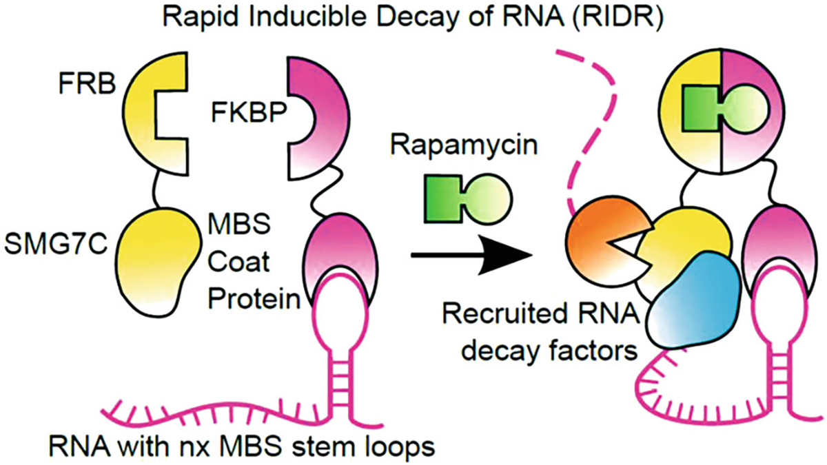
Modulating RNA decay is a powerful tool to investigate RNA degradation dynamics. Here, we describe a protocol to inducibly recruit protein factors to regulate target RNA metabolism, called Rapid Inducible Decay of RNA (RIDR). RIDR induces fast and synchronous decay of target mRNAs within minutes and enables direct visualization of mRNA decay dynamics and subcellular kinetics in living cells. Here, we provide detailed procedures to make stable cell lines, conduct fixed- and live-cell measurements, and perform data analysis. We discuss the potential pitfalls and make RIDR applicable to a general biology lab.
Keywords: Fluorescent in situ hybridization; Immunofluorescence; Kinetics; RNA decay; Single molecule imaging.
© 2025. The Author(s), under exclusive license to Springer Science+Business Media, LLC, part of Springer Nature.
Figures


Similar articles
-
A rapid inducible RNA decay system reveals fast mRNA decay in P-bodies.Nat Commun. 2024 Mar 28;15(1):2720. doi: 10.1038/s41467-024-46943-z. Nat Commun. 2024. PMID: 38548718 Free PMC article.
-
A Rapid Inducible RNA Decay system reveals fast mRNA decay in P-bodies.bioRxiv [Preprint]. 2023 Apr 26:2023.04.26.538452. doi: 10.1101/2023.04.26.538452. bioRxiv. 2023. Update in: Nat Commun. 2024 Mar 28;15(1):2720. doi: 10.1038/s41467-024-46943-z. PMID: 37162943 Free PMC article. Updated. Preprint.
-
Single-cell analysis of RNase L-mediated mRNA decay.Methods Enzymol. 2023;692:157-175. doi: 10.1016/bs.mie.2023.04.016. Epub 2023 May 14. Methods Enzymol. 2023. PMID: 37925178
-
RNA voyeurism: A coming of age story.Methods. 2016 Apr 1;98:10-17. doi: 10.1016/j.ymeth.2015.11.024. Epub 2015 Nov 27. Methods. 2016. PMID: 26638774 Review.
-
Mechanisms of deadenylation-dependent decay.Wiley Interdiscip Rev RNA. 2011 Mar-Apr;2(2):167-83. doi: 10.1002/wrna.40. Epub 2010 Sep 15. Wiley Interdiscip Rev RNA. 2011. PMID: 21957004 Free PMC article. Review.
References
MeSH terms
Substances
Grants and funding
LinkOut - more resources
Full Text Sources
Miscellaneous
