. 2024 Nov 15;10(46):eadq8773.
doi: 10.1126/sciadv.adq8773. Epub 2024 Nov 13.

Channel width modulates the permeability of DNA origami-based nuclear pore mimics

Affiliations

Channel width modulates the permeability of DNA origami-based nuclear pore mimics

Qingzhou Feng et al. Sci Adv. .

Abstract

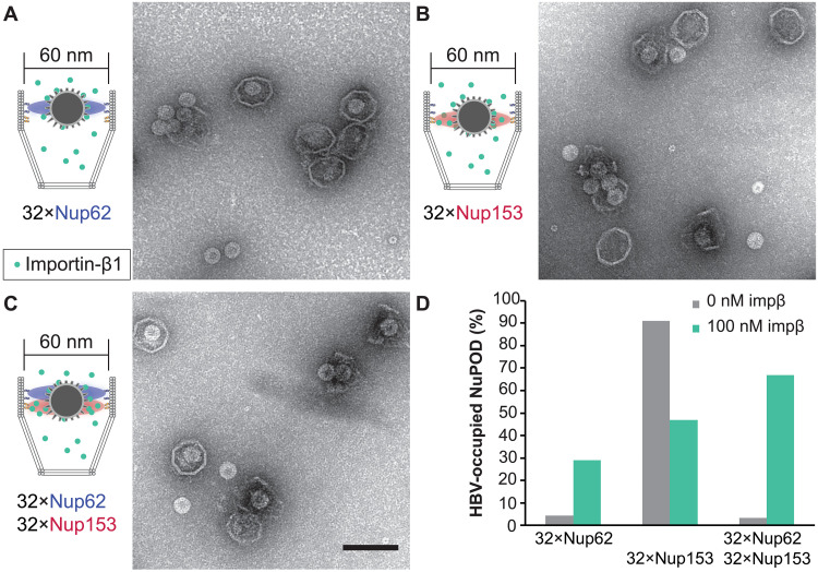
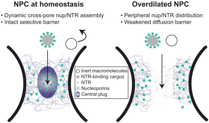
Nucleoporins (nups) in the nuclear pore complex (NPC) form a selective barrier that suppresses the diffusion of most macromolecules while enabling rapid transport of nuclear transport receptor (NTR)-bound cargos. Recent studies have shown that the NPC may dilate and constrict, but how altering the NPC diameter affects its selective barrier properties remains unclear. Here, we build DNA nanopores with programmable diameters and nup arrangements to model the constricted and dilated NPCs. We find that Nup62 proteins form a dynamic cross-channel barrier impermeable to hepatitis B virus (HBV) capsids when grafted inside 60-nm-wide nanopores but not in 79-nm pores, where Nup62 cluster locally. Furthermore, importin-β1 substantially changes the dynamics of Nup62 assemblies and facilitates the passage of HBV capsids through the 60-nm NPC mimics containing Nup62 and Nup153. Our study shows that transport channel width is critical to the permeability of nup barriers and underscores NTRs' role in dynamically remodeling nup assemblies and mediating the nuclear entry of viruses.


Figures

Fig. 1.
Fig. 1.. DNA origami nanostructures used to build NuPODs.
(A) A 60-nm-wide channel built by homodimerizing DNA nanopores with shape-complementary docking interfaces (blue and red). (B) A 79-nm-wide channel built by homodimerizing DNA nanopores with shape-complementary interfaces. (C) Small DNA basket with a docking interface (blue) compatible with the 60-nm channel. (D) Large DNA basket with a top docking interface (blue) compatible with the 79-nm channel and a bottom interface (red) compatible with the small basket. (E) A 60-nm channel capped by the small basket on one end. (F) A 79-nm channel capped by the two baskets on one end. For each panel, a cartoon model (DNA double helices are represented by rods) is shown next to representative TEM images. The experiment was repeated three times (technical replicates) with similar folding results. Scale bars, 100 nm.
Fig. 2.
Fig. 2.. Morphology of Nup62 inside DNA origami nanopores of different widths.
(A) Schematic diagrams showing the process of attaching nups inside DNA nanopores to form NuPODs. h, hours. (B) Empty 60-nm DNA channel. (C) Empty 79-nm DNA channel. (D) A 60-nm NuPOD with 32 copies of Nup62 grafted on 21-nt handles. (E) A 79-nm NuPOD with 32 copies of Nup62 grafted on 21-nt handles. (F) A 79-nm NuPOD with 80 copies of Nup62 grafted on 21-nt handles. (G) A 79-nm NuPOD with 32 copies of Nup62 grafted on 51-nt handles. (H) A 79-nm NuPOD with 32 copies of Nup62 grafted on 38-nt handles. For (B) to (H), (left) schematics showing an interior face (top; nup-grafting handle positions denoted by black dots) and the top view (bottom; handle/anti-handle pairs shown as double helices) of a DNA channel; (middle) class average negative-stain TEM image (top) and intensity profile across the center of the DNA channel (red line); (right) representative TEM images of the DNA channel. All images are 120 by 120 nm2.
Fig. 3.
Fig. 3.. Protein dynamics inside Nup62 NuPODs of different widths.
(A) Empty 60-nm DNA channel. (B) A 60-nm Nup62 NuPOD. (C) A 60-nm Nup62 NuPOD with 100 nM importin-β1 (impβ). In (A) to (C), (top row) representative AFM images, scale bars, 20 nm; (middle row) representative kymographs derived from HS-AFM line scans (1.875 ms per line), with movement tracking of the central plug–like cluster overlaid (black line) for the 100 nM impβ condition. (D) Histogram summarizing the positions of the central plug–like cluster in the 60-nm Nup62 NuPOD with 100 nM impβ. (E) Height profiles of empty nanopores and NuPODs with different concentrations of importin-β1. Solid curves and shadows represent the means and SDs of the line scan height profiles, respectively. Number of nanopores measured: 6 (empty pore), 10 (NuPOD), 7 (NuPOD+10 nM impβ), 8 (NuPOD+100 nM impβ), and 8 (NuPOD+1 μM impβ). (F to J) Same as (A) to (E), except for the 79-nm pores. Number of nanopores measured: 12 (empty pore), 7 (NuPOD), 6 (NuPOD+10 nM impβ), 12 (NuPOD+100 nM impβ), and 11 (NuPOD+1 μM impβ). DNA handles are 21-nt long (~7 nm upon hybridization with anti-handles).
Fig. 4.
Fig. 4.. HBV capsid interactions with NuPODs of different widths.
(A) HBV capsids mixed with capped 60-nm Nup153 NuPODs. (B) HBV capsids mixed with capped 79-nm Nup153 NuPODs. (C) HBV capsids mixed with capped 60-nm Nup62-Nup153 NuPODs. (D) HBV capsids mixed with capped 79-nm Nup62-Nup153 NuPODs. For (A) to (D), schematic diagrams of the binding experiments are shown next to representative TEM images. Scale bar, 100 nm. (E) Percentages of NuPODs occupied by HBV capsids in experiments (A) to (D). The experiments were repeated two to three times (technical replicates) with similar results. NuPODs counted in each experiment are (from left to right) 168, 247, 279, and 215.
Fig. 5.
Fig. 5.. Importin-mediated HBV capsid interaction with NuPODs.
(A) HBV capsids mixed with capped 60-nm Nup62 NuPODs in the presence of 100 nM importin-β1 (impβ). (B) HBV capsids mixed with capped 60-nm Nup153 NuPODs in the presence of 100 nM impβ. (C) HBV capsids mixed with capped 60-nm Nup62-Nup153 NuPODs in the presence of 100 nM impβ. For (A) to (C), schematic diagrams of the binding experiments are shown next to representative TEM images. The experiments were repeated twice (technical replicates) with similar results. Scale bar, 100 nm. (D) Percentages of NuPODs occupied by HBV capsids with (green) or without (gray) 100 nM impβ. NuPODs counted in each experiment (from left to right): 288, 206, 168, 380, 279, and 203. Occupancies of importin-free Nup153 and Nup62-Nup153 NuPODs (Fig. 4E) are shown here for comparison.
Fig. 6.
Fig. 6.. Proposed model of diameter-modulated NPC selectivity.
(Left) Cells maintain a range of nuclear pore sizes, under which a dynamic central plug consisting of nups and NTRs define the selectivity of nucleocytoplasmic transport. (Right) When nuclear pores dilate beyond the homeostatic range, the central channel of the NPC contains less gatekeeping molecules (nups and NTRs), leading to a weaker barrier that allows nonselective transport events.

Update of

References

    1. Hampoelz B., Andres-Pons A., Kastritis P., Beck M., Structure and assembly of the nuclear pore complex. Annu. Rev. Biophys. 48, 515–536 (2019). - PubMed
    1. Lin D. H., Hoelz A., The structure of the nuclear pore complex (an update). Annu. Rev. Biochem. 88, 725–783 (2019). - PMC - PubMed
    1. Frey S., Gorlich D., A saturated FG-repeat hydrogel can reproduce the permeability properties of nuclear pore complexes. Cell 130, 512–523 (2007). - PubMed
    1. Hulsmann B. B., Labokha A. A., Gorlich D., The permeability of reconstituted nuclear pores provides direct evidence for the selective phase model. Cell 150, 738–751 (2012). - PubMed
    1. Rout M. P., Aitchison J. D., Magnasco M. O., Chait B. T., Virtual gating and nuclear transport: The hole picture. Trends Cell Biol. 13, 622–628 (2003). - PubMed

LinkOut - more resources