. 2024 Nov 15;10(46):eadp7658.
doi: 10.1126/sciadv.adp7658. Epub 2024 Nov 13.

The vacuolar K+/H+ exchangers and calmodulin-like CML18 constitute a pH-sensing module that regulates K+ status in Arabidopsis

Affiliations

The vacuolar K+/H+ exchangers and calmodulin-like CML18 constitute a pH-sensing module that regulates K+ status in Arabidopsis

Miguel Daniel-Mozo et al. Sci Adv. .

Abstract

Shifts in cytosolic pH have been recognized as key signaling events and mounting evidence supports the interdependence between H+ and Ca2+ signaling in eukaryotic cells. Among the cellular pH-stats, K+/H+ exchange at various membranes is paramount in plant cells. Vacuolar K+/H+ exchangers of the NHX (Na+,K+/H+ exchanger) family control luminal pH and, together with K+ and H+ transporters at the plasma membrane, have been suggested to also regulate cytoplasmic pH. We show the regulation of vacuolar K+/H+ exchange by cytoplasmic pH and the calmodulin-like protein CML18 in Arabidopsis. The crystal structure and physicochemical properties of CML18 indicate that this protein senses pH shifts. Interaction of CML18 with tonoplast exchangers NHX1 and NHX2 was favored at acidic pH, a physiological condition elicited by K+ starvation in Arabidopsis roots, whereas excess K+ produced cytoplasmic alkalinization and CML18 dissociation. These results imply that the pH-responsive NHX-CML18 module is an essential component of the cellular K+- and pH-stats.


Figures
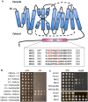
Fig. 1.
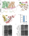
Fig. 1.. The CML18-binding domain is essential for the activity of NHX1.
(A) Top: Schematic representation of NHX1 topology. Blue barrels represent transmembrane (TM) segments. The semi-helices TM5 and TM12 crossed over TM6 constitute the catalytic core known as NhaA-fold. Dashed lines represent the protein loops connecting TM11, TM12, and TM13, not drawn to scale. The CBD is in the cytosolic C-terminal portion of NHX1. Bottom: Alignment of the putative CBDs of the endosomal NHX proteins of A. thaliana (NHX1 to NHX6). The conserved His, Trp, and Asp residues analyzed in this work are highlighted in red. (B) Yeast two-hybrid assay. NHX1 with the CBD deleted (NHX1ΔCBD, residues S486 to G516) was fused to the GAL4 binding domain in plasmid vector pGBKT7 (V1). CML18 was fused to the GAL4 activation domain in vector pGADT7 (V2). Plasmid-encoding pSV40 and p53 were used as positive controls, whereas empty vectors (V1 + V2) served as negative controls. Five-microliter drops of 10-fold serial dilutions of cells transformed with the indicated plasmids were spotted on selective YNB media without His for selection of interaction. Plates were photographed after 2 days at 28°C. Two independent transformed clones are shown for each construct. (C) Complementation assay. The cDNAs of wild-type (WT) NHX1 and NHX2 and mutant NHX1∆CBD lacking the CBD (residues from S486 to G516) were cloned into the yeast expression vector pDR195 and transformed into the AXT3K strain (Δena1-4 Δnha1 Δnhx1). Samples were processed as in (B) and platted onto yeast extract, peptone, and dextrose (YPD) plates with and without hygromycin B (+HygB; 50 μg/ml). Two independent transformed clones are shown for each plasmid in the top panel.
Fig. 2.
Fig. 2.. CML18 binding to NHX vacuolar proteins.
(A and B) Yeast two-hybrid assays. The entire C-terminal cytosolic portion of NHX1 (residues T435 to A538), NHX2 (residues G434 to P546), NHX3 (residues T438 to P552), and NHX4 (residues T437 to C529) was fused to the GAL4 binding domain in plasmid vector pGBKT7 (V1). CML18 was fused to the GAL4 activation domain in vector pGADT7 (V2). Plasmid-encoding pSV40 and p53 were used as positive controls, whereas empty vectors (V1 + V2) served as negative controls. Five-microliter drops of 10-fold serial dilutions of cells transformed with the indicated plasmids were spotted on selective YNB media without His or Ade for selection of interaction. Plates were photographed after 2 days at 28°C. (C) Bimolecular fluorescence complementation assay. Fluorescence and bright-field confocal images of N. benthamiana leaf sections expressing NHX1 or NHX2 fused to the N-terminal fragment of the eYFP in pSPYNE173, in combination with CML18 fused to the C-terminal fragment of the eYFP protein in pSPYCE(M). An empty vector expressing the C-terminal fragment of the eYFP was used as a negative control. (D) Interaction between NHX1 and CML18 takes place through the CBD. Confocal images of N. benthamiana leaf sections expressing NHX1 or NHX1ΔCBD fused to the N-terminal fragment of the eYFP in combination with NHX2 or CML18 fused to the C-terminal fragment of the eYFP protein. An empty vector pSPYCE(M) was used as a negative control. Scale bars, 20 µm.
Fig. 3.
Fig. 3.. Conserved residues in TM9 and CBD are essential for the activity of NHX1 and the interaction with CML18.
(A) Side view of the structure of the NHX1 homodimer as predicted by the AlphaFold software. Protomers are in green and bronze colors. The CBD (pink) packs against the cytosolic side of the core TM domain forming the transporting pore. (B) Cytoplasmic view of an NHX1 protomer structure with the TM helices labeled; the CBD, TM7, and TM9 are highlighted; the dashed line represents the cytoplasmic loop connecting TM13 and CBD. (C) Intramolecular interactions between the CBD and TM9. α Helices are shown with their surface at 80% transparency, residues as thin lines, and relevant amino acid residues as green-colored backbone sticks. (D) Interaction between TM9 and CBD measured by reconstitution of nanoluciferase luminescence. The protein loop separating the CBD and TM13 (L453-N480) was used as a negative control. Shown are the means and SE of two independent experiments with two technical replicates each (P = 2.4 × 10⁻¹⁸ in unpaired t test). AU, arbitrary units. (E) Complementation test. AXT3K cultures expressing the WT NHX1 and the indicated single mutants were normalized to OD600 = 0.5, and aliquots (5 μl) of serial decimal dilutions were spotted onto YPD plates with and without HygB (50 μg/ml). Cells were grown at 30°C for 3 days. (F) Yeast two-hybrid assay. WT NHX1 and mutants W502A and D506A were cloned in pGBKT7 (V1) and tested for interaction with CML18 in vector pGADT7 (V2). Plasmids were transformed in strain AH109 and plated in selective YNB media with and without supplemental histidine. Empty vectors and p53/pSV40 were used as negative and positive controls, respectively. Plates were incubated at 30°C for 2 days.
Fig. 4.
Fig. 4.. Binding of CML18 is essential for AtNHX1 activity in yeast.
(A) The cDNAs of NHX1 and the mutant alleles at the indicated residues were cloned into the yeast vector pDR195 and transformed into the AXT3K strain. Overnight cultures were normalized to OD600 = 0.5, and aliquots (5 μl) of serial decimal dilutions were spotted onto YPD plates with and without HygB (50 μg/liter). Cells were grown at 30°C for 3 days. Shown are two independent colonies for each genotype. (B) The vacuolar pH of the WT (W303), untransformed nhx1 mutant (AXT3K), and AXT3K expressing the indicated mutant proteins was measured by BCECF-AM. Two independent colonies (biological replicas) were used to measure vacuolar pH four times each (technical replicas). The experiment was repeated twice with similar results, and one representative measurement was presented. Data were grouped according to the biological samples tested together in the same experiment. Different letters indicate statistically significant differences in pairwise comparison by Tukey’s post hoc test (n = 8; P < 0.01 in the top panel, P < 0.05 in the bottom panel). (C) Y2H assay. Ten-fold serial dilutions of yeast AH109, transformed with WT NHX1 and the indicated mutants in plasmid pGBKT7 (V1), and CML18 in plasmid pGADT7 (V2) were spotted on selective YNB media without histidine or adenine for selection of interaction. Plates were photographed after 4 days at 28°C. (D) Drops were spotted on selective YNB media without histidine and supplemented with 0.1 and 0.5 mM 3-amino-1,2,4-triazone (3AT). Plates were photographed after 4 days at 28°C.
Fig. 5.
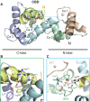
Fig. 5.. CML18 conformation is sensitive to pH.
(A) A cartoon representation of the crystal structure of CML18 in a complex with Ca2+ atoms at pH 5.6. The structure of CML18 comprises two domains, each formed by a pair of EF-hand motifs that bind two Ca2+ atoms. The folding produces two hydrophobic cavities that oppose the Ca2+-binding sites at each domain. The zoom represents a detailed view of the interactions at the N- and C-lobes interface. Notably, interactions involving residues D161 and W164 and the C-terminal carboxylate group of G165 at F4 and S75 at helices E1 and R22 at helix F2 stabilize the C-terminal domain in the interface between the two lobes. (B) Far-ultraviolet (UV) circular dichroism (CD) spectra of CML18 dialyzed against pHs ranging from 6.3 to 8.3. The inset represents the CD signal at 222 nm versus pH. (C) Far-UV CD spectra of CML18 at pH 6.3 and 8.3 in the presence of either 180 μM Ca2+ or 18 μM EGTA. (D) The ratio of the fluorescence signal at 350/330 nm (Trp164 exposure) of CML18 and D161N mutant at both pH 6.3 and 8.3 in the presence or absence of Ca2+. D161N mutation mimics a perma-exposed Trp164.
Fig. 6.
Fig. 6.. pH-dependent interaction between CBD peptide and CML18.
Far-UV CD spectra of CML18, CBD peptide, and a mixture of them at both pH 6.3 (left) and 8.3 (right). The expected spectrum for noninteracting samples, calculated as the sum of both spectra, is represented in purple.
Fig. 7.
Fig. 7.. Model showing the interaction of the CBD peptide with CML18.
(A) Structural model of the CBD peptide (in pale yellow) bound to the large cavity of CML18. (B and C) Detailed view of the interaction between the CBD peptide and CML18 showing the interaction pattern of D506 (B) and H499 with EF2 (C).
Fig. 8.
Fig. 8.. Changes in cytosolic pH and free Ca2+ concentration elicited by differential K+ availability.
Fluorescence emitted by roots of the R-GECO1–E2GFP line was captured 20 min after transferring the seedlings from imaging buffer (IB) with 5 mM KCl to IB without K+ (−KCl) or with 50 mM KCl (+KCl). Top: Ratiometric fluorescence of E2GFP was converted to pH values, whereas the intensiometric fluorescence of R-GECO1 is given as arbitrary fluorescence units [F(Ca2+)]. The box plots present quartiles Q1 to Q3. The X symbols are the means. Data are of 33 to 35 individual seedlings per treatment, compiled from two independent experiments. Outlier data points were not excluded from statistical analyses. Letters indicate statistical significance at P < 0.01 (two-tailed t test). Bottom: Representative seedlings of each treatment.
Fig. 9.
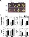
Fig. 9.. Enhanced growth of cml17 cml18 mutant plants under high potassium.
Plants of genotype Col-0 and cml17 cml18 were grown for 2 weeks in hydroponic culture with LAK medium and then transferred to fresh media with the indicated concentrations of K2SO4 for 2 weeks before harvesting. (A) Two representative plants of each genotype and condition at harvest. (B) Means and SE of fresh weight and K+ concentration in the cell sap of shoots and roots. The P values for WT versus mutant comparisons are given (two-tailed t test, n = 5 to 6 plants per genotype and treatment).
Fig. 10.
Fig. 10.. Model for the regulation of vacuolar K+/H+ exchangers by the pH-sensitive calmodulin-like protein CML18.
Working model representing the role of CML18 in the regulation of NHX1. In the NHX1 protomer, the helices forming the ion-conducting pore are shown in two conformations, inward facing and outward facing. The active center is located at the junction between TM5 and TM12, with NHX1 exchanger activity depending on the transition between these conformations. NHX1 pumps K+ into the vacuole and H+ into the cytoplasm. K+ deficiency leads to cytosol acidification, which triggers a CML18 conformational change that facilitates the interaction with NHX1 via the CBD and the subsequent inhibition of K+ transport into the vacuole. Disruption of the H285-D506 interaction in NHX1 disables ion transport. The two lobes of CML18 are depicted in blue and orange. The residues involved in the pH-responsive conformation CML18 at low pH (D161 and W164) are indicated.

References

    1. Chanroj S., Wang G., Venema K., Zhang M. W., Delwiche C. F., Sze H., Conserved and diversified gene families of monovalent cation/H+ antiporters from algae to flowering plants. Front. Plant Sci. 3, 25 (2012). - PMC - PubMed
    1. Pardo J. M., Cubero B., Leidi E. O., Quintero F. J., Alkali cation exchangers: Roles in cellular homeostasis and stress tolerance. J. Exp. Bot. 57, 1181–1199 (2006). - PubMed
    1. Isayenkov S. V., Dabravolski S. A., Pan T., Shabala S., Phylogenetic diversity and physiological roles of plant monovalent cation/H+ antiporters. Front. Plant Sci. 11, 573564 (2020). - PMC - PubMed
    1. Venema K., Quintero F. J., Pardo J. M., Donaire J. P., The Arabidopsis Na+/H+ exchanger AtNHX1 catalyzes low affinity Na+ and K+ transport in reconstituted liposomes. J. Biol. Chem. 277, 2413–2418 (2002). - PubMed
    1. Barragan V., Leidi E., Andres Z., Rubio L., De Luca A., Fernandez J., Cubero B., Pardo J., Ion exchangers NHX1 and NHX2 mediate active potassium uptake into vacuoles to regulate cell turgor and stomatal function in Arabidopsis. Plant Cell 24, 1127–1142 (2012). - PMC - PubMed

MeSH terms