. 2024 Nov 28;128(47):10202-10212.
doi: 10.1021/acs.jpca.4c05628. Epub 2024 Nov 13.

From Gas to Solution: The Changing Neutral Structure of Proline upon Solvation

Affiliations

From Gas to Solution: The Changing Neutral Structure of Proline upon Solvation

Bruno Credidio et al. J Phys Chem A. .

Abstract

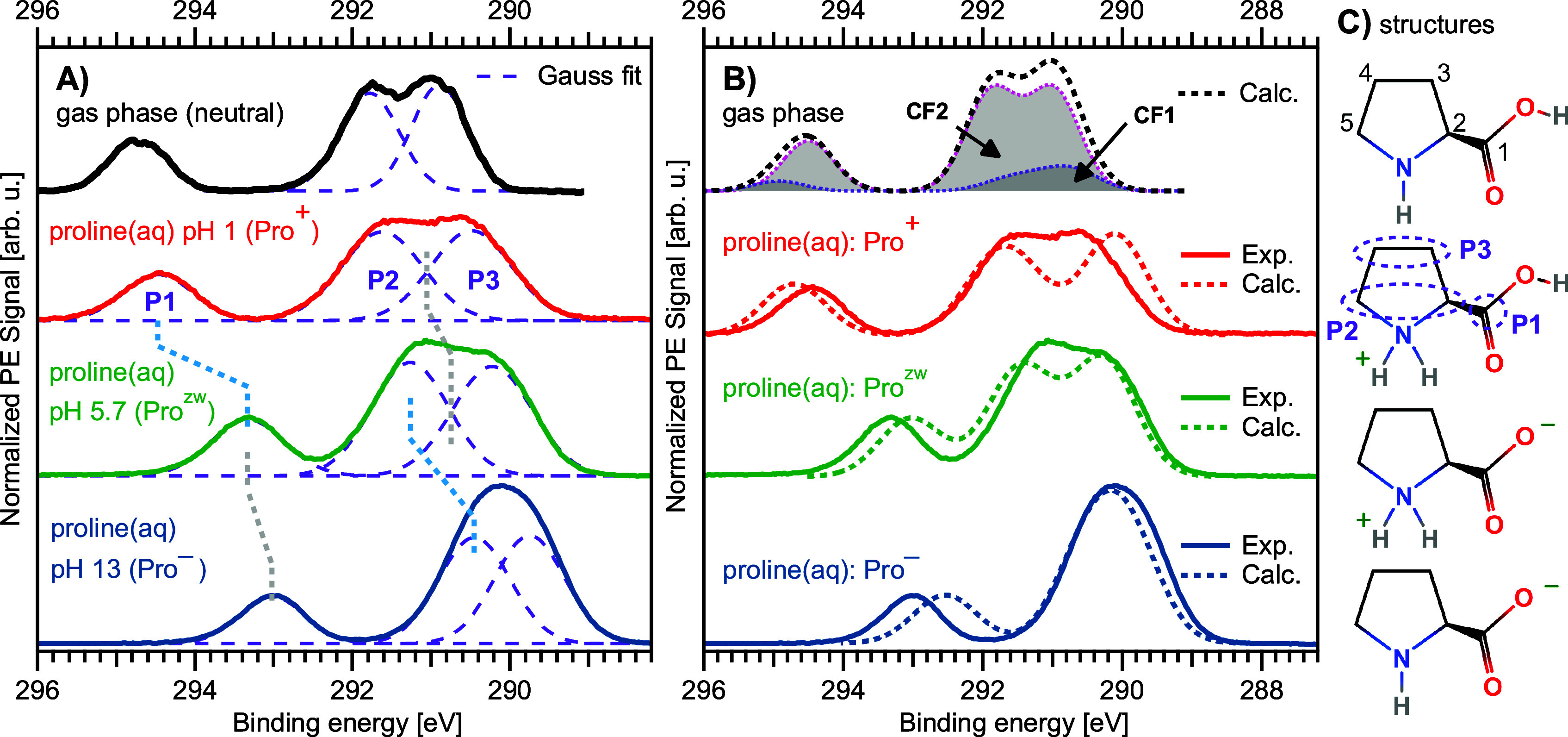
Liquid-jet photoelectron spectroscopy (LJ-PES) and electronic-structure theory were employed to investigate the chemical and structural properties of the amino acid l-proline in aqueous solution for its three ionized states (protonated, zwitterionic, and deprotonated). This is the first PES study of this amino acid in its biologically relevant environment. Proline's structure in the aqueous phase under neutral conditions is zwitterionic, distinctly different from the nonionic neutral form in the gas phase. By analyzing the carbon 1s and nitrogen 1s core levels as well as the valence spectra of aqueous-phase proline, we found that the electronic structure is dominated by the protonation state of each constituent molecular site (the carboxyl and amine groups) with small yet noticeable interference across the molecule. The site-specific nature of the core-level spectra enables the probing of individual molecular constituents. The valence photoelectron spectra are more difficult to interpret because of the overlapping signals of proline with the solvent and pH-adjusting agents (HCl and NaOH). Yet, we are able to reveal subtle effects of specific (hydrogen-bonding) interaction with the solvent on the electronic structure. We also demonstrate that the relevant conformational space is much smaller for aqueous-phase proline than for its gas-phase analogue. This study suggests that caution must be taken when comparing photoelectron spectra for gaseous- and aqueous-phase molecules, particularly if those molecules are readily protonated/deprotonated in solution.


Conflict of interest statement

The authors declare no competing financial interest.

Figures

Figure 1
Figure 1
Sketches of the proline molecule: (Top) gas phase, Pro0, in its two possible conformers CF1 and CF2, which are each composed of two energy-degenerate forms due to ring puckering. (Bottom) aqueous phase: Pro+, Prozw, and Pro. pKa values are taken from ref. (19).
Figure 2
Figure 2
A) N 1s spectra of 1 M proline(aq) in the protonated (pH 1, red), zwitterionic (pH 6.7, green), and deprotonated (pH 13, blue) states measured at a photon energy of 499.47 eV. 10% signal contribution of the zwitterionic species was subtracted from the spectrum of the protonated species (see Methods and SI for details). The gas-phase spectrum from Plekan et al., measured at 495 eV photon energy, is plotted in black at the top. A split peak is observed, originating from the two conformers CF1 and CF2 in the gas phase with slightly different BEs, here indicated by two contributions (dotted lines) as a guide to the eye, which are approximated with a Gaussian (CF1) and Exponentially Modified Gaussian (CF2) peak shape, respectively. B) Corresponding calculated spectra shown as dashed lines, where the spectra of the protonated (red), zwitterionic (green), and deprotonated (blue) species have been shifted by −0.42, 0.24, and 0.79 eV, respectively, analogous to Figure 3; these shifts were extracted from the C 1s spectral comparison. The gaseous contributions of conformers CF1 and CF2 have been added in the same 1:1.12 ratio as in the experiment.
Figure 3
Figure 3
A) C 1s spectra of 1 M proline(aq) in its protonated (pH 1, red), zwitterionic (pH 5.7, green), and deprotonated (pH 13, blue) state. A 10% signal contribution of the zwitterionic species was subtracted from the spectrum of the protonated species (see Methods and SI for details). The spectrum in black is from gaseous proline from ref. (16). Purple dashed lines are Gaussian fits to the experimental spectra, which were constrained to yield equal areas for peaks P2 and P3. B) Liquid-phase experimental spectra in comparison to their theoretical counterparts. The theoretical spectra of the protonated (dotted red), zwitterionic (dotted green), and deprotonated (dotted blue) species have been shifted by −0.42, 0.24, and 0.79 eV, respectively; this matches the centroid of each spectrum with the experimental one and is compensating for an over/under-estimation of the polarization screening in the model (these are the same shifts applied to the N 1s theory in Figure 2). The spectrum in black at the top is a computation of the neutral molecule in the gas phase, which consists of contributions from the two conformers CF1 and CF2 as indicated by the labels; both contributions were summed with a ratio of 1:4, which does not match that found in the nitrogen data but best fits the experimental gas-phase spectrum. C) A sketch of the molecule in each respective state (reproduced from Figure 1) is shown next to the corresponding spectra; we enumerate each carbon site in the topmost sketch. The labels P1, P2, and P3 are the three spectral features from the aqueous phase, as introduced in the main text, along with their correspondence to the molecule’s carbon atoms as indicated in one of the molecular sketches by the purple ovals.
Figure 4
Figure 4
Valence PE spectra of aqueous-phase proline at pH 1 (red), 5.7 (green), and 13 (blue), measured with a photon energy of 403.08 eV (the same as used for C 1s). The purple spectrum is from neat water, where 50 mM NaCl was dissolved to maintain conductivity. Intensities are normalized to water’s 1b1 band. The black spectrum is from gaseous proline from ref. (16) measured with a photon energy of 99 eV. Note that the liquid-phase spectra are dominated by the solvent PE signal and are thus not directly comparable to the gas phase. HCl or NaOH was added to yield a solution with pH 1 or 13, which introduces additional Cl or OH anion signal contributions, respectively. The BEs of Cl 3p and OH 2p, 9.6 and 9.2 eV, respectively, are indicated as vertical dashed lines., The box (orange dotted line) indicates the energy region shown in Figure 5.
Figure 5
Figure 5
A) Valence photoelectron spectra from Figure 4 of proline(aq) at pH 1 (red), pH 5.7 (green), and pH 13 (blue) after subtraction of a reference neat-water spectrum, as well as isolated OH(aq) and Cl(aq) signal contributions for the pH = 1 and pH = 13 spectra, respectively, to reveal only the aqueous-proline signal contributions. We compare the results again with the gas-phase spectrum from Plekan et al. (black, top). This gas-phase spectrum has been fitted with Gaussian functions to reveal individual conformer contributions (dotted lines) in the ratio 1:1.12 as used for the N 1s spectra. B) Corresponding theoretical spectra of gaseous proline, again consisting of the two conformers CF1 and CF2 (ratio 1:1.12) as indicated, and protonated (red), zwitterionic (green), and deprotonated (blue) proline in aqueous solution; all calculated liquid-phase spectra have been uniformly shifted by 0.5 eV toward higher BE.

Similar articles

Cited by

  • Photoelectron circular dichroism of aqueous-phase alanine.
    Stemer D, Thürmer S, Trinter F, Hergenhahn U, Pugini M, Credidio B, Malerz S, Wilkinson I, Nahon L, Meijer G, Powis I, Winter B. Stemer D, et al. Chem Sci. 2025 Apr 7;16(20):8637-8647. doi: 10.1039/d5sc00167f. eCollection 2025 May 21. Chem Sci. 2025. PMID: 40255964 Free PMC article.

References

    1. Morris A. L.; MacArthur M. W.; Hutchinson E. G.; Thornton J. M. Stereochemical quality of protein structure coordinates. Proteins 1992, 12, 345–364. 10.1002/prot.340120407. - DOI - PubMed
    1. Karna E.; Szoka L.; Huynh T. Y. L.; Palka J. A. Proline-dependent regulation of collagen metabolism. Cell. Mol. Life Sci. 2020, 77, 1911–1918. 10.1007/s00018-019-03363-3. - DOI - PMC - PubMed
    1. Patriarca E. J.; Cermola F.; D’Aniello C.; Fico A.; Guardiola O.; De Cesare D.; Minchiotti G. The multifaceted roles of proline in cell behavior. Front. Cell Dev. Biol. 2021, 9, 728576.10.3389/fcell.2021.728576. - DOI - PMC - PubMed
    1. Kavi Kishor P. B.; Suravajhala P.; Rathnagiri P.; Sreenivasulu N. Intriguing role of proline in redox potential conferring high temperature stress tolerance. Front. Plant Sci. 2022, 13, 867531.10.3389/fpls.2022.867531. - DOI - PMC - PubMed
    1. Liang X.; Zhang L.; Natarajan S. K.; Becker D. F. Proline mechanisms of stress survival. Antioxid. Redox Signaling 2013, 19, 998–1011. 10.1089/ars.2012.5074. - DOI - PMC - PubMed