

Objective analysis of orbital rim fracture CT images using curve and area measurement
- PMID: 39537669
- PMCID: PMC11561094
- DOI: 10.1038/s41598-024-76818-8
Objective analysis of orbital rim fracture CT images using curve and area measurement
Abstract
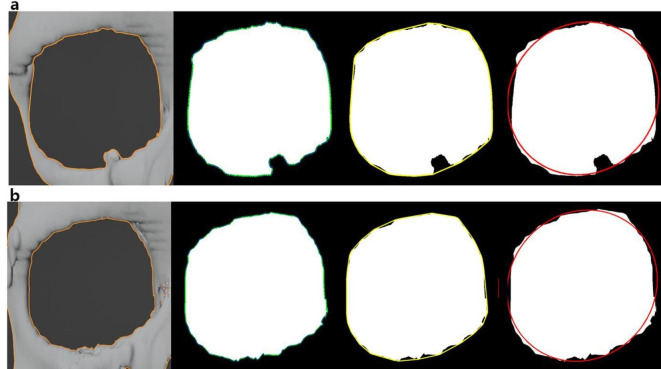
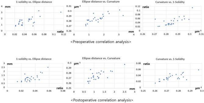
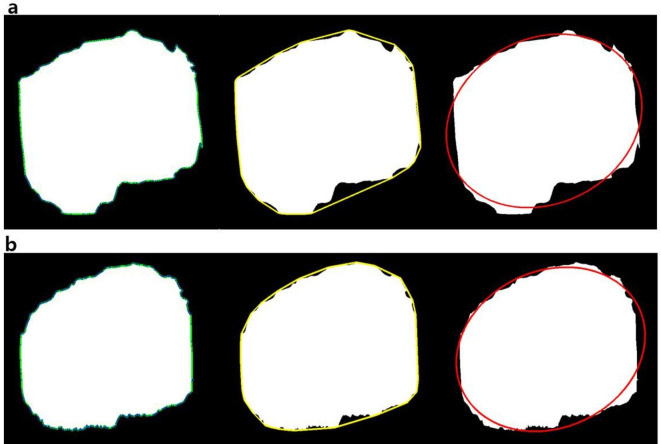
The orbital bone presents a closed curve, and fracture results in disfigurement. An image analysis procedure was developed to examine before and after corrective surgery. An ellipse and circumscribed contour embodied the closed curve. Three-dimensional (3D) computed tomography (CT) images were collected from 25 patients. Orbital rim data were generated, and binary images were created to facilitate closed curve analysis. Various indices, including the solidity value (closed curve area/convex hull area) and ellipse distance (discrepancy between the closed curve and the ellipse traversing the curve), were utilized. The ratios of various indices-including the number of vertices, solidity value, and ellipse distance-between the affected and unaffected sides showed postoperative values that were closer to 1, which would indicate perfect symmetry, than the preoperative measurements (P < 0.05). The solidity value increased, while both the ellipse distance and curvature values decreased, reflecting the transformation of bends into smooth contours following reduction surgery (P < 0.05). Significant correlations were observed between 1-solidity, ellipse distance, and curvature using the Pearson correlation test (P < 0.05). This study validated postoperative changes in various indices and established correlations among multiple values, specifically solidity, ellipse distance, and curvature. Employing multiple indices with mutual complements has provided objective information confidently.
Keywords: Area; Computed tomography; Curve; Fracture; Orbital bone.
© 2024. The Author(s).
Conflict of interest statement
Figures


Similar articles
-
Measurements of orbital volume change using computed tomography in isolated orbital blowout fractures.Arch Facial Plast Surg. 2009 Nov-Dec;11(6):395-8. doi: 10.1001/archfacial.2009.77. Arch Facial Plast Surg. 2009. PMID: 19917900
-
Possibility of Using Standard Plates for Orbital Fracture.J Craniofac Surg. 2016 Oct;27(7):1735-1737. doi: 10.1097/SCS.0000000000002980. J Craniofac Surg. 2016. PMID: 27513779
-
Intraoperative imaging with a 3D C-arm system after zygomatico-orbital complex fracture reduction.J Oral Maxillofac Surg. 2013 May;71(5):894-910. doi: 10.1016/j.joms.2012.10.031. Epub 2013 Jan 23. J Oral Maxillofac Surg. 2013. PMID: 23352428
-
[Measurement of the globe proptosis by CT and its accuracy in patients with unilateral orbital fracture].Zhonghua Zheng Xing Wai Ke Za Zhi. 2009 May;25(3):169-72. Zhonghua Zheng Xing Wai Ke Za Zhi. 2009. PMID: 19803193 Chinese.
-
Blowout fractures - clinic, imaging and applied anatomy of the orbit.Dan Med J. 2018 Mar;65(3):B5459. Dan Med J. 2018. PMID: 29510812 Review.
References
-
- Mehta, D., Daleo, N. S., Dörfler, F. & Hauenstein, J. D. Algebraic geometrization of the Kuramoto model: Equilibria and stability analysis. Chaos. 25, 053103. 10.1063/1.4919696 (2015). - PubMed
-
- Hahn, L. & Klein, P. The impact of multiple representations on students’ understanding of vector field concepts: Implementation of simulations and sketching activities into lecture-based recitations in undergraduate physics. Front. Psychol.13, 1012787. 10.3389/fpsyg.2022.1012787 (2022). - PMC - PubMed
MeSH terms
LinkOut - more resources
Full Text Sources
Medical
