. 2024 Nov 13;14(1):27867.
doi: 10.1038/s41598-024-79217-1.

Characterization of variably protease-sensitive prionopathy by capillary electrophoresis

Affiliations

Characterization of variably protease-sensitive prionopathy by capillary electrophoresis

Jennifer Myskiw et al. Sci Rep. .

Abstract

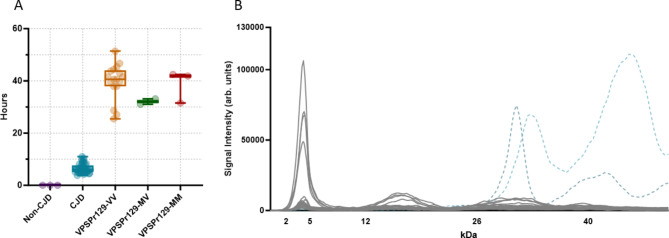
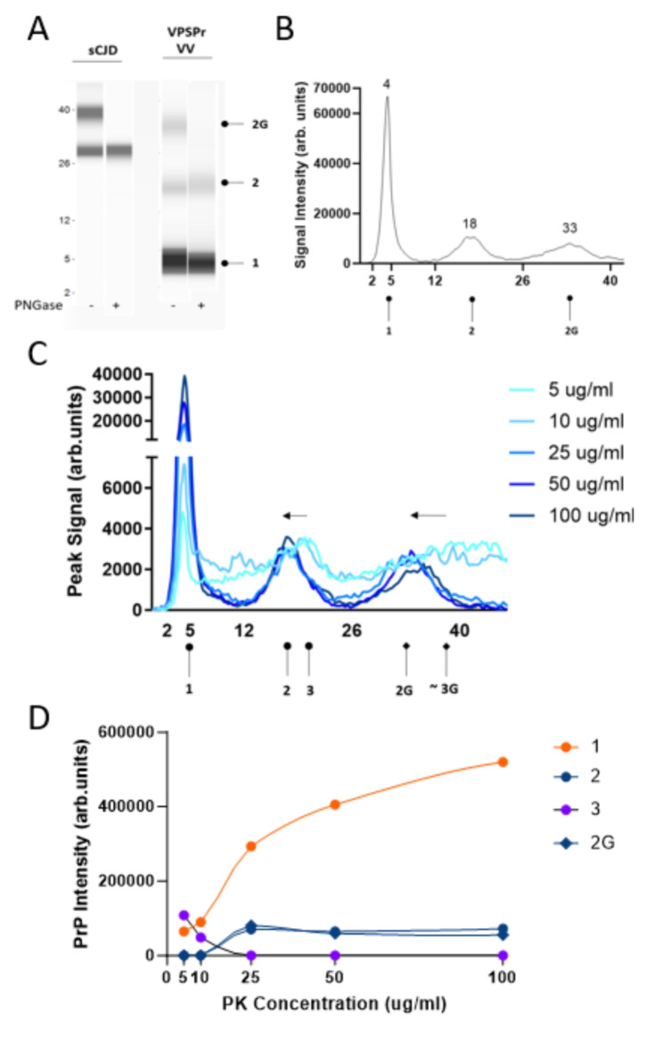
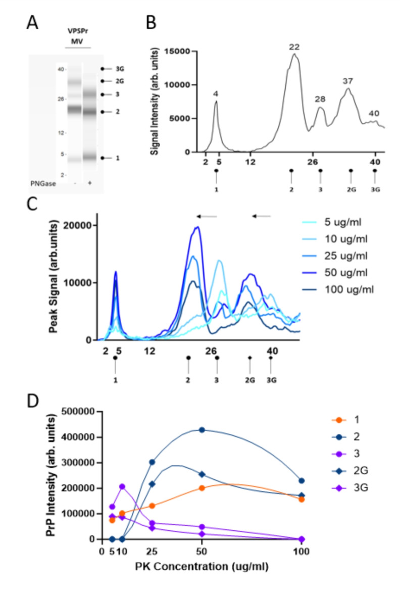
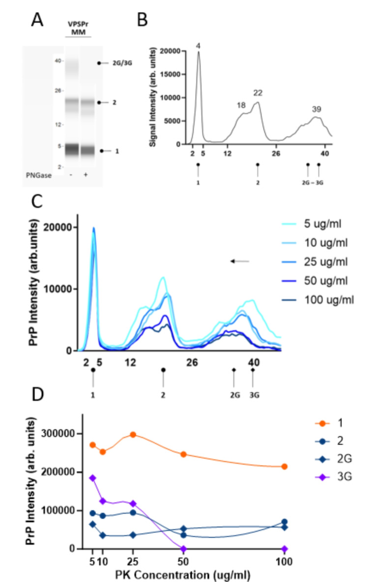
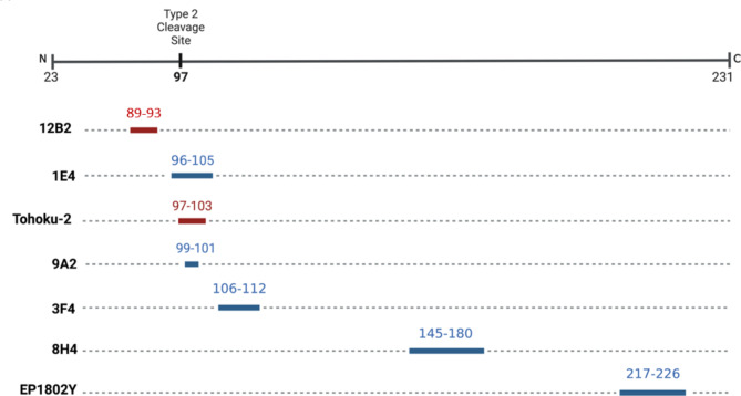
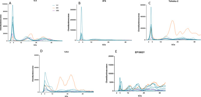
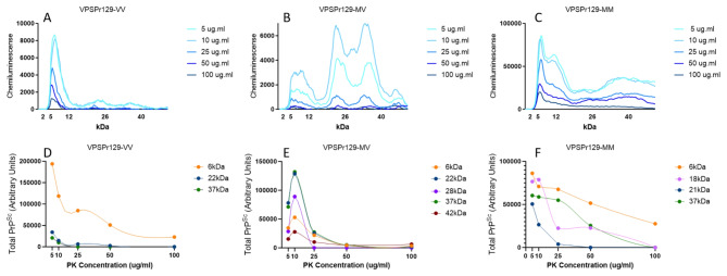
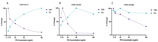
Variably Protease Sensitive Prionopathy (VPSPr) is a rare human prion disease that, like Creutzfeldt-Jakob disease (CJD), results in the deposition of abnormally folded prion protein aggregates in the brain and is ultimately fatal. Neuropathology and clinical features of VPSPr are heterogeneous. However, the key discriminating feature is the relative sensitivity of the pathological prion protein to proteinase digestion compared to that typically seen in other human prion cases. Three major fragments of 23, 17 and 7 kDa are characteristic of the disease following digestion with proteinase K. We recently reported the utility of the highly adaptive and reproducible ProteinSimple™ capillary electrophoresis (CE) system to perform protein separation of PK digested prion protein in CJD. Consequently, we explored capillary-based electrophoresis (CE) technology as a sensitive method to detect and characterize VPSPr in a cohort of 29 cases. The unique 7 kDa fragment has high intensity, particularly in cases with the codon 129 VV genotype, but can be missed by regular Western blotting due to the small size. However, this fragment is readily detected by CE in all cases. In addition, the flexibility of CE produced highly reproducible, semi-quantitative data for determining relative proteinase K sensitivity and epitope mapping of representative cases from each codon 129 genotype (VV, MV and MM).


Conflict of interest statement

Declarations Competing interests The authors declare no competing interests.

Figures

Fig. 1
Fig. 1
Seeding lag phase and electrophoretogram of VPSPr PrPSc immunoprobed with antibody 3F4. (A) Seeding lag phases of PrPSc from non-CJD, sCJD, and VPSPr (all genotypes) 10% (w/v) brain homogenates at a 10− 4 dilution were seeded with recombinant full-length hamster PrPC using the real-time quaking-induced conversion (RT-QuIC) assay. Time from the assay start time to when a pre-defined amyloid formation threshold was reached (lag phase time) was calculated and plotted along with bars showing the min–max values. sCJD (n=33) displayed with a lag phase time ranging from 5 to 11 hours. VPSPr (n=20) displayed a longer lag phase, ranging from 25 to over 50 hours to reach the threshold. (B) Overlay of electrophoretograms from 29 VPSPr Cases (grey) and individual representative type 1 and 2 CJD cases (light and dark blue dotted lines, respectively) PK-digested (25 µg/ml) and immunoprobed with antibody 3F4 using the CE immunoassay.
Fig. 2
Fig. 2
Electrophoretic profiles of VPSPr129-VV fragments and their sensitivity to proteinase-K digestion. (A) Mock “western blot view” of sCJD and VPSPr129-VV digested with 25 µg/ml of PK treated with or without PNGase using the CE assay. Circle heads and labels are used to annotate VPSPr fragments: “1” is the C-terminal truncated unglycosylated fragment, “2” is the larger C-terminal truncated unglycosylated fragment, “2G” is the monoglycosylated form of “2”, “3” is the full-length unglycosylated fragment (not detected at 25 µg/ml, shown in Panel C), and “3G” is the monoglycosylated form of “3” (not detected at 25 µg/ml, shown in Panel C) (B) Electrophoretogram of VPSPr129-VV digested with 25 µg/ml of PK (C) Electrophoretogram of PK-titration assay of a VPSPr129-VV case. The arrows demonstrate the shift from a higher molecular weight to a lower molecular weight due to the truncation of fragments as PK concentrations increase. While we did observe a rough signal where the band “3G” was anticipated to be located, the signal was too faint to indicate an exact molecular weight. (D) Graph showing the PrP signal intensity change of each VPSPr fragment throughout the PK titration assay.
Fig. 3
Fig. 3
Electrophoretic profiles of VPSPr129-MV fragments and their sensitivity to proteinase-K digestion. (A) Mock “western blot view” of VPSPr129-MV digested with 25 µg/ml of PK treated with or without PNGase using the CE assay. Fragments are labelled according to convention described in Figure 2. (B) Electrophoretogram of VPSPr129-MV digested with 25 µg/ml of PK (C) Electrophoretogram of PK-titration assay of a VPSPr129-MV case. The arrows demonstrate the shift from a higher molecular weight to a lower molecular weight due to the truncation of fragments as PK concentrations increase (3 → 2 and 3G → 2G). (D) Graph showing the PrP signal intensity change of each VPSPr fragment throughout the PK titration assay.
Fig. 4
Fig. 4
Electrophoretic profiles of VPSPr129-MM fragments and their sensitivity to proteinase-K digestion. (A) Mock “western blot view” of VPSPr129-MM digested with 25 µg/ml of PK treated with or without PNGase using the CE assay. Fragments are labelled according to convention described in Figure 2. (B) Electrophoretogram of VPSPr129-MM digested with 25 µg/ml of PK (C) Electrophoretogram of PK-titration assay of a VPSPr129-MM case. The arrows demonstrate the shift from a higher molecular weight to a lower molecular weight due to the truncation of fragments as PK concentrations increase (3G → 2G) (D) Graph showing the PrP signal intensity change of each VPSPr fragment throughout the PK titration assay.
Fig. 5
Fig. 5
PrP-Specific Antibody Epitope Binding Regions to the Human Prion Protein. Antibodies recognizing PrP epitopes depicted in red are sCJD type-specific, 12B2 is type-1 CJD specific and Tohoku-2 is type-2 CJD specific. Antibodies recognizing epitopes depicted in blue have a binding affinity to both Type 1 and 2 CJD.
Fig. 6
Fig. 6
Epitope Mapping of VPSPr Cases with PrP-specific Antibodies. VPSPr129-VV, -MV and -MM cases were PTA precipitated and PK digested at a final concentration of 50 µg/ml before being probed with PrP-specific antibodies using the CE assay. Electrophoretogram data was exported and graphed on Graphpad Prism to provide signal overlays (orange MV, blue VV, and purple MM) (A) 1E4 (residues 96-105), (B) 3F4 (residues 106-112), (C) Tohoku-2 (residues 97-103), (D) 12B2 (residues 89-93), and (E) EP1802Y (residues 217-226).
Fig. 7
Fig. 7
Analysis from PK-Titration of PrPSc Fragments from VPSPr129-VV, -MV, and -MM Genotypes Immunoprobed with PrP-Antibody 12B2. Equal concentrations of PTA-precipitated 10% brain homogenate were treated with PK concentrations ranging from 5 µg/ml to 100 µg/ml followed by capillary-electrophoresis to analyze conformational stability of the different VPSPr fragments from (A) VPSPr129-VV, (B) VPSPr129-MV and (C) VPSPr-MM. (D), (E), & (C) Signal intensity changes for each VPSPr fragment was evaluated for each genotype.
Fig. 8
Fig. 8
Rate of Change During PK-Titration of Low Molecular Weight VPSPr Fragments Bound to PrP-Specific 1E4 and 12B2. Rates of change were calculated for the smallest VPSPr fragment for (A) VPSPr129-VV (B) VPSPr129-MV and (C) VPSPr129-MM. Each graph shows the rates of change when the smallest molecular weight VPSPr fragment was immunoprobed with 1E4 (residues 96–106) and 12B2 (89–93) for a single representative sample.
Fig. 9
Fig. 9
Schematic Diagram of VPSPr-Associated PK-resistant PrPSc Fragments, Adapted from Zhang et al. The gray bar at the top represents full length human PrP (23–231). The purple bars represent fragments bound to N-terminal antibody 1E4 (96–105), the green bars represent fragments bound to C-terminal antibody EP1802Y (217–226), and the red fragment represents the fragment exclusively bound to N-terminal antibody 12B2 (89–93). VPSPr fragments with known cleavage sites are labelled with an amino acid cleavage site number, whereas dotted lines represent the regions of currently unknown cleavage sites.

Similar articles

References

    1. Sitammagari, K. K. & Masood, W. Creutzfeldt Jakob Disease, In: StatPearls, StatPearls Publishing, Treasure Island (FL), http://www.ncbi.nlm.nih.gov/books/NBK507860/ (Accessed 25 July 2023). - PubMed
    1. Kupfer, L., Hinrichs, W., Groschup, M. H. & Misfolding, P. P. Curr. Mol. Med.9 826–835. 10.2174/156652409789105543. (2009). - PMC - PubMed
    1. Requena, J. R. & Wille, H. The structure of the infectious prion protein. Prion8, 60–66. 10.4161/pri.28368 (2014). - PMC - PubMed
    1. Hill, A. F. et al. Molecular classification of sporadic Creutzfeldt-Jakob disease. Brain J. Neurol.126, 1333–1346. 10.1093/brain/awg125 (2003). - PubMed
    1. Gambetti, P., Kong, Q., Zou, W., Parchi, P. & Chen, S. G. Sporadic and familial CJD: classification and characterisation. Br. Med. Bull.66, 213–239. 10.1093/bmb/66.1.213 (2003). - PubMed

Substances

LinkOut - more resources