

Dual-encoder architecture for metal artifact reduction for kV-cone-beam CT images in head and neck cancer radiotherapy
- PMID: 39537735
- PMCID: PMC11561079
- DOI: 10.1038/s41598-024-79305-2
Dual-encoder architecture for metal artifact reduction for kV-cone-beam CT images in head and neck cancer radiotherapy
Abstract
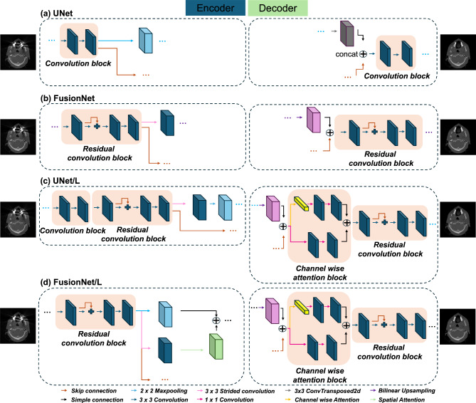
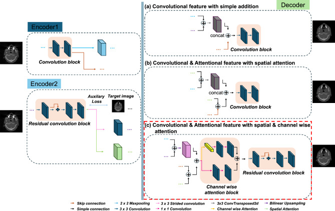
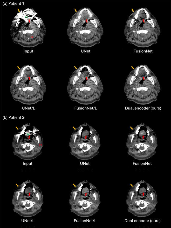
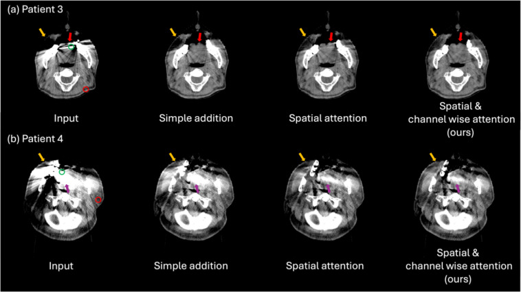
During a radiotherapy (RT) course, geometrical variations of target volumes, organs at risk, weight changes (loss/gain), tumor regression and/or progression can significantly affect the treatment outcome. Adaptive RT has become the effective methods along with technical advancements in imaging modalities including cone-beam computed tomography (CBCT). Planning CT (pCT) can be modified via deformable image registration (DIR), which is applied to the pair of pCT and CBCT. However, the artifact existed in both pCT and CBCT is a vulnerable factor in DIR. The dose calculation on CBCT is also suggested. Missing information due to the artifacts hinders the accurate dose calculation on CBCT. In this study, we aim to develop a deep learning-based metal artifact reduction (MAR) model to reduce the metal artifacts in CBCT for head and neck cancer RT. To train the proposed MAR model, we synthesized the kV-CBCT images including metallic implants, with and without metal artifacts (simulated image data pairs) through sinogram image handling process. We propose the deep learning architecture which focuses on both artifact removal and reconstruction of anatomic structure using a dual-encoder architecture. We designed four single-encoder models and three dual-encoder models based on UNet (for an artifact removal) and FusionNet (for a tissue restoration). Each single-encoder model contains either UNet or FusionNet, while the dual-encoder models have both UNet and FusionNet architectures. In the dual-encoder models, we implemented different feature fusion methods, including simple addition, spatial attention, and spatial/channel wise attention. Among the models, a dual-encoder model with spatial/channel wise attention showed the highest scores in terms of peak signal-to-noise ratio, mean squared error, structural similarity index, and Pearson correlation coefficient. CBCT images from 34 head and neck cancer patients were used to test the developed models. The dual-encoder model with spatial/channel wise attention showed the best results in terms of artifact index. By using the proposed model to CBCT, one can achieve more accurate synthetic pCT for head and neck patients as well as better tissue recognition and structure delineation for CBCT image itself.
© 2024. The Author(s).
Conflict of interest statement
Figures


Similar articles
-
Quantifying the accuracy of deformable image registration for cone-beam computed tomography with a physical phantom.J Appl Clin Med Phys. 2019 Oct;20(10):92-100. doi: 10.1002/acm2.12717. Epub 2019 Sep 21. J Appl Clin Med Phys. 2019. PMID: 31541526 Free PMC article.
-
Comparison of CBCT-based dose calculation methods in head and neck cancer radiotherapy: from Hounsfield unit to density calibration curve to deep learning.Med Phys. 2020 Oct;47(10):4683-4693. doi: 10.1002/mp.14387. Epub 2020 Aug 11. Med Phys. 2020. PMID: 32654160
-
Streaking artifact reduction for CBCT-based synthetic CT generation in adaptive radiotherapy.Med Phys. 2023 Feb;50(2):879-893. doi: 10.1002/mp.16017. Epub 2022 Oct 18. Med Phys. 2023. PMID: 36183234
-
[Advances in low-dose cone-beam computed tomography image reconstruction methods based on deep learning].Sheng Wu Yi Xue Gong Cheng Xue Za Zhi. 2025 Jun 25;42(3):635-642. doi: 10.7507/1001-5515.202409021. Sheng Wu Yi Xue Gong Cheng Xue Za Zhi. 2025. PMID: 40566788 Free PMC article. Review. Chinese.
-
The Effect of Changes in the Angular Position of Implants on Metal Artifact Reduction in Cone-Beam Computed Tomography Images: A Scoping Review.Radiol Res Pract. 2023 Jul 31;2023:5539719. doi: 10.1155/2023/5539719. eCollection 2023. Radiol Res Pract. 2023. PMID: 37554657 Free PMC article.
References
-
- Hussain, A. & Muhammad, W. Treatment planning in radiation therapy. In An Introduction to Medical Physics 63–129 (2017).
-
- Son, J., Lee, S. Y., Choi, C. H., Park, J. M. & Kim, J. I. Impact of 0.35 T magnetic field on dose calculation for non-small cell lung cancer stereotactic radiotherapy plans. J. Radiat. Prot. Res.48, 117–123 (2023).
-
- Abuhaimed, A. & Martin, C. J. Assessment of organ and size-specific effective doses from cone beam CT (CBCT) in image-guided radiotherapy (IGRT) based on body mass index (BMI). Radiat. Phys. Chem.208, 110889 (2023).
-
- Giacometti, V., Hounsell, A. R. & McGarry, C. K. A review of dose calculation approaches with cone beam CT in photon and proton therapy. Phys. Med.76, 243–276 (2020). - PubMed
MeSH terms
Substances
Grants and funding
LinkOut - more resources
Full Text Sources
Medical
