. 2024 Nov 13;7(1):1501.
doi: 10.1038/s42003-024-07213-2.

Cryo-EM of human rhinovirus reveals capsid-RNA duplex interactions that provide insights into virus assembly and genome uncoating

Affiliations

Cryo-EM of human rhinovirus reveals capsid-RNA duplex interactions that provide insights into virus assembly and genome uncoating

David Gil-Cantero et al. Commun Biol. .

Abstract

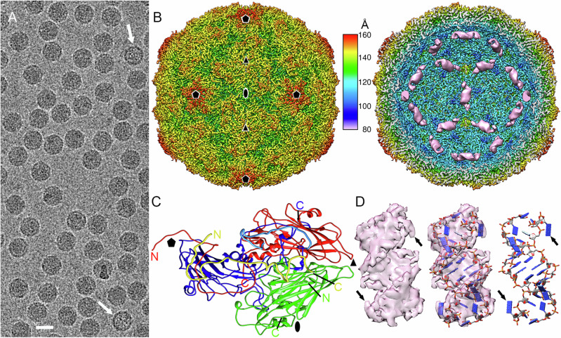
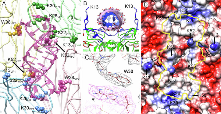
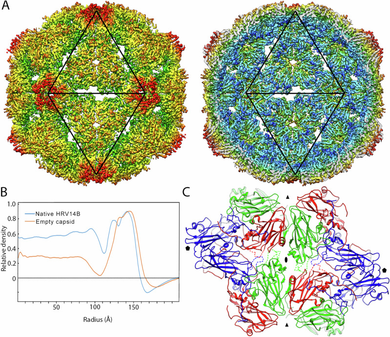
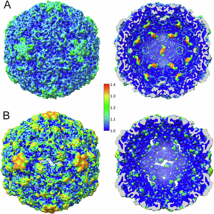
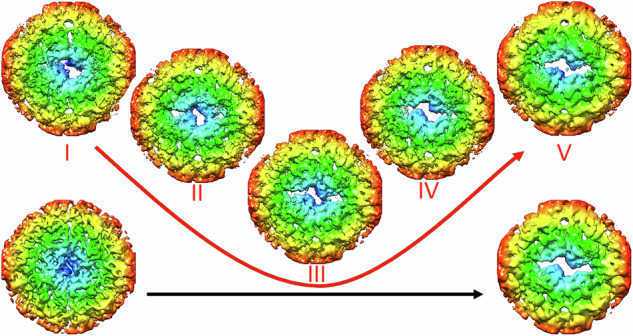
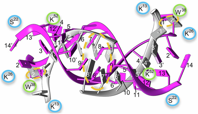
The cryo-EM structure of the human rhinovirus B14 determined in this study reveals 13-bp RNA duplexes symmetrically bound to regions around each of the 30 two-fold axes in the icosahedral viral capsid. The RNA duplexes (~12% of the ssRNA genome) define a quasi-dodecahedral cage that line a substantial part of the capsid interior surface. The RNA duplexes establish a complex network of non-covalent interactions with pockets in the capsid inner wall, including coulombic interactions with a cluster of basic amino acid residues that surround each RNA duplex. A direct comparison was made between the cryo-EM structure of RNA-filled virions and that of RNA-free (empty) capsids that resulted from genome release from a small fraction of viruses. The comparison reveals that some specific residues involved in capsid-duplex RNA interactions in the virion undergo remarkable conformational rearrangements upon RNA release from the capsid. RNA release is also associated with the asynchronous opening of channels at the 30 two-fold axes. The results provide further insights into the molecular mechanisms leading to assembly of rhinovirus particles and their genome uncoating during infection. They may also contribute to development of novel antiviral strategies aimed at interfering with viral capsid-genome interactions during the infectious cycle.


Conflict of interest statement

Competing interests The authors declare no competing interests.

Figures

Fig. 1
Fig. 1. Cryo-EM structure of RV-B14 virions at 2.9 Å resolution.
A Cryo-EM image of RV-B14. Arrows indicate empty capsids. Bar = 250 Å. B Radially color-coded surface representation of RV-B14 outer (left) and inner (right) surfaces viewed along a two-fold axis. Ordered, genomic RNA at the inner surface of the RV-B14 capsid beneath the two-fold axes (pink). The color key (center) indicates the radial distance (in Å) from the particle center. In these images the capsid density is sharpened, whereas dsRNA density is unsharpened. Symbols indicate icosahedral symmetry axes. C A 5S protomer (or asymmetric unit) viewed from the inside of the capsid, showing the ordered regions of VP1 in blue, VP2 in green, VP3 in red, and VP4 in yellow. Ordered N and C termini are indicated. The N-terminal segment of VP1 Glu7-Lys30 is highlighted in light blue. D Sharpened cryo-EM density of a dsRNA helical segment (left), fitted with an atomic model of a 13bp-dsRNA segment and two pendant bases (blue slabs for bases) (center & right images). Arrows indicate the two terminal unpaired nucleotides at each end of the dsRNA.
Fig. 2
Fig. 2. Capsid protein-dsRNA interactions in RV-B14 virions.
A Interactions of Trp38 and Lys52 of VP2, and Lys13, Ser22, Lys26, and Lys 30 of VP1 with the genomic RNA duplex (pink) viewed from the inside of the capsid. These interacting residues are highlighted (spheres). The four 5S protomers that interact with a dsRNA segment are colored in pale yellow, green, blue, and pink. Protomers in yellow and blue belong to the same pentamer, and protomers in green and pink to an adjacent pentamer. B Transverse view of the structure (rotated 90° relative to A). The RNA duplex backbone is shown as pink coils with blue slabs for the bases. The side-chains of two Lys13 residues, one from each of the neighboring VP1 act as “guide posts” for duplex placement. C The stacking contact between the aromatic rings of Trp38 in VP2 and the purine at the end of the dsRNA segment. D The RV-B14 capsid inner surface is represented as an electrostatic potential surface, showing the distribution of negative (red) and positive (blue) charges, around a two-fold position. The yellow line indicates the outline for a bound dsRNA duplex, indicating the basic residues that interact with it.
Fig. 3
Fig. 3. Cryo-EM structure of empty RV-B14 capsids at 3.8 Å resolution.
A Radially color-coded surface representation of empty RV-B14 outer (left) and inner (right) surfaces viewed along a two-fold axis. Black triangles outline two adjacent icosahedral facets and indicate the icosahedral symmetry axes that expand to create pores. B Comparison of radial density profiles of the cryo-EM maps of virions (blue) and empty capsids (orange). Ordered RNA is located at radii < 113 Å in the virion. C Atomic models around the two-fold, Z-shaped pores viewed from the outside of the capsid. VP1 is shown in blue, VP2 in green, and VP3 in red. Highly flexible (or invisible) loops are indicated by dashed lines in the conformation of closed virions. Symbols indicate icosahedral symmetry axes.
Fig. 4
Fig. 4. Relative local resolution of cryo-EM maps of full and empty RV-B14 maps.
Relative local resolution assessment for full virion (A) and empty capsid (B) RV-B14 maps was calculated by dividing the absolute local resolution values by the global (averaged), FSC-derived resolution of the map. In the virion the density corresponding to RNA duplexes at each 2-fold axis has been included. Surface-shaded representations of the RV-B14 particle outer (left) and inner (right) surfaces viewed along an icosahedral two-fold axis (scale color at center). Cryo-EM density regions with relative local resolution values > 1.1 (i.e., lower resolution) indicate high conformational dynamics. Regions with the highest resolution (relative local resolution values < 1.1; deep blue shades) correspond to low dynamic conformational states.
Fig. 5
Fig. 5. Analysis of pores in empty RV-B14 capsids.
Asymmetric classification of pores at the two-fold axes in the five classes of the expanded virus particles, termed I−V (red), viewed from the inside. Localized reconstructions were calculated around the two-fold axes within a radius of 81 Å. Aperture sizes increase from left to right, starting with a closed structure (left, bottom) and leading to a capsid state in which the two-fold axis is fully opened (right, bottom). These conformational states were derived from virions and empty capsid particles, respectively, after applying icosahedral symmetry; arrows indicate aperture sizes in each case.
Fig. 6
Fig. 6. Comparisons of different organization states for the ordered dsRNA in RV-B14.
Ordered RNA regions in two initial states of the RV-B14 virion. Our model of dsRNA (pink,13 bp indicated with black numbers) is superimposed onto PDB 7BG6 (gray, 8 bp indicated using white numbers). Similar areas are highlighted by orange dashed ovals: two paired bps at the center and the non-matched base interacting with Trp38 of VP2 at the end of the RNA segments. Some interacting residues of VP1 and VP2 are shown (encircled in blue and green, respectively).

References

    1. Weston, M. & Frieman, M. B. In Encyclopedia of microbiology (ed. Schmidt, T.) 4th edn 85-101 (Elsevier, 2019).
    1. Andrup, L., Krogfelt, K. A., Hansen, K. S. & Madsen, A. M. Transmission route of rhinovirus - the causative agent for common cold. A systematic review. Am. J. Infect. Control51, 938–957 (2023). - PubMed
    1. Jacobs, S. E. et al. Clinical and molecular epidemiology of human rhinovirus infections in patients with hematologic malignancy. J. Clin. Virol.71, 51–58 (2015). - PMC - PubMed
    1. Hershenson, M. B. In The Picornaviruses (eds Ehrenfeld, E., Domingo, E., & Roos, R.) 369-381 (ASM Press, 2010).
    1. Jacobs, S. E., Lamson, D. M., St George, K. & Walsh, T. J. Human rhinoviruses. Clin. Microbiol. Rev.26, 135–162 (2013). - PMC - PubMed

MeSH terms