Clinical Trial
. 2025 Jan;637(8046):708-715.
doi: 10.1038/s41586-024-08171-9. Epub 2024 Nov 13.

Intravenous and intracranial GD2-CAR T cells for H3K27M+ diffuse midline gliomas

Affiliations
Clinical Trial

Intravenous and intracranial GD2-CAR T cells for H3K27M+ diffuse midline gliomas

Michelle Monje et al. Nature. 2025 Jan.

Erratum in

  • Author Correction: Intravenous and intracranial GD2-CAR T cells for H3K27M+ diffuse midline gliomas.
    Monje M, Mahdi J, Majzner R, Yeom KW, Schultz LM, Richards RM, Barsan V, Song KW, Kamens J, Baggott C, Kunicki M, Rietberg SP, Lim AS, Reschke A, Mavroukakis S, Egeler E, Moon J, Patel S, Chinnasamy H, Erickson C, Jacobs A, Duh AK, Tunuguntla R, Klysz DD, Fowler C, Green S, Beebe B, Carr C, Fujimoto M, Brown AK, Petersen AG, McIntyre C, Siddiqui A, Lepori-Bui N, Villar K, Pham K, Bove R, Musa E, Reynolds WD, Kuo A, Prabhu S, Rasmussen L, Cornell TT, Partap S, Fisher PG, Campen CJ, Grant G, Prolo L, Ye X, Sahaf B, Davis KL, Feldman SA, Ramakrishna S, Mackall C. Monje M, et al. Nature. 2024 Dec;636(8043):E6. doi: 10.1038/s41586-024-08452-3. Nature. 2024. PMID: 39613972 Free PMC article. No abstract available.

Abstract

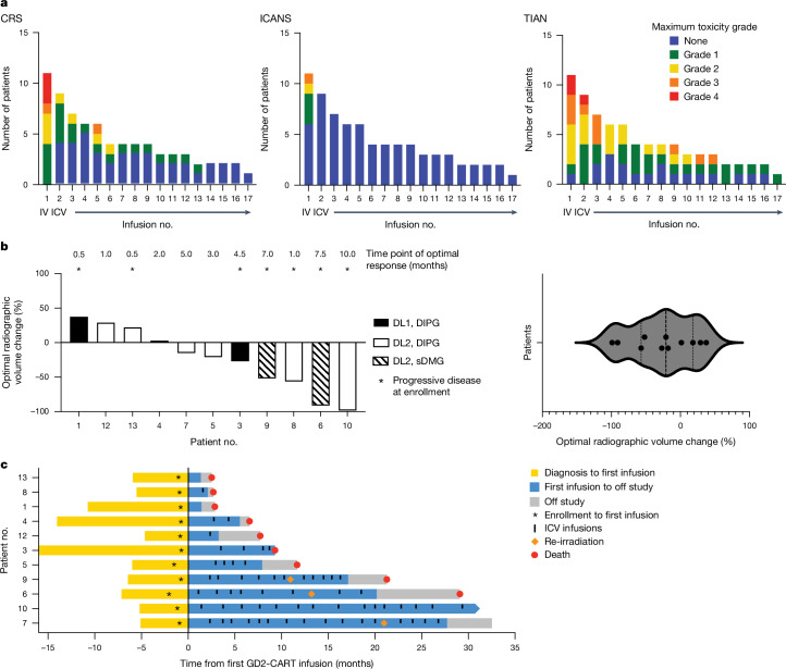
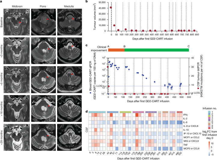
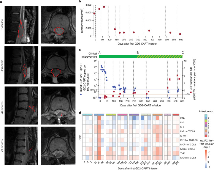
H3K27M-mutant diffuse midline gliomas (DMGs) express high levels of the disialoganglioside GD2 (ref. 1). Chimeric antigen receptor-modified T cells targeting GD2 (GD2-CART) eradicated DMGs in preclinical models1. Arm A of Phase I trial no. NCT04196413 (ref. 2) administered one intravenous (IV) dose of autologous GD2-CART to patients with H3K27M-mutant pontine (DIPG) or spinal DMG (sDMG) at two dose levels (DL1, 1 × 10⁶ kg-1; DL2, 3 × 10⁶ kg-1) following lymphodepleting chemotherapy. Patients with clinical or imaging benefit were eligible for subsequent intracerebroventricular (ICV) intracranial infusions (10-30 × 106 GD2-CART). Primary objectives were manufacturing feasibility, tolerability and the identification of maximally tolerated IV dose. Secondary objectives included preliminary assessments of benefit. Thirteen patients enroled, with 11 receiving IV GD2-CART on study (n = 3 DL1 (3 DIPG); n = 8 DL2 (6 DIPG, 2 sDMG)). GD2-CART manufacture was successful for all patients. No dose-limiting toxicities occurred on DL1, but three patients experienced dose-limiting cytokine release syndrome on DL2, establishing DL1 as the maximally tolerated IV dose. Nine patients received ICV infusions, with no dose-limiting toxicities. All patients exhibited tumour inflammation-associated neurotoxicity, safely managed with intensive monitoring and care. Four patients demonstrated major volumetric tumour reductions (52, 54, 91 and 100%), with a further three patients exhibiting smaller reductions. One patient exhibited a complete response ongoing for over 30 months since enrolment. Nine patients demonstrated neurological benefit, as measured by a protocol-directed clinical improvement score. Sequential IV, followed by ICV GD2-CART, induced tumour regressions and neurological improvements in patients with DIPG and those with sDMG.


Conflict of interest statement

Competing interests: M.M., R.M. and C. Mackall are coinventors on a patent for the use of GD2-CAR T cells in regard to H2K27M gliomas, coordinated through Stanford University. C. Mackall is a coinventor on patents for the use of dasatinib and other small molecules to modulate CAR function and control CAR-associated toxicity, and on several patents related to CAR T cell therapies. C. Mackall holds equity in CARGO Therapeutics, Link Cell Therapies and GBM Newco, which are developing CAR-based therapies, and consults for CARGO, Link, Immatics, Ensoma, GBM NewCo and Red Tree Capital. She receives research funding from Lyell and Tune Therapeutics. R.M. is a cofounder of, and holds equity in, Link Cell Therapies and CARGO Therapeutics, and is a consultant for Lyell Immunopharma, NKarta, Arovella Pharmaceuticals, Innervate Radiopharmaceuticals, Aptorum Group, Gadeta, FATE Therapeutics (Data and Safety Monitoring Board) and Waypoint Bio. S.A.F. holds several patents in the field of cellular immunotherapy. V.B. is an investor and Director at Umoja Biopharma and Arsenal Bio. S.P.R. holds equity in Lyell Immunopharma. M.M. holds equity in MapLight Therapeutics and CARGO Therapeutics. The other authors declare no competing interests.

Figures

Fig. 1
Fig. 1. Toxicity and response measures.
a, Number of patients experiencing CRS (left), ICANS (middle) and TIAN (right) following each infusion. Grade of maximal toxicity is indicated by colour in legend. Infusion no. 1 was administered intravenously, infusions 2–17 intracerebroventricularly. b, Left, waterfall plot depicting best volumetric change in tumour volume following GD2-CART therapy from baseline measured prior to the first infusion. Asterisks (*) indicate those patients with documented disease progression at the time of first GD2-CAR T cell infusion. The time point at which best radiographic change was measured is noted above the waterfall plot for each patient. Black, patients with DIPG treated at DL1 (n = 3 patients); white, patients with DIPG treated at DL2 (n = 8 patients); hatched markings, patients with sDMG treated at DL2. Right, violin plot of best volumetric change in tumour volume from baseline, illustrating the normal (Gaussian) distribution of responses; see also Extended Data Fig. 4a for Q–Q plot demonstrating normal distribution of these data. Each point represents one patient (n = 11 patients). c, Swimmer plot depicting patient survival. Each bar represents the time from diagnosis to first treatment (yellow), time on trial (blue), time until death (red dot) or data cut-off for individual patients (n = 11 patients). Patients remained on trial (blue) until disease progression that was unresponsive to CAR T cell therapy; time elapsing between removal from study for disease progression and death is depicted in grey. Vertical marks indicate each ICV infusion, asterisks indicate time of trial enrolment; first treatment indicated on the y axis at time 0. Pause in infusions for focal therapy was allowed per protocol, and three patients (nos. 006, 007 and 009) received re-irradiation, as indicated by orange diamonds. Imaging and clinical benefit in patient nos. 003 and 004 were previously reported; note that, in the previous report, our patient no. 001 was described as DIPG Patient 1, patient no. 003 as DIPG Patient 2 and patient no. 004 as DIPG Patient 3. Source Data
Fig. 2
Fig. 2. Therapeutic response and correlative findings for patient no. 010.
a, T2-weighted, axial MRI images of tumour at midbrain, pons and medulla levels at baseline and at 3, 5, 16 and 29 months following first infusion. Red arrowheads indicate T2 signal abnormality. At baseline, extensive tumour involving the midbrain (left more than right), pons and medulla is evident, with mass obscuring the fourth ventricle at baseline resolving by 3 months. At 3 months, return of CSF around the brainstem is evident as the size of the brainstem normalizes, and T2 signal normalizes throughout much of the brainstem. At 3 months, T2 signal abnormality, probably represening tumour, remains around the area of the biopsy tract in the pons (red arrowhead) and resolves by 5 months. At 5, 16 and 29 months, the arrowheads indicate stable T2 signal abnormality of the biopsy tract and stable area of subtle T2 hyperintensity of unclear significance in the medulla (as standard for axial MRI images, the patient’s left is the reader’s right). b, Tumour volume as a function of days following first GD2-CAR T infusion. c, Overlay of clinical change, CAR T cell persistence and tumour cell-free DNA. Clinical improvement is depicted as a bar at the top of the panel: red, clinical worsening from baseline; green, clinical improvement from baseline. CAR T cell persistence in blood, as measured by CAR construct quantitative PCR (qPCR, blue data points, left y axis). Cell-free tumour DNA (H3K27M) in CSF by digital droplet PCR (ddPCR, red data points, right y axis). GD2-CART infusions indicated as dotted vertical lines. Each qPCR data point represents the mean of three technical replicates; each ddPCR data point represents the mean of four technical replicates; error bars represent s.e.m. d, CSF cytokine levels following each infusion, expressed as log2 fold change (FC) from day 0 before the first infusion. Time points are days from first infusion (first infusion administered intravenously, subsequent infusions administered intracerebroventricularly); infusions are separated by vertical white spacing; infusion number indicated by coloured bar above each heatmap; each square of the heat map represents one biological replicate. LLOQ, lower level of quantitation. Source Data
Fig. 3
Fig. 3. Therapeutic response and correlative findings for patient no. 006.
a, T2-weighted, sagittal (left) and axial (right) MRI images of spinal cord DMG at baseline and at 2, 6 and 9 months following first infusion. Red outline indicates T2 signal abnormality (tumour). At baseline, tumour centred at T11/12 spinal level diffusely involves the spinal cord and expands the cord to fill the entire spinal canal, with no CSF visualized around cord. Tumour infiltration of the cord progressively improves until it is of normal calibre and T2 signal abnormality is minimal (red arrow). b, Tumour volume as a function of time (days) following first GD2-CART infusion. c, Overlay of clinical change, CAR T cell persistence and tumour cell-free DNA. Clinical improvement is depicted as a bar at the top of the panel, with solid green indicating clinical improvement from baseline and hatched green indicating improvement from pretreatment baseline but worsened from peak improvement. CAR T cell persistence in blood, as measured by qPCR of the CAR construct, is denoted by blue circles (left y axis); cell-free tumour DNA (H3K27M) in CSF, as measured by ddPCR, is indicated by red squares (right y axis). Each qPCR data point represents the mean of three technical replicates; each ddPCR data point represents the mean of four technical replicates; error bars represent s.e.m. GD2-CART infusions indicated as dotted vertical lines. Note that, as CAR T cells become undetectable in blood by qPCR, cell-free tumour DNA elevates; this inflection point correlates with the beginning of disease progression. d, CSF cytokine levels following each infusion, expressed as log2 fold change from day 0 before the first infusion. Time points represent days from first infusion (first infusion administered intravenously, subsequent infusions administered intracerebroventricularly); infusions separated by vertical white spacing; infusion number indicated by coloured bar above each heatmap; each square of the heatmap represents one biological replicate. Source Data
Extended Data Fig. 1
Extended Data Fig. 1. GD2-CAR T cell manufacturing process and drug product characterization.
a. GD2-CART manufacturing workflow from patient apheresis collection to final drug product harvest across 7 days. b. Schematic representing GD2.4-1BB.CD3ζ CAR containing 14G2α binding domain, CD8α hinge and transmembrane (TM) domain, 4-1BB co-stimulatory domain, and a CD3ζ domain. c. Flow cytometric immunophenotyping of apheresis, CD4+/CD8+ enriched cells, and final drug product demonstrates a predominance of CD3 + T cells and depletion of other subsets. d. The CD4:CD8 ratio was 1.63 ± 1.19 (SD) at apheresis, and 2.48 ± 0.06 (SD) in the GD2-CART final drug product. e. Phenotyping of T-cell subsets shows a predominance of central memory (CM) T cells (CD45RACCR7+), followed by effector memory (EM) cells (CD45RACCR7), in both CD8+ (left) and CD4+ (right) subsets in the final cell product. f. Exhaustion marker expression on CAR+ and CAR populations reveal no significant differences in CD39, LAG3, and TIM3 expression, while a significantly greater percentage (two-tailed unpaired t-test) of CAR+ cells express of PD-1. For all graphs, n = 19 GD2-CART products across 12 patients. Data includes 7 additional GD2-CART products re-manufactured for these patients to enable additional infusions. One point = 1 GD2-CART product. Schematics in a and b created using BioRender (https://biorender.com). Source Data
Extended Data Fig. 2
Extended Data Fig. 2. CONSORT diagram.
83 patients were assessed for eligibility. Of these, 34 were not eligible, 13 were enrolled, and 36 patients either chose other trials, chose not to enroll on a clinical trial, or progressed to the point of ineligibility while on the waitlist. Source Data
Extended Data Fig. 3
Extended Data Fig. 3. Functional assessment of GD2-CAR T cells in vitro.
a. Inset: GD2 expression on NALM6-GD2 cell line (acute lymphoblastic leukemia cells engineered to express GD2) showing 38,379 and 0.5 molecules per cell, for stained (red) and unstained (blue) populations, respectively. GD2-CAR T cell reactivity to NALM6-GD2 tumor cells measured by intracellular cytokine secretion (ICS) shows upregulation of CD69, CD107a and secretion of interferon-γ (IFNγ), interleukin-2 (IL-2) and tumor necrosis factor-α (TNFα). b. Incucyte based cytotoxic activity (E:T 1:1), showed that each patient drug product-controlled tumor cell growth within 96 h of co-culture. Each data point is the mean of three technical replicates; error bars, standard error of the mean. c. Transduction efficiency for each patient product plotted as a function of VCN per CAR+ cell (R2 = 0.1910). d. No significant difference (ns = p > 0.05, two-tailed unpaired t-test) was observed in GD2-CAR transduction efficiency when manufacturing runs were executed using fresh or frozen patient apheresis material. e. CAR mean fluorescence intensity (MFI) values for CAR T cell expression. For all graphs, n = 19 GD2-CART products across 12 patients. Data includes 7 additional products re-manufactured for these patients to enable additional infusions. For a and c, one point = one GD2-CART product. For b, one line = one GD2-CART product. Source Data
Extended Data Fig. 4
Extended Data Fig. 4. Tumor volumetric change over time and overall survival.
a. QQ plot of Shapiro-Wilk Test Normality Test for Gaussian Distribution (significance level alpha = 0.05). b. Tumor volume as a function of days following first GD2-CART infusion is shown using the same methods used in Figs. 2 and 3. Red dots represent timing of measurements; dashed line denotes IV GD2-CART infusion; dotted lines denote ICV GD2-CART infusions. Volumetric data for patients 1, 3 and 4 were presented previously in, denoted in that report as DIPG Patient #1, DIPG Patient #2 and DIPG Patient #3, respectively. Volumetric data for patients 6 and 10 are presented in Figs. 2 and 3. c. Kaplan-Meier survival curves depicting time from diagnosis to death or data cutoff for all patients (left graph) and those patients with DIPG (right graph, excluding the two patients with spinal cord DMGs). Brookmeyer-Crowley method was used to calculate confidence intervals of median survival time. Source Data
Extended Data Fig. 5
Extended Data Fig. 5. GD2-CART levels in blood and CSF, and tumor cell-free DNA in CSF.
a. GD2-CART copy number was detected using a validated qPCR method and based on a standard curve for GD2-CAR. The average GD2-CAR copy number were normalized to average albumin copies by plate. Copies per 100 ng DNA were depicted as mean ± standard deviation is depicted in blue. We quantified the H3K27M mutation copies per ml of cell free CSF using the ddPCR protocol established previously; the mutation number/ml ± standard deviation is depicted in red. The dashed vertical lines designate infusion time points. The bold dashed vertical line designates the initial infusion. Each qPCR data point represents the mean of three technical replicates; each ddPCR data point represents the mean of four technical replicates; error bars, standard error of the mean. b. Percent of CD3 + T cells in CSF that are GD2-CAR-positive T cells. Flow cytometry (FACS) for GD2-CAR, using an anti-idiotype antibody (clone 1A7), demonstrated presence of GD2-CAR T cells (threshold of detection > 0.1% GD2-CART of CD3% T cells) in patient CSF following CAR T cell infusions in CSF samples with sufficient cells to assess by CAR-FACS. Source Data
Extended Data Fig. 6
Extended Data Fig. 6. Plasma and CSF cytokine levels following GD2-CART infusions.
CSF cytokine levels after each infusion, expressed as log2 fold change from Day 0 prior to the first infusion for the indicated patient. Timepoints expressed as days from first infusion; (first infusion always administered i.v., subsequent infusions always administered i.c.v.); infusions are separated by a vertical white spacing to indicate the beginning of a new infusion cycle. Infusion number indicated by solid bar above each heatmap. Each square represents one measurement. Source Data
Extended Data Fig. 7
Extended Data Fig. 7. Correlates of toxicity and response.
a. Correlates of cytokine release syndrome (CRS). Higher plasma MCP-1 and IL2 levels correlate with grade 2 or higher CRS. (CRS grade 2+, n = 7 patients; CRS grade 0-1, n = 4 patients) b. Correlates of tumor inflammation-associated neurotoxicity (TIAN). Higher CSF levels of MCP-1, IL10 and TNF-alpha correlate with higher grade TIAN (grade 2 and higher). (TIAN grade 2+, n = 15 patients; TIAN grade 0-1, n = 33 patients) c. CD4:CD8 ratios were calculated based on manufacturing characterization and compared in patients based on best response (responder = decreased or stable tumor volume at best response and clinical improvement, n = 8 patients; non-responder = increased tumor volume at best response, n = 3 patients, as shown in Fig. 1b). d. Correlates of Response. Higher plasma levels of IL2 were found in patients who responded to GD2-CAR T cell therapy (n = 8 patients) compared to non-responders (n = 3 patients). e. Peak levels of IP10, a chemokine secreted in response to interferon-gamma signaling, were decreased at the time of tumor progression compared to timepoints during response in the same patients; dotted line indicates paired values for individual patients (n = 3 patients with paired samples) at time of response (first ICV infusion) and time of progression. Cytokine/chemokine data is represented in pg ml−1 based on standard curves. For statistical analysis, data were grouped by patient, infusion, sample type, and cytokine. The maximum cytokine level per infusion for each patient was compared across a. CRS grading (Grade 0-1 vs Grade 2+), b. TIAN grading (Grade 0-1 vs Grade 2+), c-d. best response (responder = decreased or stable tumor volume at best response; non-responder = increased tumor volume at best response), and e. progression (responding = first ICV infusion; progressing = first infusion after which tumor progression was evident) using a two-tailed Mann-Whitney U Test. As these analyses were exploratory and sample size was limited, no multiple comparison analysis was conducted. Source Data
Extended Data Fig. 8
Extended Data Fig. 8. CSF TGF-beta cytokine levels following GD2-CART infusions.
a. CSF TGF-beta cytokine levels after each infusion, expressed as log2 fold change from Day 0 prior to the first infusion for the indicated patient. Timepoints expressed as days from first infusion; (first infusion always administered i.v., subsequent infusions always administered i.c.v.); infusions are separated by a vertical white spacing to indicate the beginning of a new infusion cycle. Infusion number indicated by solid bar above each heatmap. Each square represents one measurement. b. Absolute TGF-beta1 levels per patient over time. Dotted line represents each GD2-CART infusion for each patient. Data for Patient 012 is missing due to limited CSF sampling for that individual. c. TGF-beta1 levels before and after GD2-CAR T cell therapy (left, n = 10 patients) and at early timepoints following GD2-CART infusion before (“responding”, n = 3 patients) and after progression (right, n = 4 patients). Statistical comparison by two-tailed Mann-Whitney U Test. Source Data
Extended Data Fig. 9
Extended Data Fig. 9. Clinical Improvement Scores.
The clinical improvement scale (CIS) is simple tool to quantify changes in the neurological exam in which each item tested in a comprehensive neurological exam is assigned one point if improved, and −1 point if worse than pre-infusion baseline for each infusion. Clinical improvement scores are assessed one month after each infusion, and compared to the pre-infusion baseline for that infusion. The clinical improvement scale was not assessed if the patient required corticosteroid therapy until at least 7 days from cessation of corticosteroids to avoid confounding effects on neurological function. Each GD2-CAR T cell infusion is treated independently, such that the changes reported compared to the pre-infusion baseline for that infusion. A score of zero (represented as white here) means that there was no clinical change, or that there were an equal number of improved and worsened symptoms/signs following that infusion. Please note that, because the score reflects change after a given infusion, neurological improvement after a previous cycle, with continued benefit but no further improvement, would be annotated with white (no change) in this heatmap. Similarly, improvement in one domain with decreased function in another domain, would also be annotated with white (no change) in this heatmap. Green = better than pre-infusion baseline for that infusion, red = worse than pre-infusion baseline for that infusion. An X indicates that the CIS was not evaluable due to corticosteroid use. Source Data
Extended Data Fig. 10
Extended Data Fig. 10. Representative gating for flow cytometry of patient samples.
a. Representative gating of immune subsets throughout manufacturing process. b. Representative gating of phenotypic and functional analyses of manufactured GD2-CART following manufacturing. c. Representative gating of a patient CSF sample by flow cytometry (identical gating was used for PBMC obtained from blood). Source Data

References

    1. Mount, C. W. et al. Potent antitumor efficacy of anti-GD2 CAR T cells in H3-K27M(+) diffuse midline gliomas. Nat. Med.24, 572–579 (2018). - PMC - PubMed
    1. Majzner, R. G. et al. GD2-CAR T cell therapy for H3K27M-mutated diffuse midline gliomas. Nature603, 934–941 (2022). - PMC - PubMed
    1. Maude, S. L. et al. Tisagenlecleucel in children and young adults with B-cell lymphoblastic leukemia. N. Engl. J. Med.378, 439–448 (2018). - PMC - PubMed
    1. Schultz, L. M. et al. Disease burden affects outcomes in pediatric and young adult B-cell lymphoblastic leukemia after commercial tisagenlecleucel: a pediatric real-world chimeric antigen receptor consortium report. J. Clin. Oncol.40, 945–955 (2022). - PMC - PubMed
    1. Neelapu, S. S. et al. Axicabtagene ciloleucel CAR T-cell therapy in refractory large B-cell lymphoma. N. Engl. J. Med.377, 2531–2544 (2017). - PMC - PubMed

Publication types

MeSH terms

LinkOut - more resources