. 2024 Nov 13;7(1):1499.
doi: 10.1038/s42003-024-07178-2.

Artemisinin-resistant Plasmodium falciparum Kelch13 mutant proteins display reduced heme-binding affinity and decreased artemisinin activation

Affiliations

Artemisinin-resistant Plasmodium falciparum Kelch13 mutant proteins display reduced heme-binding affinity and decreased artemisinin activation

Abdur Rahman et al. Commun Biol. .

Abstract

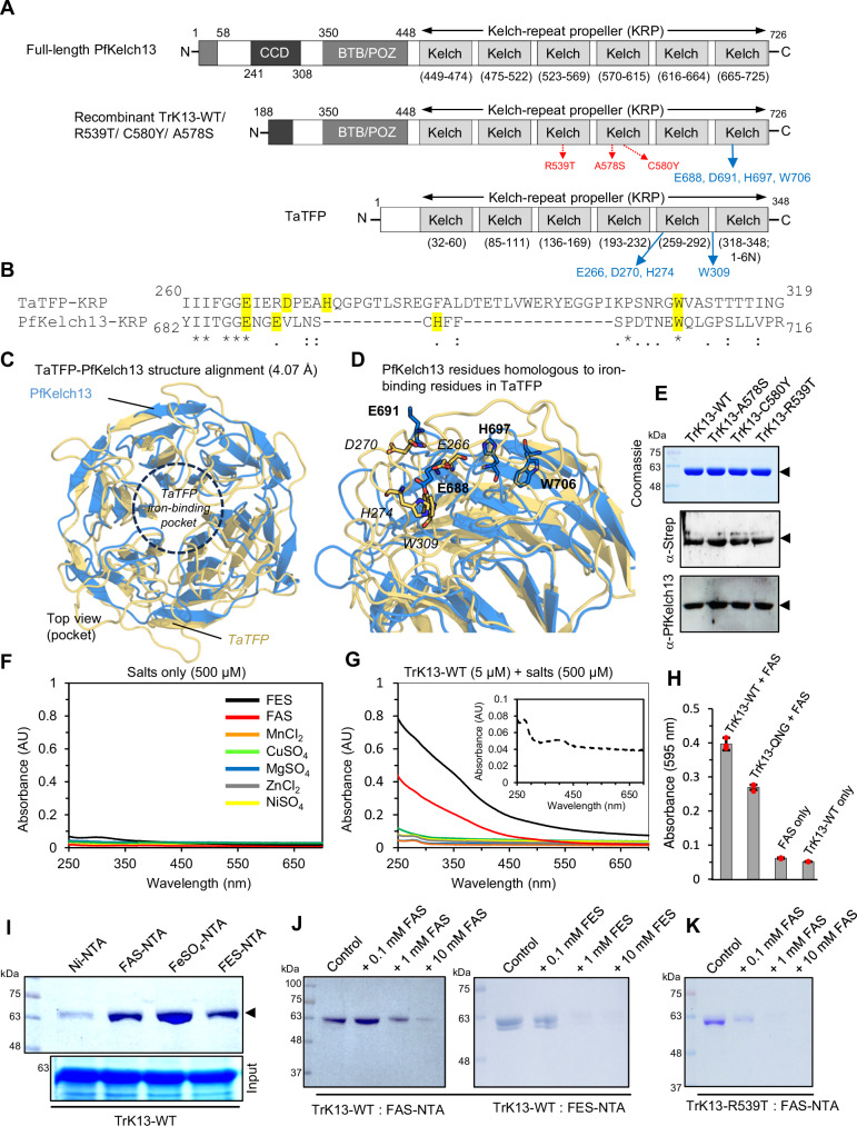
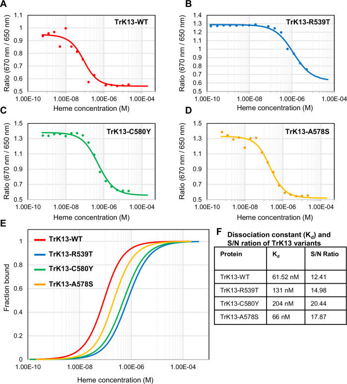
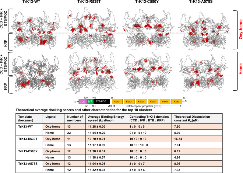
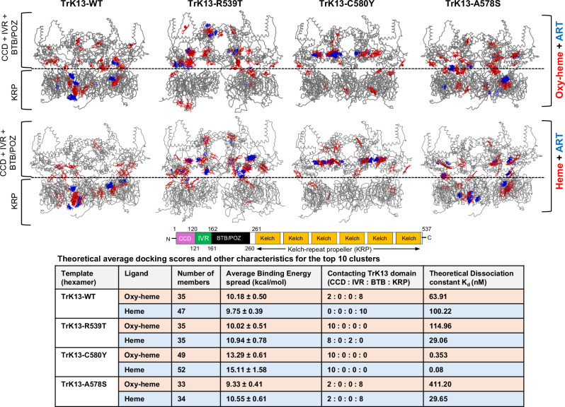
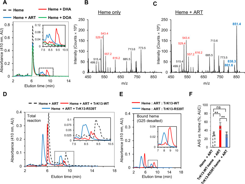
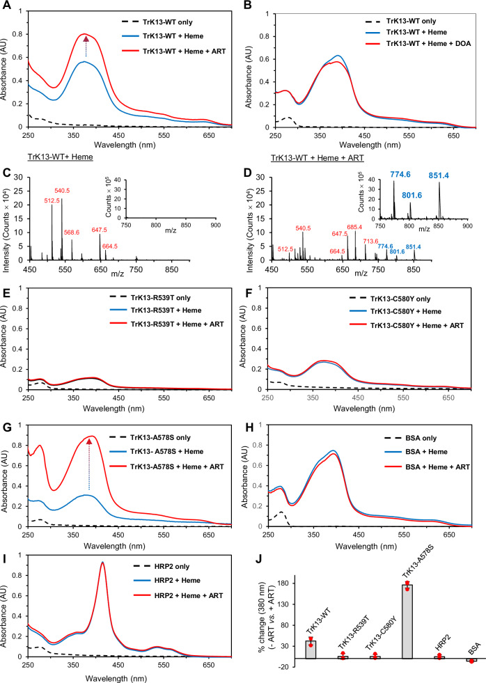
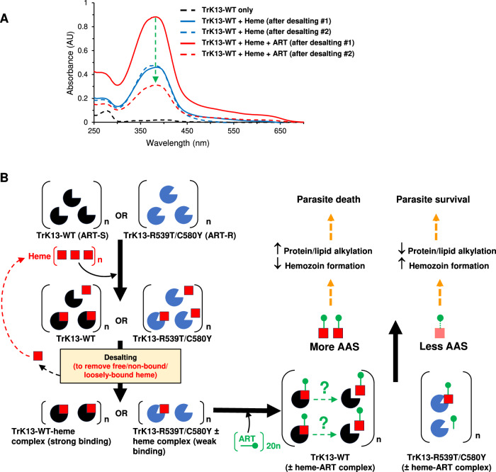
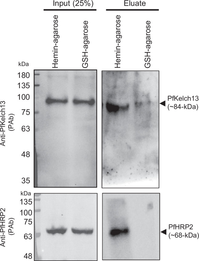
The potency of frontline antimalarial drug artemisinin (ART) derivatives is triggered by heme-induced cleavage of the endoperoxide bond to form reactive heme-ART alkoxy radicals and covalent heme-ART adducts, which are highly toxic to the parasite. ART-resistant (ART-R) parasites with mutations in the Plasmodium falciparum Kelch-containing protein Kelch13 (PfKekch13) exhibit impaired hemoglobin uptake, reduced yield of hemoglobin-derived heme, and thus decreased ART activation. However, any direct involvement of PfKelch13 in heme-mediated ART activation has not been reported. Here, we show that the purified recombinant PfKelch13 wild-type (WT) protein displays measurable binding affinity for iron and heme, the main effectors for ART activation. The heme-binding property is also exhibited by the native PfKelch13 protein from parasite culture. The two ART-R recombinant PfKelch13 mutants (C580Y and R539T) display weaker heme binding affinities compared to the ART-sensitive WT and A578S mutant proteins, which further translates into reduced yield of heme-ART derivatives when ART is incubated with the heme molecules bound to the mutant PfKelch13 proteins. In conclusion, this study provides the first evidence for ART activation via the heme-binding propensity of PfKelch13. This mechanism may contribute to the modulation of ART-R levels in malaria parasites through a novel function of PfKelch13.


Conflict of interest statement

Competing interests The authors declare no competing interests.

Figures

Fig. 1
Fig. 1. Structural and functional similarities in the iron-binding properties between the KRP domains of PfKelch13 and TaTFP.
A Schematic representation of the domain organization in full-length PfKelch13 (top), recombinant TrK13-WT (middle), and TaTFP (bottom), showing the respective spans of the coiled-coil domain (CCD), intervening region (IVR), bric-à-brac; (BTB)/poxvirus and zinc-finger (POZ) domain (BTB/POZ), and the Kelch-repeat propeller (KRP) domain. The three specific mutations in the full-length PfKelch13: R539T, C580Y and A578S are indicated in red text. The iron binding residues in TaTFP and corresponding homologous amino acids in TrK13 are shown in blue text. B Sequence alignment between a part of the KRP domains of TaTFP and PfKelch13. Residues highlighted in yellow represent positional conservation between PfKelch13 and those in TaTFP involved in Fe2+ binding. C–D Structural alignment of the PfKelch13 propeller (blue) and TaTFP (gold) proteins. Protein structures are shown as cartoons. Iron-binding amino acid residues in TaTFP (italic) and homologous sites in PfKelch13 (bold) are shown as sticks (in D). E SDS-PAGE and Western blot profiles showing affinity-purified TrK13 proteins of ~61 kDa (arrowheads) and their specific recognition by antibodies. F–G UV-visible spectroscopy scans showing the increase in absorbance profiles of TrK13-WT only in the presence of FAS and FES, but not with other metal salts (G). The corresponding spectrum of free salts in the absence of any TrK13-WT protein is shown in F. The spectrum profile of TrK13-WT protein without any metal salt is shown as an inset. The graphs shown are best representative of three independent experiments. H. Graph showing the relatively higher binding of the TrK13-WT protein to FAS, as measured by absorbance at 595 nm using the Ferene S assay, compared to the TrK13-QNG mutant protein. Data are shown with their respective standard errors from three independent experiments. I. SDS-PAGE showing higher affinity of TrK13-WT protein (arrowhead) for FAS-, FeSO4- and FES-NTA beads compared to Ni-NTA beads. Input samples are shown below. J-K. Inhibition of TrK13-WT (J) or TrK13-R539T (K) binding to FAS-NTA or FES-NTA beads with stepwise increase of free FAS or FES concentrations. Molecular weight standards (in kDa) for all SDS-PAGE gels are as shown.
Fig. 2
Fig. 2. The recombinant TrK13-WT protein shows specific interactions with heme/FPIX. A.
Native PAGE of TrK13-WT shows a bluish-green TMBZ-positive band (arrow) only in the heme-preincubated sample (right lane) but not in the absence of heme (left lane). Corresponding Coomassie-stained SDS-PAGE of TrK13-WT protein with or without preincubated heme are shown in the left gel. B–C UV-visible spectroscopy scans showing increase in the relative absorbance profiles of G-25 desalted TrK13-WT (red) and positive control BSA (blue), but not negative control lysozyme (green), in the presence of heme (C) compared to their absorbance profiles without heme (B). No soret peak of free heme (black) is detected in C at this concentration (10 μM), indicating effective heme removal by the G25 desalting column. D–E Differential absorption spectra of TrK13-WT protein (10 µM) titrated with heme (0-26 µM) in the wavelength region 250–700 nm (D) and double reciprocal plot of 1/ΔA versus 1/[heme] (E), showing the variation of difference in absorbance of TrK13-WT-heme complex with heme concentrations. The Kd and R2 values are as indicated. F Far-UV circular dichroism (CD) spectra of TrK13-WT protein (10 μM) titrated with heme (0–30 μM) in the wavelength region 200-260 nm. G UV-visible spectroscopy scans showing the increase in the relative absorbance profiles of G-25 desalted TrK13-WT protein (10 μM) in the presence of 10 μM heme/FPIX (solid red line) or its analogs such as Zn-PPIX (solid blue line) or PPIX (solid green line) compared to the control (solid black line). The spectral profiles of the remaining unbound heme variants after the G-25 desalting step are shown as corresponding colored dotted lines and indicate efficient removal as no soret peak was visible. H-I. SDS-PAGE (H) and graph (I) showing the relative abundance of TrK13-WT protein (arrow) pulled down with hemin-agarose beads in the absence or presence of excess free heme, PPIX, or Zn-PPIX. Molecular weight standards (in kDa) for all SDS-PAGE images are as shown.
Fig. 3
Fig. 3. Heme-induced fluorescence quenching of tryptophan residues in the four variants of TrK13 proteins without profound changes in the circular dichroism profile.
A–D Fluorescence spectra of TrK13-WT (A), TrK13-R539T (B), TrK13-C580Y (C), and TrK13-A578S (D) incubated in the absence (dotted lines) or presence of heme at final concentrations between 0.5-5.0 μM heme (gradual light to dark shading in each color) and showing a successive decrease in fluorescence intensity. E Changes in tryptophan fluorescence quenching in the four variants of TrK13 as deduced from A to D. F Plots of log[(F0-F)/F] versus log[heme] for the interaction of TrK13-WT protein and heme deduced from A to D. The Stern-Volmer quenching constants (KSV), association constants (Ka) and the number of binding sites (n) were calculated from the equations F0/F = Ksv [Q] + 1 and log [(F0-F/F] = logKa + nLog[Q], where F0 = fluorescence intensity in the absence of heme, F = fluorescence intensity in the presence of heme, [Q] = concentration of quencher (heme). Curves are the average of three replicates. G Tabular representation of Ksv, Ka and n.
Fig. 4
Fig. 4. Quantitative evaluation of TrK13- heme binding by MST.
A–D Dose-response curves showing negative amplitude of response for TrK13-WT (A), TrK13-R539T (B), TrK13-C580Y (C) and TrK13-A578S (D) titrated against increasing concentrations of heme. MST experiments were performed at medium MST power at 25 °C. LED power was set to 60% (A and D) or 40% (B and C) excitation. E Comparative dose-response curves for the binding interaction between the four TrK13 variants and heme, as indicated. The protein concentration was kept constant at either 50 nM (TrK13-WT or TrK13-A578S) or 25 nM (TrK13-R539T or TrK13-C580Y), while the ligand heme concentration varied from 0-5 µM. F Table showing the decreasing affinity scale: TrK13-WT > TrK13-A578S > TrK13-R539T > TrK13-C580Y and signal-to-noise (S/N) ratio.
Fig. 5
Fig. 5. Docking clusters of oxygenated (oxy-heme) and deoxygenated heme in the hexameric assemblies of the TrK13-WT and mutant structures.
SAXS-derived hexameric assemblies (grey ribbon; derived from https://www.sasbdb.org) of the ART-sensitive (TrK13-WT and TrK13-A578S), and ART-resistant (TrK13-R539T and TrK13-C580Y) variants showing the 38 docked oxy-heme (top panel) and heme (bottom panel) clusters (red). Dotted lines indicate separation between the KRP/β-propeller domain and the CCD + IVR + BTB/POZ domains in the TrK13 structures. Theoretical average theoretical docking scores and other characteristics for the top 10 clusters are shown in the table below. The CCD (purple), IVR (green), BTB/POZ (black) and the KRP domain with six β-propeller regions (orange) are colored accordingly and shown as a schematic above the table. In cases where the oxy-/heme cluster contacted interface between two domains, numbers were assigned to both the domains of TrK13.
Fig. 6
Fig. 6. Docking clusters of ART in the pre-docked oxy-/heme assemblies of TrK13-WT and mutant structures.
Oxy-heme (red; top panel) and heme (red; bottom panel) clusters (red) complexes with the SAXS-derived hexameric assemblies (grey ribbon; derived from https://www.sasbdb.org) of the ART-sensitive (TrK13-WT and TrK13-A578S), and ART-resistant (TrK13-R539T and TrK13-C580Y) variants showing the 38 docked ART clusters (blue). Dotted lines indicate separation between the KRP/β-propeller domain and the CCD + IVR + BTB/POZ domains in the TrK13 structures. Theoretical average docking scores and other characteristics for the top 10 clusters are shown in the table below. The CCD (purple), IVR (green), BTB/POZ (black) and the KRP domain with six β-propeller regions (orange) are colored accordingly and shown as a schematic above the table. In cases where the ART cluster contacted interface between two domains, numbers were assigned to both the domains of TrK13.
Fig. 7
Fig. 7. Detection of AAS by HPLC and mass spectrometry.
A HPLC elution profiles at 410 nm for the total reaction mixtures of heme and the corresponding peaks for free heme (tall peaks at 6–7 min) and AAS (smaller peaks at 8–10 min elution time; boxed and magnified in the inset) in the presence of either ART (blue) or DHA (red) or the inactive DOA analog (green). The elution profile of free heme is indicated by the dotted black line. B MS data of the eluted samples from A (dotted box) showing the characteristic heme intensity peaks (red text) at m/z 529.4, 543.4, 616.2 and 557.2 and several other peaks generated by in-source fragmentation of heme and oxidation-reduction processes during electrospray ionization (black text). C Mass spectra of the two-component mixture of heme-ART from the eluted samples of A (dotted box), showing additional AAS peaks at m/z 801.6, 838.3, and 851.4 (blue text). Peaks at these m/z were absent in the heme alone sample shown in B. D–E HPLC elution profiles showing the heme (tall peaks at 5–7 min elution time) and AAS peaks (short peaks at 8–10 min elution time) in TrK13-WT (red) or TrK13-R539T (blue) containing reaction mixtures with heme (D) or in the protein-bound heme (after G-25 desalting; E). The AAS peaks are framed by dotted lines and magnified in the inset. F Percentage of AAS versus heme, as measured by area under the curve (AUC) for the respective sample peaks and expressed as a percentage. Data are mean ± SD of three independent experiments, comparison was performed by unpaired t-test, * p < 0.05, ** p < 0.005, ns not significant (p > 0.05).
Fig. 8
Fig. 8. Reduced AAS formation in ART-R TrK13 mutants compared to ART-S variants. A, E-G.
UV-visible spectroscopy scans showing further hyperchromic shift (red lines; increase indicated by blue to red gradient dotted arrow) in the absorbance profiles at 380 nm in the heme-bound ART-S TrK13-WT (A) and TrK13-A578S (G) proteins, but not in the ART-R TrK13-R539T (E) and TrK13-C580Y (F) proteins, when incubated in the presence of ART for 30 min. The Soret peaks at 380 nm (red lines) and absorption profiles of the respective proteins in the absence of heme (dotted black lines) are as indicated. B UV-visible scan showing no hyperchromic shift on the Soret peak of heme-bound TrK13-WT in the presence of the inactive ART analog DOA. C–D MS data of the heme-bound TrK13-WT samples (after G-25 desalting) incubated in the absence (C) or presence (D) of ART. While the peaks corresponding to heme (in red text) were visible in both samples, the peaks at m/z 801.6 and 851.4 (blue text) were only visible in the presence of ART (D) and are indicative of AAS. All samples were treated as described earlier13. The inset shows the expanded m/z region between 750 and 900. H–I UV-visible spectroscopy scan showing no AAS formation by the heme-binding proteins BSA and HRP2. J Graphical representation of the percentage increase in the Soret peak at 380 nm for the data in A, B, E–I Error bars are from three independent experimental replicates.
Fig. 9
Fig. 9. Heme bound to TrK13-WT protein is released after AAS formation.
A UV-visible spectroscopy scans showing the heme absorbance peak at 378 nm due to the TrK13-WT-bound heme (solid blue line) after desalting step #1 and its hyperchromic shift upon addition of ART (solid red line). The scan profile of TrK13-WT without heme is shown as a dotted black line. After desalting step #2, a steep decrease (green dotted arrow) of the heme absorbance maximum peak at 380 nm is observed in the ART-treated sample (dotted red line), indicating the release of AAS from the TrK13-WT protein and its removal by the second desalting step. The resultant peak at 380 nm could be due to residual bound heme that did not form AAS. No decrease in λmax at 380 nm is observed in the TrK13-WT-heme-bound sample processed similarly in the absence of ART (dotted blue line), thus excluding the possibility of random heme release during the incubation period. B Schematic representation of the molecules at various stages of the experiment. Recombinant ART-S (TrK13-WT; black pac-man) or ART-R (TrK13-R539T or –C580Y; blue pac-man) proteins with higher or lower heme-binding affinity (solid red square), respectively were desalted (desalting step #1) to remove the excess free heme or loosely-bound heme. Following incubation with 20-times excess ART (green ball pin), desalting step #2 was performed to remove any AAS released from its TrK13-bound state (green question mark). The orange dotted arrow symbolizes the proposed role of liberated AAS in alkylating parasite protein targets or inhibiting hemozoin formation during the intra-erythrocytic stages of infection. Reaction mixtures at each step were analyzed by UV-visible spectrophotometry spanning 200–700 nm wavelengths.
Fig. 10
Fig. 10. Pulldown of the native PfKelch13 protein from P. falciparum total protein extracts using hemin-agarose beads.
3D7 parasites were isolated by saponin treatment from asynchronized culture and proteins were solubilized with RIPA buffer. Protein extracts were incubated with hemin-agarose or glutathione (GSH)-agarose beads. After extensive washing, bound proteins were eluted with Laemmli buffer, separated by SDS-PAGE, and blotted against PfKelch13 antibodies. PfKelch13 antibodies were custom-generated using a PfKelch13 peptide. Molecular weight standards (in kDa) are as indicated.

References

    1. WHO. World Health Organization. https://www.who.int. (2022).
    1. Meshnick, S. R. Artemisinin: Mechanisms of action, resistance and toxicity. Int. J. Parasitol.32, 1655–1660 (2002). - PubMed
    1. Robert, A., Coppel, Y. & Meunier, B. Alkylation of heme by the antimalarial drug artemisinin. Chem. Commun.5, 414–415 (2002). - PubMed
    1. Laurent, S. A.-L., Robert, A. & Meunier, B. C10-Modified Artemisinin Derivatives: Efficient Heme-Alkylating Agents. Angew. Chem.117, 2060–2063 (2005). - PubMed
    1. Ismail, H. M. et al. Artemisinin activity-based probes identify multiple molecular targets within the asexual stage of the malaria parasites Plasmodium falciparum 3D7. Proc. Natl Acad. Sci. Usa.113, 2080–2085 (2016). - PMC - PubMed

LinkOut - more resources