Biomechanical impact of progressive meniscal extrusion on the knee joint: a finite element analysis
- PMID: 39538324
- PMCID: PMC11562606
- DOI: 10.1186/s13018-024-05249-y
Biomechanical impact of progressive meniscal extrusion on the knee joint: a finite element analysis
Abstract
Background: While measuring meniscal extrusion quantitatively is an early risk factor for knee osteoarthritis (KOA), the biomechanics involved in this process are not well understood. This study aimed to investigate the effects of varying degrees of medial and lateral meniscal extrusion and their material softening on knee osteoarthritis progression.
Methods: Finite element analysis (FEA) was utilized to simulate varying degrees of meniscal extrusion (1-5 mm) in 72 knee joint models, representing progressive meniscal degeneration and material softening due to injury. Changes in von Mises stress of the cartilage and menisci and the load distribution on the tibial plateau's meniscus and cartilage were studied under balanced standing posture in both healthy and injured knees, and statistical analysis was performed using Spearman correlation.
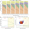
Results: Compared to healthy knees, peak stress in medial compartment tissues increased by over 40% with 4 mm of medial meniscus extrusion, and in lateral compartment tissues with 2 mm of lateral meniscus extrusion. Meniscus extrusion reduced the contact load between the meniscus and femoral cartilage but increased it between the tibial and femoral cartilages, with a maximum increase up to fivefold. Spearman correlation analysis indicated that meniscal extrusion significantly affected peak stress and contact loads in the respective knee compartment (p < 0.001), with a lesser impact on the opposite compartment. Notably, medial meniscal extrusion also significantly increased peak stress in the lateral tibial cartilage (p < 0.05).
Conclusions: The quantitative analysis revealed that meniscal extrusion significantly affected the biomechanics of soft tissues within the same compartment, with limited impact on the opposite side. Specifically, Medial extrusion beyond 4 mm significantly affected the biomechanics of the medial compartment, while lateral extrusion over 2 mm had a similar impact on the lateral compartment. Meniscal softening, without altering joint contact characteristics, primarily affected the biomechanics of the meniscus itself, with minimal impact on other soft tissues.
Keywords: Correlation analysis; Finite element analysis; Load distribution; Meniscal extrusion; Von-Mises stress.
© 2024. The Author(s).
Conflict of interest statement
Figures


Similar articles
-
[Biomechanical study of knee joint based on coronal plane alignment of the knee].Zhongguo Xiu Fu Chong Jian Wai Ke Za Zhi. 2024 Dec 15;38(12):1466-1473. doi: 10.7507/1002-1892.202408048. Zhongguo Xiu Fu Chong Jian Wai Ke Za Zhi. 2024. PMID: 39694836 Free PMC article. Chinese.
-
[Finite element analysis of impact of bone mass and volume in low-density zone beneath tibial plateau on cartilage and meniscus in knee joint].Zhongguo Xiu Fu Chong Jian Wai Ke Za Zhi. 2025 Mar 15;39(3):296-306. doi: 10.7507/1002-1892.202412018. Zhongguo Xiu Fu Chong Jian Wai Ke Za Zhi. 2025. PMID: 40101904 Free PMC article. Chinese.
-
Utilization of Transtibial Centralization Suture Best Minimizes Extrusion and Restores Tibiofemoral Contact Mechanics for Anatomic Medial Meniscal Root Repairs in a Cadaveric Model.Am J Sports Med. 2019 Jun;47(7):1591-1600. doi: 10.1177/0363546519844250. Epub 2019 May 15. Am J Sports Med. 2019. PMID: 31091129
-
Meniscus Centralization and Its Effects on Meniscal Extrusion and Knee Biomechanics: A Systematic Review.Sports Med Arthrosc Rev. 2025 Jun 1;33(2):61-68. doi: 10.1097/JSA.0000000000000421. Epub 2025 Jan 15. Sports Med Arthrosc Rev. 2025. PMID: 40424168
-
Medial meniscal extrusion: Detection, evaluation and clinical implications.Eur J Radiol. 2018 May;102:115-124. doi: 10.1016/j.ejrad.2018.03.007. Epub 2018 Mar 6. Eur J Radiol. 2018. PMID: 29685524 Review.
Cited by
-
Compliant buffer layer achieves native-like cartilage stress in talar prosthesis: a finite element analysis study.J Orthop Surg Res. 2025 Jul 8;20(1):620. doi: 10.1186/s13018-025-05996-6. J Orthop Surg Res. 2025. PMID: 40629372 Free PMC article.
-
A Geometric Deep Learning Model for Real-Time Prediction of Knee Joint Biomechanics Under Meniscal Extrusion.Ann Biomed Eng. 2025 Jul 15. doi: 10.1007/s10439-025-03798-9. Online ahead of print. Ann Biomed Eng. 2025. PMID: 40663282 Review.
-
Influence of preoperative medial meniscus extrusion and subchondral bone marrow edema on outcomes after medial opening wedge high tibial osteotomy.J Orthop Surg Res. 2025 Apr 9;20(1):353. doi: 10.1186/s13018-025-05656-9. J Orthop Surg Res. 2025. PMID: 40205393 Free PMC article.
References
-
- Xu D, van der Voet J, Waarsing JH, Oei EH, Klein S, Englund M, Zhang F, Bierma-Zeinstra S, Runhaar J. Are changes in meniscus volume and extrusion associated to knee osteoarthritis development? A structural equation model. Osteoarthritis Cartilage. 2021;29(10):1426–31. - PubMed
-
- Krych AJ, Bernard CD, Leland DP, Camp CL, Johnson AC, Finnoff JT, Stuart MJ. Isolated meniscus extrusion associated with meniscotibial ligament abnormality. Knee Surg Sports Traumatol Arthrosc. 2020;28(11):3599–605. - PubMed
-
- Swamy N, Wadhwa V, Bajaj G, Chhabra A, Pandey T. Medial meniscal extrusion: detection, evaluation and clinical implications. Eur J Radiol. 2018;102:115–24. - PubMed
MeSH terms
Grants and funding
LinkOut - more resources
Full Text Sources

